This website uses cookies to ensure you get the best experience on our website.


- Table of Contents
Plan TMEM107 paraffin-section IHC using the cytoplasmic staining reported across most tissues (HPA tissue IHC). Adrenal glandular cells show medium staining, while adipocytes have no detected staining and can serve as a comparison (HPA tissue IHC).

Expected localisation, validated protocols, controls and antibodies — the at-a-glance facts below, then the full design guide.
| Expected localisation | Cytoplasmic in tissue IHC (HPA tissue IHC); ciliary transition zone at the molecular level (UniProt) | |
| Staining pattern | Cytoplasmic staining in most tissues (HPA tissue IHC) | |
| Antigen retrieval | Citrate pH 6 HIER, heat-mediated (datasheet A04966) | |
| Positive control | Adrenal gland+4 more · see all | |
| Negative control | Adipose tissue+3 more · see all |
| Fixation | Keep fixation consistent across paraffin sections. (standard IHC practice; not target-specific) | |
| Caveat | Tissue staining awaits external verification (HPA tissue IHC) | |
| Regulation | Expression regulation is not annotated (UniProt) | |
| Isoform / epitope | 4 isoforms; check epitope coverage and loop orientation (UniProt) |
The catalog antibody’s IHC-P protocol (datasheet A04966) is accompanied by one published TMEM107 tissue staining protocol (PMC7779196).
| Sample | Paraffin-embedded human intestinal cancer tissues; fixative not specified (datasheet A04966) |
| Fixation | Image fixative and duration unreported (datasheet A04966); verify before use. |
| Sectioning | 4–5 µm sections on charged slides (standard) |
| Deparaffinisation | Xylene, graded ethanol series to water (standard) |
| Antigen retrieval | Heat retrieval: Citrate pH 6, 20 min (datasheet A04966) |
| Peroxidase block | 3% H2O2, 10 min, room temperature (standard) |
| Blocking | 10% goat serum (datasheet A04966) |
| Primary antibody | Rabbit anti-TMEM107, 0.5-1μg/ml (datasheet A04966) |
| Primary incubation | Overnight at 4 °C (datasheet A04966) |
| Detection | Streptavidin-biotin complex (SABC), DAB chromogen (datasheet A04966) |
| Counterstain | Hematoxylin, blue, dehydrate and mount (standard) |
| Expected result | TMEM107-positive staining in glandular cells of adrenal gland (HPA tissue IHC: Medium). HPA tissue profile: Cytoplasmic expression in most tissues. No signal in the no-primary control. |
TMEM107 is a four-pass membrane protein at the ciliary transition zone (UniProt Q6UX40: topology and location). In paraffin-section IHC, HPA reports cytoplasmic staining in most tissues, including medium staining in several glandular, neuronal and endothelial cell populations (HPA tissue IHC). Treat that slide-level pattern as the practical expectation; the HPA tissue profile is Approved, with external verification pending (HPA tissue IHC: reliability).
| Cytoplasmic staining in glandular cells of breast, appendix or endometrium, or neurons of cerebral cortex. | These named populations have medium staining in HPA tissue IHC. Compare the signal with neighboring structures and the negative control; HPA's Approved rating still has external verification pending (HPA tissue IHC: cell levels and reliability). |
| A sharply nuclear or exclusively extracellular signal, without the expected cytoplasmic pattern. | That compartment disagrees with HPA's cytoplasmic tissue pattern and UniProt's membrane and ciliary location. Review morphology and detection background before calling it TMEM107 (HPA tissue IHC: profile; UniProt Q6UX40: location). |
| Strong staining in adipocytes, cholangiocytes, smooth muscle cells or spleen red-pulp cells. | HPA lists those specific cell populations as not detected. Unexpected staining warrants checks for cross-reactivity or endogenous detection activity; their surrounding tissues are not whole-section negative controls (HPA tissue IHC: not-detected cells; general IHC practice). |
| Diffuse color across many cell types, stroma and blank areas, with weak cellular contrast. | The distribution is poorly aligned with HPA's cell-level observations. Inspect the no-primary control and reagent background before interpreting intensity; diffuse color alone cannot establish TMEM107 expression (HPA tissue IHC: profile; general IHC practice). |
| No detectable staining in an otherwise well-preserved breast glandular or cerebral-cortex neuronal sample. | HPA records medium staining in those cells, so examine the positive control and detection workflow. A single absent signal cannot establish biological absence, especially while external verification remains pending (HPA tissue IHC: cell levels and reliability; general IHC practice). |
| Slide-level localization | HPA describes cytoplasmic IHC in most tissues, while UniProt places TMEM107 at the ciliary transition zone. Routine chromogenic IHC may show cellular staining without resolving that small structure; do not require visible ciliary puncta to score the HPA pattern (HPA tissue IHC: profile; UniProt Q6UX40: location; general IHC practice). |
| Membrane topology and unknown antibody epitope | UniProt annotates four transmembrane segments at residues 7–27, 53–73, 83–103 and 113–133. The payload gives no antibody epitope, so topology cannot identify which part the IHC antibody recognizes or predict a retrieval condition (UniProt Q6UX40: topology). |
| Isoforms and glycosylation | UniProt lists four isoforms and one glycosylation site at residue 79. Their effects on this antibody's staining are unreported here; do not attribute a cell-specific difference or absent signal to either feature without separate evidence (UniProt Q6UX40: isoforms and glycosylation). |
| Cell-specific reference levels | HPA reports medium staining in named glandular, neuronal and endothelial populations, but low staining in bronchial respiratory epithelium and kidney tubules. Select a medium-staining cell population when assessing whether a weak run reflects the assay (HPA tissue IHC: positive and low cell levels). |
| Validation and RNA evidence | The listed antibody HPA052555 has Approved IHC status, while the tissue profile awaits external verification. HPA's choroid-plexus RNA enhancement is transcript evidence; this payload supplies no matching choroid-plexus protein-staining level (HPA antibodies: IHC status; HPA tissue IHC: reliability and RNA specificity). |
| Situation | Likely cause | Next action |
|---|---|---|
| The intended positive cells have no signal. | The detection run or staining conditions may be inadequate; HPA reports medium staining in breast glandular and cerebral-cortex neuronal cells (HPA tissue IHC: positive cells; general IHC practice). | Check a documented positive tissue section and detection controls, then review the chosen IHC antibody dilution and retrieval procedure. No TMEM107-specific retrieval setting or dilution is supplied here (general IHC practice). |
| The entire section has diffuse chromogen. | Nonspecific reagent binding or detection-system activity may obscure cell-level contrast (general IHC practice). | Inspect the no-primary control, blocking step and detection reagents; interpret only cellular signal that separates from background (general IHC practice). |
| Nuclei dominate the stain. | A nuclear-dominant pattern conflicts with the reported cytoplasmic IHC profile and membrane/ciliary annotation (HPA tissue IHC: profile; UniProt Q6UX40: location). | Review the counterstain and no-primary control, then reassess localization in an HPA medium-staining cell population (HPA tissue IHC: positive cells; general IHC practice). |
| An HPA not-detected cell population stains strongly. | Cross-reactivity or endogenous detection activity is possible; HPA reports no staining in adipocytes and smooth muscle cells, among other named populations (HPA tissue IHC: not-detected cells; general IHC practice). | Compare cell identity and the no-primary control; assess whether the signal follows tissue morphology or background before assigning it to TMEM107 (general IHC practice). |
| A low-staining sample looks negative. | HPA reports low staining in bronchial respiratory epithelial cells and kidney tubular cells; a faint result is harder to distinguish from background (HPA tissue IHC: low cell levels; general IHC practice). | Judge that sample alongside a medium-staining reference and matched negative control rather than treating one faint section as proof of absence (HPA tissue IHC: cell levels; general IHC practice). |
| IF shows no clear ciliary focus despite acceptable IHC. | UniProt assigns TMEM107 to the ciliary transition zone, but HPA supplies no ICC-IF image or established main subcellular location for comparison (UniProt Q6UX40: location; HPA subcellular: image and location fields). | Keep the IHC interpretation tied to HPA's tissue pattern; evaluate IF localization in its separate guide with appropriate imaging and controls (HPA tissue IHC: profile; HPA subcellular: no ICC-IF images; general IF practice). |
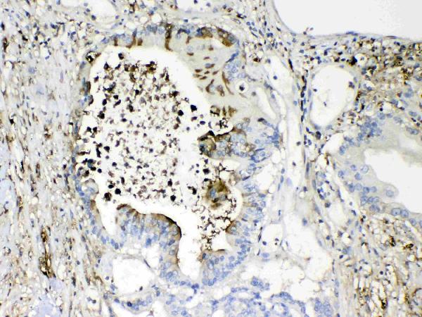
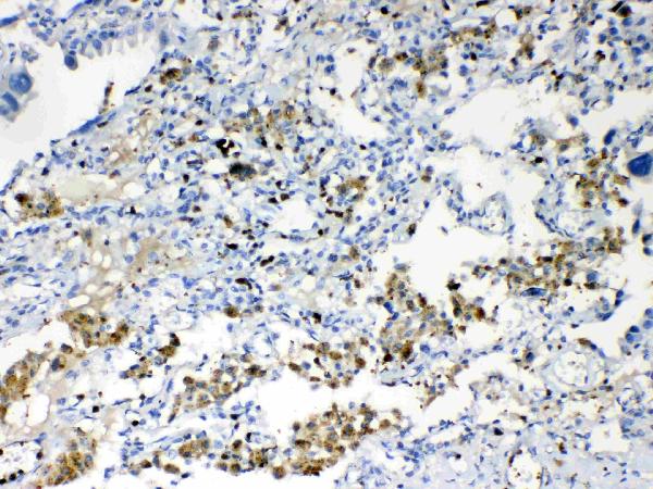
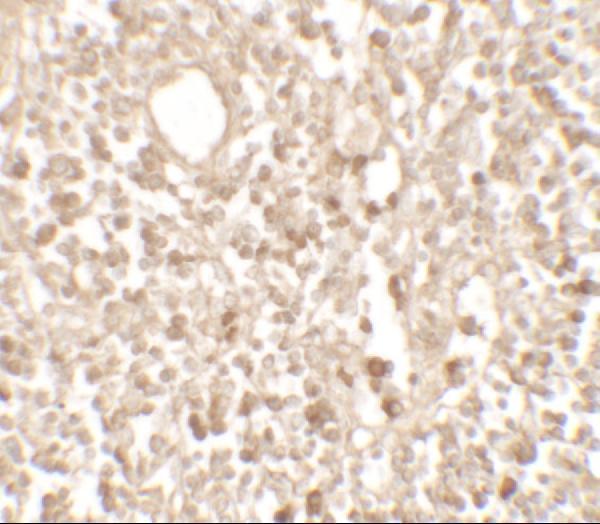
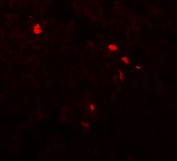


Comprehensive Human Protein Atlas IHC scoring per tissue (reliability: Approved — Pending external verification.). Rows are taken directly from the HPA tissue chart — click any row's HPA link to view the source.
| Tissue | Cell type | Level | Evidence | Source |
|---|---|---|---|---|
| Adrenal gland | Glandular cells | Medium | Protein (IHC) | HPA → |
| Appendix | Glandular cells | Medium | Protein (IHC) | HPA → |
| Breast | Glandular cells | Medium | Protein (IHC) | HPA → |
| Caudate | Neuronal cells | Medium | Protein (IHC) | HPA → |
| Cerebral cortex | Neuronal cells | Medium | Protein (IHC) | HPA → |
| Tissue | Cell type | Level | Evidence | Source |
|---|---|---|---|---|
| Adipose tissue | Adipocytes | Not detected | Protein (IHC) | HPA → |
| Liver | Cholangiocytes | Not detected | Protein (IHC) | HPA → |
| Smooth muscle | Smooth muscle cells | Not detected | Protein (IHC) | HPA → |
| Spleen | Cells in red pulp | Not detected | Protein (IHC) | HPA → |
Troubleshoot TMEM107 staining in paraffin sections by checking retrieval, compartment, controls and scoring before interpreting chromogenic signal.
Two human-reactive anti-TMEM107 antibodies have IHC tissue images; one also has an IF tissue image (catalog applications and image captions).
A04966 has IHC images from paraffin sections of human intestinal and lung cancer tissue (A04966 IHC captions). A04966-1 has IHC and IF images from human lymph node tissue (A04966-1 image captions).
Which to pick: For paraffin-section IHC, choose A04966 when a documented starting concentration is useful: its captions report 1 μg/mL in paraffin sections, while the fixative is unreported (A04966 IHC captions). For IF, choose A04966-1; its application list includes IF and its human lymph node IF caption reports 20 μg/mL, but ICC validation is unreported (A04966-1 catalog applications and IF caption). Neither SKU has documented cross-species reactivity; both list human only, and A04966 is identified as polyclonal while A04966-1 has no clonality stated (catalog reactivity and antibody details).