This website uses cookies to ensure you get the best experience on our website.


- Table of Contents
Plan chromogenic IHC-P for TMEM18 using the IHC-validated antibody (datasheet: IHC-P). Compare observed cytoplasmic tissue staining (HPA tissue IHC) with the nuclear membrane location annotated for the protein (UniProt), and account for medium consistency between staining and RNA data (HPA tissue IHC).

Expected localisation, validated protocols, controls and antibodies — the at-a-glance facts below, then the full design guide.
| Expected localisation | Cytoplasmic staining (HPA tissue IHC); nuclear membrane expected (UniProt) | |
| Staining pattern | Cytoplasmic staining, including glandular cells (HPA tissue IHC) | |
| Antigen retrieval | Tris-EDTA pH 9.0 HIER, 95–98 °C, 20 min (rule: nuclear antigen) | |
| Positive control | Epididymis+4 more · see all | |
| Negative control | Adipose tissue+4 more · see all |
| Fixation | Keep fixation consistent across sections (standard IHC practice; not target-specific) | |
| Caveat | Medium staining–RNA consistency; verify signal (HPA tissue IHC) | |
| Regulation | No staining-linked regulation annotated (UniProt) | |
| Isoform / epitope | 2 isoforms; epitope coverage is unknown (UniProt) |
Compare the catalog antibody’s IHC-P protocol (datasheet) with the published FFPE prostate carcinoma tissue microarray protocol (PMC12203752).
| Sample | Tissue sections; selected-image fixative not specified (standard IHC workflow) |
| Fixation | Image fixative and duration unreported (datasheet A05347); verify before use. |
| Sectioning | 4–5 µm sections on charged slides (standard) |
| Deparaffinisation | Xylene, graded ethanol series to water (standard) |
| Antigen retrieval | Heat-induced epitope retrieval in Tris-EDTA buffer, pH 9.0, 20 min at 95–98 °C (standard rule: nuclear antigen) |
| Peroxidase block | 3% H2O2, 10 min, room temperature (standard) |
| Blocking | 10% normal serum of the secondary host, 30 min, room temperature (standard) |
| Primary antibody | Rabbit anti-TMEM18, 2.5 μg/mL (datasheet A05347) |
| Primary incubation | Overnight at 4 °C (standard) |
| Detection | HRP-polymer secondary, DAB chromogen 5–10 min (standard) |
| Counterstain | Hematoxylin, blue, dehydrate and mount (standard) |
| Expected result | TMEM18-positive staining in glandular cells of epididymis (HPA tissue IHC: High). HPA tissue profile: Cytoplasmic expression in most tissues. No signal in the no-primary control. |
For paraffin-section IHC, expect predominantly cytoplasmic staining in many tissues, with high staining reported in selected glandular and trophoblastic cells (HPA tissue IHC). UniProt also places TMEM18 at the nuclear membrane and describes 3 transmembrane segments (UniProt Q96B42 topology). Treat the tissue pattern as a guide: HPA rates its IHC reliability Approved, with medium agreement between staining and RNA data and external verification pending (HPA tissue IHC).
| Clear cytoplasmic stain in pancreatic exocrine glandular cells, with little diffuse background. | This fits a reported high-staining cell population and the predominant tissue-IHC compartment (HPA tissue IHC). Compare cell-associated signal with a no-primary control before scoring; chromogen across empty spaces does not establish TMEM18 staining (general IHC practice). |
| A nuclear rim, isolated nuclear stain, or another compartment dominates while cytoplasmic staining is weak. | A nuclear rim is plausible from the annotated nuclear-membrane location, but it is not the predominant reported tissue-IHC pattern (UniProt Q96B42 subcellular location; HPA tissue IHC). Isolated nuclear stain should prompt a control and localization review before interpretation (general IHC practice). |
| Strong stain appears mainly in adipocytes, skeletal myocytes, or esophageal squamous cells. | These cell populations are listed as Not detected, so the result conflicts with the HPA reference pattern (HPA tissue IHC). Check cell identification and controls; cross-reactivity or endogenous detection activity are possible explanations, not established causes (general IHC practice). |
| Uniform haze covers multiple cell types, stroma, or tissue-free regions. | The expected reference is cell-associated cytoplasmic staining, not an indiscriminate field-wide signal (HPA tissue IHC; general IHC practice). Review the no-primary control and detection background before assigning positive cells (general IHC practice). |
| No convincing signal appears in an otherwise interpretable pancreatic exocrine compartment. | Pancreatic exocrine glandular cells are reported High, making them a useful comparator, though one negative section cannot prove antibody failure (HPA tissue IHC). Check tissue identity, controls, reagent performance, and staining conditions before calling the sample negative (general IHC practice). |
| Tissue and cell choice | HPA reports High staining in epididymal, salivary, seminal-vesicle, and stomach glandular cells; pancreatic exocrine glandular cells; and placental trophoblastic cells (HPA tissue IHC). Appendix and breast glandular cells are Medium. Use the named cell population, not the entire organ, when judging a control (HPA tissue IHC; general IHC practice). |
| Reference reliability | The tissue-IHC profile is Approved but has medium agreement with RNA expression and awaits external verification (HPA tissue IHC). Low tissue RNA specificity does not mean every cell should stain equally; interpret a local result against its named HPA cell population and appropriate controls (HPA tissue IHC; general IHC practice). |
| Membrane topology and compartment | UniProt annotates nuclear membrane and cytoplasm, with 3 transmembrane segments and regions facing the nucleoplasm or perinuclear space (UniProt Q96B42 topology). These annotations support considering a nuclear-rim pattern, but do not establish the catalog antibody’s epitope, antigen-retrieval needs, or a fixation effect (UniProt Q96B42 topology). |
| Isoforms and processing | UniProt lists 2 isoforms, a single chain spanning residues 1–140, and no annotated signal peptide, propeptide, glycosylation site, or modified residue (UniProt Q96B42). Without an epitope map, these entries cannot establish which isoforms an antibody detects or explain a particular section’s staining intensity (UniProt Q96B42). |
| Q: Should IF/ICC show the same compartment as tissue IHC? | A: HPA’s ICC-IF summary reports mainly mitochondria, with additional endoplasmic-reticulum localization, and rates those locations Approved; its tissue IHC instead reports predominantly cytoplasmic expression (HPA subcellular ICC-IF; HPA tissue IHC). Interpret each application against its own evidence and controls; the discrepancy remains unresolved by these records. |
| Situation | Likely cause | Next action |
|---|---|---|
| A reported High tissue has no visible chromogen in the named cell population. | The result diverges from HPA’s reference; tissue identity, reagent failure, or staining conditions could account for it (HPA tissue IHC; general IHC practice). | Confirm the named cell type on the section, inspect a positive control, and review the recorded IHC staining steps before changing one condition at a time (general IHC practice). |
| The whole section has diffuse brown haze. | Nonspecific detection or residual endogenous activity can obscure cell-associated staining (general IHC practice). | Compare a no-primary section, check blocking and detection controls, then score only signal distinguishable from that background (general IHC practice). |
| Nuclear stain dominates, without a convincing cytoplasmic pattern. | UniProt allows nuclear-membrane localization, but HPA tissue IHC describes predominantly cytoplasmic expression; the observed compartment needs verification (UniProt Q96B42; HPA tissue IHC). | Inspect whether staining follows the nuclear rim or fills nuclei, and compare controls and the HPA tissue pattern before assigning localization (general IHC practice; HPA tissue IHC). |
| Adipocytes or skeletal myocytes stain strongly. | Both are listed as Not detected in HPA tissue IHC; cross-reactivity or detection background is possible but unproven (HPA tissue IHC; general IHC practice). | Verify the cell type, inspect a no-primary control, and compare an HPA-reported High population in the same run (HPA tissue IHC; general IHC practice). |
| A weak signal appears in cerebral-cortex glial cells or bronchial ciliated-cell bodies. | HPA reports these populations at Low, so weak cell-associated staining may fit the reference (HPA tissue IHC). | Compare with controls and a reported High population; avoid treating Low as either a required strong positive or a definitive negative (HPA tissue IHC; general IHC practice). |
| An IF/ICC image seems inconsistent with the tissue-IHC call. | HPA reports mainly mitochondrial and additional endoplasmic-reticulum localization in ICC-IF, while tissue IHC is described as predominantly cytoplasmic (HPA subcellular ICC-IF; HPA tissue IHC). | Record the application and compartment separately, then judge each against its application-specific HPA description and controls (HPA subcellular ICC-IF; HPA tissue IHC; general IHC practice). |
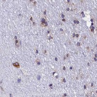

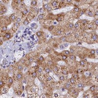
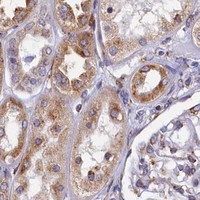


Comprehensive Human Protein Atlas IHC scoring per tissue (reliability: Approved — Medium consistency between antibody staining and RNA expression data. Pending external verification.). Rows are taken directly from the HPA tissue chart — click any row's HPA link to view the source.
| Tissue | Cell type | Level | Evidence | Source |
|---|---|---|---|---|
| Epididymis | Glandular cells | High | Protein (IHC) | HPA → |
| Pancreas | Exocrine glandular cells | High | Protein (IHC) | HPA → |
| Placenta | Trophoblastic cells | High | Protein (IHC) | HPA → |
| Salivary gland | Glandular cells | High | Protein (IHC) | HPA → |
| Seminal vesicle | Glandular cells | High | Protein (IHC) | HPA → |
| Tissue | Cell type | Level | Evidence | Source |
|---|---|---|---|---|
| Adipose tissue | Adipocytes | Not detected | Protein (IHC) | HPA → |
| Adrenal gland | Glandular cells | Not detected | Protein (IHC) | HPA → |
| Esophagus | Squamous epithelial cells | Not detected | Protein (IHC) | HPA → |
| Hippocampus | Glial cells | Not detected | Protein (IHC) | HPA → |
| Oral mucosa | Squamous epithelial cells | Not detected | Protein (IHC) | HPA → |
Use compartment and cell type controls when interpreting TMEM18 staining: tissue and subcellular observations differ across sources (UniProt Q96B42; HPA tissue IHC; HPA subcellular).
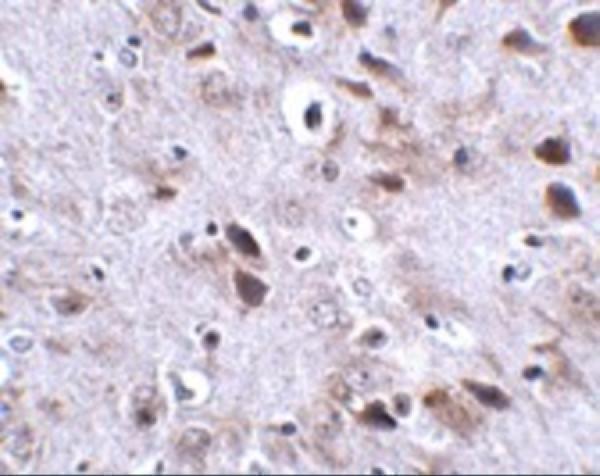
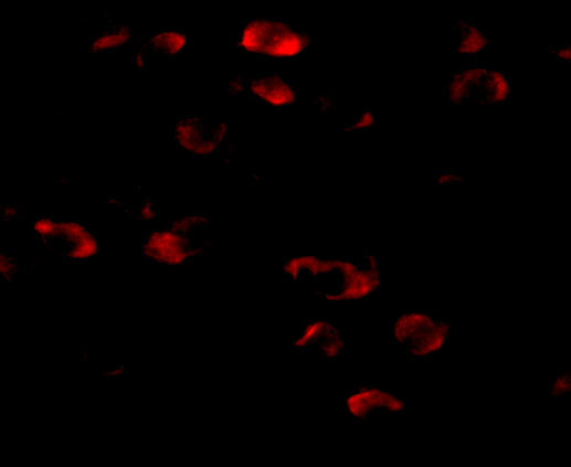
A05347 has IHC data from mouse brain tissue and IF data from mouse brain cells (catalog image captions); it lists human, mouse and rat reactivity (catalog: reactivity).
A05347 is listed for IHC-P and IF (catalog: applications), with IHC shown in mouse brain tissue at 2.5 μg/mL (IHC image caption). Its IF image shows mouse brain cells at 20 μg/mL (IF image caption); the catalog lists human, mouse and rat reactivity (catalog: reactivity).
Which to pick: Choose A05347 for paraffin-section IHC (catalog: IHC-P); its mouse brain tissue image uses 2.5 μg/mL, and the fixative is unreported (IHC image caption). Choose A05347 for IF in mouse brain cells (IF image caption: 20 μg/mL); ICC validation is unreported (catalog: applications). For cross-species work, A05347 lists human, mouse and rat reactivity (catalog: reactivity), while the supplied IHC and IF images show mouse samples (image captions); its host is rabbit and its clonality is unreported (catalog: host and clone).