TMEM88 / Transmembrane protein 88 · IHC design guide

Design Immunohistochemistry for TMEM88

This guide helps plan chromogenic TMEM88 IHC on paraffin sections, starting A12951 at 2.5 μg/mL (datasheet: IHC-P). Assess the general cytoplasmic tissue pattern and high cardiomyocyte staining (HPA tissue IHC).

Evidence assembled Oct 2026 · For research use; verify linked source records and product datasheet before use
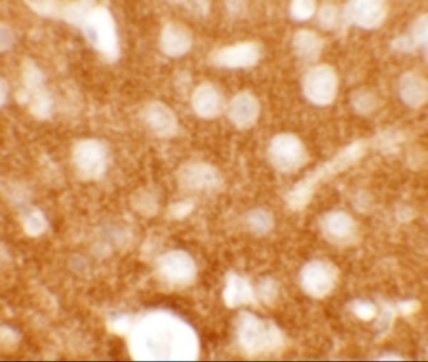
Immunohistochemistry protocol sheet for TMEM88 (IHC for TMEM88): expected localisation Cytoplasmic tissue staining (HPA tissue IHC); cell membrane localization (UniProt), antibody A12951, validated IHC image, and IHC protocol steps
Printable TMEM88 IHC protocol sheet — expected localisation Cytoplasmic tissue staining (HPA tissue IHC); cell membrane localization (UniProt), antibody A12951, controls and protocol steps. Open the full TMEM88 IHC guide →

TMEM88 Immunohistochemistry Experimental Design Guide

Expected localisation, validated protocols, controls and antibodies — the at-a-glance facts below, then the full design guide.

Must know before staining
Expected localisation Cytoplasmic tissue staining (HPA tissue IHC); cell membrane localization (UniProt)
Staining pattern General cytoplasmic staining; high in cardiomyocytes (HPA tissue IHC)
Antigen retrieval Citrate pH 6.0 HIER, 95–98 °C, 20 min (rule: cytoplasmic / membrane antigen)
Positive control ⓘ Adrenal gland+4 more · see all
Negative control ⓘ Ovary
Important caveats
Reasons your staining may differ from the expected pattern.
Fixation Matched tissue-IHC evidence does not establish the fixation claim. Validate the specimen-specific method before use. (selected-SKU IHC image A12951)
Caveat Tissue staining may look cytoplasmic despite membrane annotation (HPA tissue IHC; UniProt)
Regulation Expression regulation is not annotated (UniProt)
Isoform / epitope No isoforms annotated; epitope side is unspecified (UniProt)
Section 1

Recommended TMEM88 IHC & IF Protocols

The catalog antibody’s IHC-P protocol is accompanied by four published TMEM88 chromogenic IHC protocols (PMC12752382; PMC7339227; PMC8764240; PMC4694813).

Recommended immunohistochemistry (IHC-P) protocol parameters
SampleTissue sections; selected-image fixative not specified (standard IHC workflow)
FixationImage fixative and duration unreported (datasheet A12951); verify before use.
Sectioning4–5 µm sections on charged slides (standard)
DeparaffinisationXylene, graded ethanol series to water (standard)
Antigen retrievalHeat-induced epitope retrieval in citrate buffer, pH 6.0, 20 min at 95–98 °C (standard rule: cytoplasmic / membrane antigen)
Peroxidase block3% H2O2, 10 min, room temperature (standard)
Blocking10% normal serum of the secondary host, 30 min, room temperature (standard)
Primary antibodyRabbit anti-TMEM88, 2.5 μg/mL (datasheet A12951)
Primary incubationOvernight at 4 °C (standard)
DetectionHRP-polymer secondary, DAB chromogen 5–10 min (standard)
CounterstainHematoxylin, blue, dehydrate and mount (standard)
Expected resultTMEM88-positive staining in glandular cells of adrenal gland (HPA tissue IHC: High). HPA tissue profile: General cytoplasmic expression. No signal in the no-primary control.
💡Decision noteStart with citrate pH 6.0 retrieval at 95–98 °C for 20 min (page antigen retrieval); consider EDTA pH 9.0 for tissue microarrays (PMC12752382).
Section 2

What Is the Expected TMEM88 Staining Pattern?

TMEM88 is a two-pass cell-membrane protein (UniProt Q6PEY1 topology: residues 43–63 and 88–108). In tissue IHC, HPA reports general cytoplasmic expression, with high staining in cardiomyocytes, several epithelial cell populations, and kidney tubule cells (HPA tissue IHC: Approved, pending external verification). Interpret membrane-associated staining alongside that observed cytoplasmic pattern; topology alone does not define the appearance of a chromogenic section (UniProt Q6PEY1 topology; HPA tissue IHC).

What am I looking at on my slide?
Distinct staining in cardiomyocytes, respiratory epithelium, or kidney tubule cells.These are reported high-staining populations (HPA tissue IHC: High). Cytoplasmic signal fits the observed tissue profile (HPA tissue IHC: general cytoplasmic expression); a membrane-associated component also fits the protein annotation (UniProt Q6PEY1: cell membrane). Judge the cellular pattern against the selected tissue, rather than requiring an exclusively sharp membrane outline.
Predominantly nuclear staining, without a convincing cytoplasmic or membrane-associated pattern.A nuclear-only pattern conflicts with the reported tissue staining and location annotation (HPA tissue IHC: general cytoplasmic expression; UniProt Q6PEY1: cell membrane). Treat it as suspect and reassess controls, detection, and morphology before assigning it to TMEM88 (general IHC practice); the supplied sources do not identify a nuclear TMEM88 pattern.
Strong signal in ovarian stroma cells while an expected positive population also stains.HPA reports TMEM88 as not detected in ovarian stroma cells (HPA tissue IHC: Not detected). Unexpected staining there may reflect cross-reactivity or endogenous detection activity (general IHC practice). A positive comparator checks whether the run can reveal the reported pattern; it does not establish the identity of signal in the unexpected cells.
Diffuse chromogen across cells or tissue structures, with little distinction between populations.This lacks the cell-selective pattern needed to interpret the reported high and not-detected populations (HPA tissue IHC: High; Not detected). Consider nonspecific antibody or detection background and inspect the appropriate detection control (general IHC practice); diffuse color alone cannot establish TMEM88 localization.
No signal in cardiomyocytes or another reported high-staining population.This is discordant with the HPA tissue IHC result for that population (HPA tissue IHC: High). Check section quality, retrieval, antibody conditions, and detection controls as general IHC troubleshooting steps (general IHC practice). A failed positive control leaves a negative result in an unknown sample uninterpretable.
💡Expected TMEM88 appearanceCall a result convincing when staining is discernible in reported high-staining cells, with the observed cytoplasmic pattern and possible membrane association (HPA tissue IHC: High; general cytoplasmic expression; UniProt Q6PEY1: cell membrane); strong ovarian stromal or nuclear-only signal is suspect (HPA tissue IHC: ovarian stroma not detected; UniProt Q6PEY1: cell membrane).
How each factor affects the staining
Membrane topologyUniProt annotates two transmembrane segments at residues 43–63 and 88–108 (UniProt Q6PEY1 topology). This supports a membrane-associated interpretation, but does not identify the antibody epitope or require a crisp membrane rim in paraffin IHC.
Observed tissue localizationHPA describes general cytoplasmic expression in tissue IHC (HPA tissue IHC profile). Read that observation together with the cell-membrane annotation (UniProt Q6PEY1: cell membrane); neither source establishes that every positive cell must show both patterns.
Choice of tissue and cellsCardiomyocytes, bronchial respiratory epithelial cells, and kidney tubule cells are reported High; ovarian stroma cells are Not detected (HPA tissue IHC). Compare named cell populations within well-preserved sections rather than treating every cell in a tissue as equivalent.
Strength of validation evidenceThe tissue profile is Approved but pending external verification, and HPA052991 has IHC status Approved (HPA tissue IHC; HPA antibodies). These labels support use of the reported pattern as a reference, while leaving unexpected staining in a new specimen to be checked with controls.
RNA versus protein evidenceHeart muscle is tissue enhanced at the RNA level, while cardiomyocytes are High by tissue IHC (HPA tissue IHC). Use the protein staining observation for slide interpretation; RNA enrichment alone does not predict chromogen intensity in an individual section.
Why is my staining missing, weak or wrong?
SituationLikely causeNext action
Reported high-staining cells show no chromogen.The result conflicts with HPA's High call for the chosen cells (HPA tissue IHC); an unsuccessful IHC run is one possible explanation (general IHC practice).Review section integrity, retrieval conditions, antibody dilution, and detection controls (general IHC practice). Repeat with a reported high-staining tissue before interpreting other negative sections; the sources give no TMEM88-specific retrieval or dilution setting.
Signal appears only in nuclei.That compartment does not match the reported cytoplasmic tissue profile or membrane annotation (HPA tissue IHC; UniProt Q6PEY1).Check counterstain and detection controls, then compare the pattern with a reported high-staining cell population (general IHC practice; HPA tissue IHC: High). Do not score nuclear-only color as confirmed TMEM88.
Ovarian stroma cells stain strongly.HPA reports this population as Not detected (HPA tissue IHC). Cross-reactivity or endogenous detection activity could produce unexpected color (general IHC practice).Inspect a detection control and a reported high-staining comparator, and confirm that the stained cells are stromal (general IHC practice; HPA tissue IHC: ovarian stroma Not detected). Investigate the unexpected signal before calling it positive.
Color is widespread and cell boundaries are hard to interpret.Diffuse background may obscure the reported differences among cell populations (general IHC practice; HPA tissue IHC: High and Not detected populations).Check blocking, washing, antibody concentration, and detection controls as general IHC variables (general IHC practice). Reassess whether the reported positive cells stand out after background is reduced (HPA tissue IHC: High).
Signal is cytoplasmic but lacks a sharp membrane outline.HPA reports general cytoplasmic expression in tissue IHC, despite UniProt's cell-membrane annotation (HPA tissue IHC; UniProt Q6PEY1).Compare cell identity, intensity, and controls before rejecting the staining (general IHC practice; HPA tissue IHC: High populations). Do not infer an exact chromogenic membrane pattern from the two transmembrane segments alone (UniProt Q6PEY1 topology).
Can an IF/ICC image settle an ambiguous IHC compartment?HPA summarizes the IF location as Membrane but supplies no main location or ICC-IF cell-line images here (HPA subcellular record).Use the tissue IHC profile to interpret this paraffin-section result (HPA tissue IHC). Treat the IF summary as limited location context; it cannot resolve the appearance of the stained tissue cells without supporting images.

Sample controls for TMEM88 IHC & IF

🧪Run adrenal gland first and expect staining in its glandular cells (HPA: High in adrenal gland glandular cells). Run ovary as the negative tissue, focusing on ovarian stroma cells (HPA: Not detected in ovarian stroma cells); on the adrenal slide, compare adjacent nonglandular cells with the glandular cells, but do not assume those cells are TMEM88-negative from the HPA entry alone.
Positive control tissue: Adrenal gland (Glandular cells, HPA High)
Negative control tissue: Ovary (HPA Not detected)
ICC-IF cell lines (HPA subcellular resource): HPA carries no ICC-IF cell line for TMEM88; derive a cell-line control from the positive tissue's cell type (Glandular cells) and confirm it by RNA or western blot first.
Technical controls: Include a no-primary, secondary-only control and a host-species- and immunoglobulin-class-matched isotype control for a monoclonal primary, or nonimmune IgG from the same host for a polyclonal primary (standard IHC practice). Use TMEM88 knockout tissue or a validated immunizing-peptide block as a biological specificity control, and quench endogenous peroxidase in adrenal sections before chromogenic detection (standard IHC practice).
⚠️Feasibility: No target-specific fixation window or retrieval dependency is reported in the supplied evidence; optimize antigen retrieval empirically for paraffin sections. The selected A12951 mouse-brain IHC caption reports 2.5 µg/mL but does not state the fixative (selected A12951 tissue-IHC caption). The evidence does not establish whether frozen sections or IF are easier for TMEM88; check adrenal sections for pigment or background that could complicate chromogenic scoring (standard IHC practice).

HPA tissue IHC evidence for TMEM88

Comprehensive Human Protein Atlas IHC scoring per tissue (reliability: Approved — Pending external verification.). Rows are taken directly from the HPA tissue chart — click any row's HPA link to view the source.

Positive expression · recommended positive controls

TissueCell typeLevelEvidenceSource
Adrenal gland Glandular cells High Protein (IHC) HPA →
Bronchus Respiratory epithelial cells High Protein (IHC) HPA →
Cervix Glandular cells High Protein (IHC) HPA →
Duodenum Glandular cells High Protein (IHC) HPA →
Endometrium Glandular cells High Protein (IHC) HPA →

Undetected expression · recommended negative controls

TissueCell typeLevelEvidenceSource
Ovary Ovarian stroma cells Not detected Protein (IHC) HPA →
Section 3

Advanced TMEM88 IHC Tips

Troubleshoot TMEM88 chromogenic IHC in paraffin sections using the page retrieval method, the catalog image, and tissue and localisation references.

How should I retrieve TMEM88 in paraffin sections when staining is weak?
Start with citrate buffer at pH 6.0, heated to 95–98 °C for 20 min (page retrieval rule). Allow sections to cool in the retrieval buffer, then compare staining with an otherwise matched section processed without heat (standard IHC practice). Assess signal in cardiomyocytes or kidney tubule cells, where tissue IHC reports high expression, while checking whether background rises elsewhere (HPA: High in cardiomyocytes and kidney tubule cells). If staining remains weak, test a different retrieval condition on adjacent sections as an optimisation, documenting both signal and tissue damage (standard IHC practice). Do not attribute retrieval performance to the catalog image: its caption supplies no retrieval method (catalog image caption: A12951).
Can I adjust fixation to recover weak TMEM88 staining?
Target-specific fixation sensitivity is unknown from the supplied evidence; the mouse brain image caption does not state its fixative (catalog image caption: A12951). For a controlled IHC comparison, record fixative, fixation duration, section thickness, and retrieval conditions for each specimen, then change one variable at a time (standard IHC practice). Compare sections from the same tissue block where possible, keeping the page's citrate pH 6.0, 95–98 °C, 20 min retrieval constant (page retrieval rule). Evaluate preserved morphology alongside staining, because damaged sections can mislead interpretation (standard IHC practice). Neither tissue staining patterns nor the protein's topology establishes a TMEM88-specific fixation effect (HPA: tissue IHC profile; UniProt Q6PEY1: topology).
Should TMEM88 appear at the membrane or throughout the cytoplasm in IHC?
TMEM88 is annotated at the cell membrane and has transmembrane segments at residues 43–63 and 88–108 (UniProt Q6PEY1: location and topology). Tissue IHC nevertheless describes a general cytoplasmic expression pattern, so compare both distribution and cellular context before calling a section positive (HPA: tissue IHC profile). Examine adjacent sections at the same magnification and record whether staining outlines cells, appears cytoplasmic, or spreads across structures lacking intact cells (standard IHC practice). Include a no-primary control to identify detection-related signal (standard IHC practice). Treat an exclusively nuclear pattern as unexpected for this annotation and investigate it before reporting localisation (UniProt Q6PEY1: cell membrane).
How does the antibody epitope affect TMEM88 IHC interpretation?
The supplied record annotates a 159-residue chain with 2 transmembrane segments and no isoforms, annotated glycosylation sites, or modified residues (UniProt Q6PEY1: chain, topology, isoforms, PTMs). Check the catalog antibody's immunogen or mapped epitope before interpreting a staining pattern; the supplied image caption does not identify that epitope (catalog image caption: A12951). If its position becomes available, map it against residues 43–63 and 88–108 when choosing retrieval and assessing accessibility (UniProt Q6PEY1: topology; standard IHC practice). Keep the same antibody and processing conditions across comparison sections (standard IHC practice). The absence of annotated isoforms does not, by itself, establish antibody specificity in tissue (UniProt Q6PEY1: isoforms; standard IHC practice).
How can I investigate TMEM88 localisation by multiplex IF?
Use this as a separate IF optimisation: the supplied catalog evidence is a tissue IHC image, and no ICC/IF images are listed in the subcellular payload (catalog image caption: A12951; HPA: subcellular record). Pair TMEM88 with a validated marker for the cell type being examined, such as a cardiomyocyte marker in heart muscle, and include single-stain controls (HPA: High in cardiomyocytes; standard IF practice). Choose fluorophores after inspecting tissue autofluorescence in an unstained section, and separate channels whose signals overlap (standard IF practice). Determine whether the antibody epitope faces an intracellular compartment before adding permeabilisation, since TMEM88 spans the membrane at residues 43–63 and 88–108 (UniProt Q6PEY1: topology; standard IF practice). Verify the resulting pattern against the membrane annotation and tissue context (UniProt Q6PEY1: cell membrane).
What should I check when TMEM88 chromogenic staining is diffuse?
Run a no-primary control through the same secondary detection and chromogen steps to locate reagent-derived background (standard IHC practice). Check endogenous peroxidase blocking before DAB development, and compare tissue edges, folds, and damaged areas with intact fields (standard IHC practice). Optimise blocking, washes, and primary antibody concentration on adjacent sections while holding citrate pH 6.0, 95–98 °C, 20 min retrieval constant (page retrieval rule; standard IHC practice). The catalog mouse brain image uses 2.5 µg/mL, which is an image condition rather than a demonstrated optimum for every specimen (catalog image caption: A12951). Interpret diffuse colour cautiously because the tissue IHC profile describes general cytoplasmic expression (HPA: tissue IHC profile).
How should I score TMEM88 staining across paraffin sections? ⚠ ANSWER MARKED FOR VERIFICATION
Define the cell population and compartment before scoring: TMEM88 has a membrane annotation, while tissue IHC describes general cytoplasmic expression (UniProt Q6PEY1: location; HPA: tissue IHC profile). For comparable sections, record percent positive cells and an intensity score of 0–3, then calculate an H-score from 0–300 (standard IHC scoring practice). Alternatively, report positive-cell density per mm² of viable, annotated tissue when cell counts differ markedly (standard IHC practice). Normalise within the same cell type and tissue area, using matched staining and imaging settings across groups (standard IHC practice). Exclude folds, edges, and necrotic areas from the scoring region and document the exclusions (standard IHC practice).
How can I distinguish credible TMEM88 staining from artefact?
A credible call should agree with intact cell morphology and an interpretable cellular pattern: TMEM88 is annotated at the membrane, while tissue IHC reports general cytoplasmic expression (UniProt Q6PEY1: location; HPA: tissue IHC profile). Use cardiomyocytes or kidney tubule cells as high-expression references and ovarian stroma cells as a reported not-detected comparison, allowing for specimen and method differences (HPA: tissue IHC levels). Investigate staining confined to nuclei, section edges, folds, or necrotic regions before assigning it to TMEM88 (UniProt Q6PEY1: location; standard IHC practice). Compare any DAB colour with a no-primary control and confirm endogenous peroxidase blocking (standard IHC practice). HPA labels its tissue IHC evidence Approved but pending external verification, so report unresolved discrepancies as such (HPA: reliability).
Boster reagents

Best TMEM88 / Transmembrane protein 88 IHC Antibodies

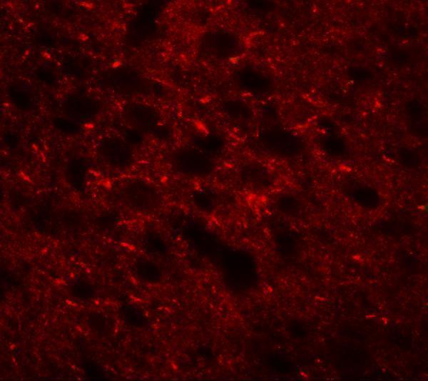
A12951 has real mouse-brain IHC and IF images (A12951 image captions). A12951-1 has a human U20S-cell ICC/IF image (A12951-1 image caption; catalog reactivity).

Real IHC data Immunohistochemistry of TMEM88 in mouse brain tissue with TMEM88 antibody at 2.5 μg/mL.
Anti-Transmembrane protein 88 TMEM88 Antibody
Cat # A12951
Real IF data IF analysis of TMEM88 using anti-TMEM88 antibody (A12951-1). TMEM88 was detected in an immunocytochemical section of U20S cells. Enzyme antigen retrieval was performed using IHC enzyme antigen retrieval reagent (AR0022) for 15 mins. The cells were blocked with 10% goat serum. And then incubated with 5 μg/mL rabbit anti-TMEM88 Antibody (A12951-1) overnight at 4°C. DyLight®488 Conjugated Goat Anti-Rabbit IgG (BA1127) was used as secondary antibody at 1:100 dilution and incubated for 30 minutes at 37°C. The section was counterstained with DAPI. Visualize using a fluorescence microscope and filter sets appropriate for the label used.
Anti-TMEM88 Antibody ®
Cat # A12951-1

A12951 lists IHC-P and IF for human, mouse, and rat reactivity, with IHC and IF images from mouse brain (catalog applications/reactivity; A12951 image captions). A12951-1 lists human ICC/IF and shows IF in U20S cells (catalog applications/reactivity; A12951-1 image caption).

Which to pick: Choose A12951 for paraffin-section tissue IHC: it lists IHC-P and shows mouse-brain staining at 2.5 μg/mL (catalog applications; A12951 IHC image caption); the caption does not report a fixative (A12951 IHC image caption). For human cell ICC/IF, choose A12951-1, a rabbit polyclonal shown staining U20S cells at 5 μg/mL (catalog host and dilution_raw; A12951-1 IF image caption). For human, mouse, or rat reactivity, A12951 has the broader species listing and includes IF; its mouse-brain IF image uses 20 μg/mL (catalog reactivity/applications; A12951 IF image caption).

Each figure is that product's own IHC / IF validation image from its datasheet.

References

  1. UniProt Consortium. UniProt entry Q6PEY1 (TMM88_HUMAN, Transmembrane protein 88).
  2. Human Protein Atlas. TMEM88 tissue IHC expression (reliability: Approved).
  3. Human Protein Atlas. TMEM88 subcellular location (ICC-IF): Membrane.
  4. Human Protein Atlas. TMEM88 antibody validation summary (1 antibodies).
  5. TMEM88 modulates the proliferation and metastasis of HCC via the GSK-3β/β-catenin pathway. BMC cancer 2025 — PMC12752382.
  6. MicroRNA-708 modulates Hepatic Stellate Cells activation and enhances extracellular matrix accumulation via direct targeting TMEM88. Journal of cellular and molecular medicine 2020 — PMC7339227.
  7. TMEM88 Modulates Lipid Synthesis and Metabolism Cytokine by Regulating Wnt/β-Catenin Signaling Pathway in Non-Alcoholic Fatty Liver Disease. Frontiers in pharmacology 2021 — PMC8764240.
  8. Cytosolic TMEM88 promotes triple-negative breast cancer by interacting with Dvl. Oncotarget 2015 — PMC4694813.
  9. PubMed PMID:15489334 — UniProt-cited evidence.
  10. PubMed PMID:21044957 — UniProt-cited evidence.
  11. PubMed PMID:23924634 — UniProt-cited evidence.