This website uses cookies to ensure you get the best experience on our website.
- Table of Contents
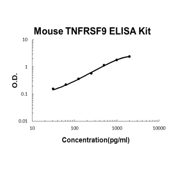
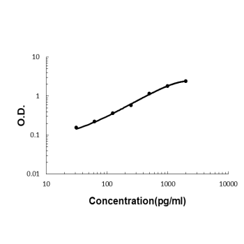
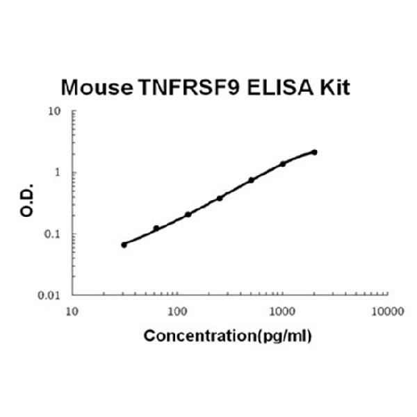
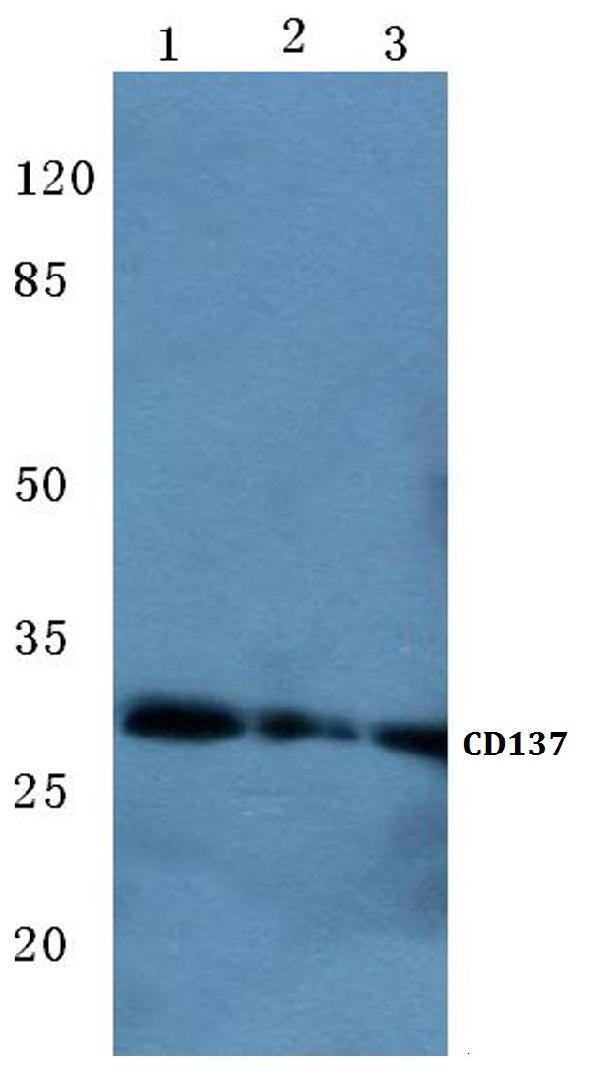
Facts about Tumor necrosis factor receptor superfamily member 9.
Receptor for TNFSF9/4-1BBL.
Maybe active during T cell activation..
Human | |
---|---|
Gene Name: | TNFRSF9 |
Uniprot: | Q07011 |
Entrez: | 3604 |
Belongs to: |
---|
No superfamily |
4-1BB ligand receptor; 41BB; 4-1BB; CD137 antigen; CD137; CD137MGC2172; CDw137; FLJ43501; ILA; ILAhomolog of mouse 4-1BB; induced by lymphocyte activation (ILA); interleukin-activated receptor, homolog of mouse Ly63; receptor protein 4-1BB; T cell antigen ILA; T-cell antigen 4-1BB homolog; T-cell antigen ILA; TNFRSF9; tumor necrosis factor receptor superfamily member 9; tumor necrosis factor receptor superfamily, member 9
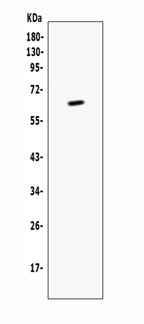
Mass (kDA):
27.899 kDA
Human | |
---|---|
Location: | 1p36.23 |
Sequence: | 1; NC_000001.11 (7915871..7941607, complement) |
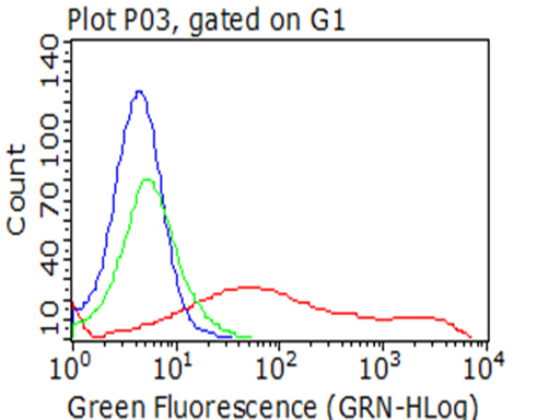
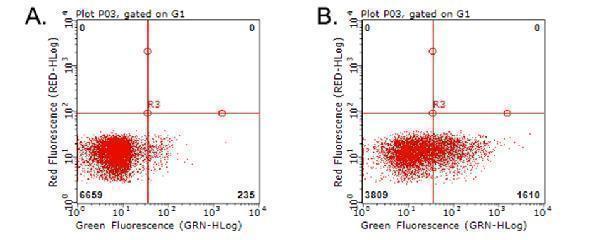
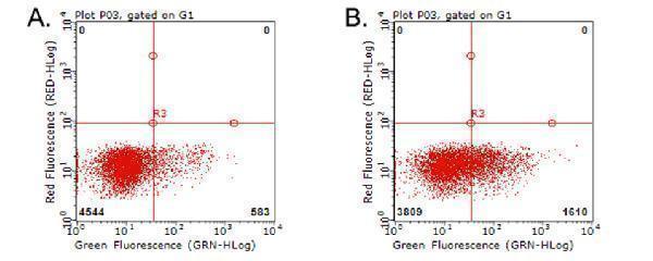
Expressed on the surface of activated T-cells.
Membrane; Single-pass type I membrane protein.
The TNFRSF9 molecular marker has been identified as highly specific and has a variety of applications. This article will discuss its typical applications as well as Methods. Learn more about Boster Microplate readers. You can also earn credits for sharing your results with others. Contact us with any questions regarding this product or if you would like to buy it.
The TNFRSF9 gene that regulates immune function in cancer cells is well-known. It regulates STAT signaling, which in turn inhibits T cell growth. Additionally, a deficiency of TNFRSF9 decreases the expression of PD-1 and inhibitory checkpoints that are involved in CD8(+) T cell exhaustion. Anti-angiogenic agents also target the VEGF-A-VEGFR pathway. TNFRSF9 is an important functional marker for HCC.
The TNFRSF9, also called tumor necrosis factor superfamily 9 is a molecule expressed on activated T cell cells. As a co-stimulatory immune checkpoint it is of special interest to immunologists. It can be costimulatory to activated T cells, and binding to its agonistic antibody enhances T cell proliferative capacity, IL-2 production and even survival. It can boost the immune system against tumors in mice.
Its presence is linked to poorer prognosis with chemotherapy for cancer. This suggests that this biomarker may play a role in prognosis and response. Additionally, TNFRSF9 expression in TI-Tregs could indicate a patient's response to immunotherapy against PD-1. These findings need further research.
T cells of TNFRSF9+CD8+ cells express a phenotype that is linked to exhaustion. Similarly, TNFRSF9+CD8+ T cells responds to ICBs with an antitumor effect. These results suggest that TNFRSF9+CD8+ T cells may be crucial in the development and use of immunotherapy for CCRC.
Preclinical models revealed significant therapeutic benefits from studies of the TNFRSF9 gene expression in the brain metastasis from melanoma. However, the initial clinical phase I/II trials were stopped due to the severe negative effects. Researchers discovered that TNFRSF9 was highly expressed in astrocytes with reactive functions in primary CNS tumors, but was largely absent in inflammatory and tumor cells. Similarly, they examined the expression of TNFRSF9 in the brain metastasis from melanoma that is an immunogenic tumor that has prominent ties to the CNS.
TNFRSF9 expression is seen on the surface cells of melanoma cells as well in the smooth muscle cells of larger vessels. The gene was previously linked with the survival of patients, and it increased in relation to the distance of tumors from blood vessels. Researchers have discovered that hypoxia may be a cause for tumors expressing this gene. The expression pattern of TNFRSF9 is not associated with the progression of the tumor.
When it comes to the markers used in AA research, TNFRSF9 has the highest association with the disease both women and men. This association is further enhanced due to the fact that the markers IL12RB1 and VEGFR2 are strongly linked to AA. This study offers an understanding of the role of TNFRSF9 in decreasing the risk of developing AA.
PMID: 8088337 by Alderson M.R., et al. Molecular and biological characterization of human 4-1BB and its ligand.
PMID: 8262389 by Schwarz H., et al. A receptor induced by lymphocyte activation (ILA): a new member of the human nerve-growth-factor/tumor-necrosis-factor receptor family.