This website uses cookies to ensure you get the best experience on our website.


- Table of Contents
Plan TNFSF12 IHC in paraffin sections using the catalog antibody’s fixation, dilution and chromogenic detection evidence (datasheet A02009-2). Compare cytoplasmic tissue staining with suitable controls while accounting for the secreted form and uncertain tissue evidence (HPA tissue IHC; UniProt).

Expected localisation, validated protocols, controls and antibodies — the at-a-glance facts below, then the full design guide.
| Expected localisation | Cytoplasmic staining (HPA tissue IHC); membrane and secreted forms (UniProt) | |
| Staining pattern | Cytoplasmic staining in most tissues; plasma positive (HPA tissue IHC) | |
| Antigen retrieval | Citrate pH 6 HIER, heat-mediated (datasheet A02009-2) | |
| Positive control | Adrenal gland+4 more · see all | |
| Negative control | Bone marrow+4 more · see all |
| Fixation | Formaldehyde-fixed paraffin sections (selected-SKU IHC image A02009-2); keep fixation consistent. (standard IHC practice; not target-specific) | |
| Caveat | Secreted variants can complicate tissue localisation (HPA tissue IHC) | |
| Regulation | Expression regulation not specified (UniProt) | |
| Isoform / epitope | 2 isoforms; cleavage and epitope location matter (UniProt) |
The catalog antibody’s IHC-P protocol is accompanied by one published TNFSF12 staining workflow (PMC13371701).
| Sample | Formaldehyde-fixed, paraffin-embedded human brain tissue (datasheet A02009-2) |
| Fixation | Image formalin-fixed; duration unreported (datasheet A02009-2); verify before use. |
| Sectioning | 4–5 µm sections on charged slides (standard) |
| Deparaffinisation | Xylene, graded ethanol series to water (standard) |
| Antigen retrieval | Heat retrieval: Citrate pH 6 (datasheet A02009-2); 20 min, 95–100 °C (standard) |
| Peroxidase block | 3% H2O2, 10 min, room temperature (standard) |
| Blocking | 10% serum (datasheet A02009-2) |
| Primary antibody | Rabbit anti-TNFSF12, 5-10 μg/mL (datasheet A02009-2) |
| Primary incubation | Overnight at 4 °C (standard) |
| Detection | HRP-polymer secondary, DAB chromogen 5–10 min (standard) |
| Counterstain | Hematoxylin, blue, dehydrate and mount (standard) |
| Expected result | TNFSF12-positive staining in glandular cells of adrenal gland (HPA tissue IHC: High). HPA tissue profile: Cytoplasmic expression in most tissues. Positivity in plasma. No signal in the no-primary control. |
TNFSF12 is a membrane protein with an extracellular region and a secreted form (UniProt O43508 topology and processing). In tissue IHC, expect predominantly cytoplasmic staining in glandular cells and muscle cells, with possible plasma positivity (HPA: tissue profile). Treat the pattern as provisional: HPA rates tissue IHC reliability Uncertain because the antibody targets proteins from more than one gene, and the secreted variant complicates location comparisons (HPA: reliability description).
| Cytoplasmic signal in heart cardiomyocytes, skeletal myocytes, or duodenal or small-intestinal glandular cells. | These are reported High-staining cells and fit the broad cytoplasmic tissue profile (HPA: tissue IHC). Compare intensity within the same run; a plausible pattern alone cannot establish TNFSF12 specificity because tissue IHC reliability is Uncertain (HPA: reliability description). |
| Predominantly nuclear staining with little cytoplasmic signal in an expected positive tissue. | A nuclear-only pattern conflicts with the reported cytoplasmic profile and membrane/secreted annotation (HPA: tissue profile; UniProt O43508 subcellular location). Consider nonspecific staining or an imaging or scoring artefact (general IHC practice). |
| Strong staining in cells reported as Not detected, such as lung alveolar cells or liver cholangiocytes. | This differs from the cited cell-level observations (HPA: lung and liver tissue IHC). Check cell identity, cross-reactivity and endogenous detection activity (general IHC practice). Given HPA's Uncertain reliability, the mismatch raises a question; it does not prove a false positive (HPA: reliability description). |
| Diffuse colour across cells and surrounding tissue, obscuring cell boundaries. | This cannot be scored confidently against HPA's cell-level pattern (HPA: tissue profile). Uneven blocking, excess detection reagent or inadequate washing can produce background (general IHC practice); a widespread signal should not be counted as TNFSF12-positive cells. |
| No signal in heart cardiomyocytes or skeletal myocytes. | Both cell types are reported High by tissue IHC, so an absent signal warrants a run-level check (HPA: heart and skeletal muscle). Confirm tissue preservation, retrieval, antibody and detection steps with appropriate controls (general IHC practice), while retaining HPA's Uncertain specificity caveat. |
| Membrane form and shedding | Residues 22–42 span the membrane; residues 43–249 are extracellular, and residues 94–249 form a secreted chain (UniProt O43508 topology and processing). Membrane association or extracellular material is biologically plausible, but the tissue IHC reference pattern remains mainly cytoplasmic (HPA: tissue profile). |
| Antibody epitope | The supplied record gives no epitope for the tissue IHC antibody. Whether it recognizes membrane and secreted forms cannot be resolved from their listed boundaries alone (UniProt O43508 processing); interpret a compartment difference cautiously. |
| Isoforms and glycosylation | UniProt lists two isoforms, including TWE-PRIL, and a glycosylation site at residue 139 (UniProt O43508 isoforms and glycosylation). The payload does not establish which forms the tissue antibody detects or any glycosylation effect on IHC staining. |
| Tissue selection | Use reported High cells to assess visibility; skin keratinocytes are Low, while bone-marrow hematopoietic cells are Not detected (HPA: tissue IHC). These categories describe observations, not absolute positive or negative controls, given the Uncertain reliability (HPA: reliability description). |
| Antibody validation | HPA052967 has Uncertain IHC validation; the overall tissue profile is also Uncertain and awaits external verification (HPA: antibody validation and reliability description). Reproduction with an independent, suitable antibody would strengthen an interpretation (general IHC practice). |
| IF/ICC Q: Should vesicle staining guide this IHC result? | A: HPA reports approved vesicle localization by ICC-IF, but cautions that its antibodies target proteins from multiple genes (HPA: subcellular summary). Use that observation as context, not as a required chromogenic tissue IHC pattern; evaluate IF/ICC on its own guide page. |
| Situation | Likely cause | Next action |
|---|---|---|
| A reported High tissue has no visible staining. | A failed staining or detection step is possible (general IHC practice); HPA's High designation does not guarantee every section will stain (HPA: tissue IHC; reliability description). | Check section quality and run controls, then review retrieval, antibody incubation and chromogen development records (general IHC practice). Do not assign a TNFSF12-specific retrieval requirement from these sources. |
| Colour spreads through nearly every cell and extracellular space. | Background from detection chemistry or insufficient washing may mask the reported cell-level pattern (general IHC practice; HPA: tissue profile). | Inspect a no-primary control, wash steps and detection conditions; score only distinguishable cellular signal (general IHC practice). |
| Blood-rich regions or plasma appear positive. | Plasma positivity is part of the reported tissue profile (HPA: tissue IHC); it can complicate assignment of colour to nearby cells (general IHC practice). | Compare the cellular compartment and section morphology before assigning positivity to a specific cell type (general IHC practice). |
| Strong colour appears in HPA Not detected cells. | Cell misidentification, cross-reactivity or endogenous detection activity is possible (general IHC practice); HPA reports no staining in those specified cells (HPA: tissue IHC). | Recheck cell identity and no-primary controls, then compare an independent antibody if available (general IHC practice); retain the HPA Uncertain caveat. |
| Staining is nuclear rather than cytoplasmic. | Nuclear staining does not match the HPA cytoplasmic profile or UniProt membrane/secreted location (HPA: tissue profile; UniProt O43508 subcellular location). | Check counterstain interpretation and controls, then review antibody and detection specificity (general IHC practice). |
| Tissue staining and reported expression seem inconsistent. | UniProt lists adult lung and spleen expression, whereas HPA reports Not detected in lung alveolar cells and spleen red-pulp cells (UniProt O43508 tissue specificity; HPA: tissue IHC). HPA warns that secreted variants complicate RNA–protein location comparisons. | Compare the named cell population and assay type, and report the discrepancy without converting tissue-wide expression into a predicted cell-level IHC result (HPA: reliability description; general IHC practice). |


Comprehensive Human Protein Atlas IHC scoring per tissue (reliability: Uncertain — Caution, targets protein from more than one gene. At least one protein variant secreted, tissue location of RNA and protein might differ and correlation is complex. Pending external verification.). Rows are taken directly from the HPA tissue chart — click any row's HPA link to view the source.
| Tissue | Cell type | Level | Evidence | Source |
|---|---|---|---|---|
| Adrenal gland | Glandular cells | High | Protein (IHC) | HPA → |
| Duodenum | Glandular cells | High | Protein (IHC) | HPA → |
| Heart muscle | Cardiomyocytes | High | Protein (IHC) | HPA → |
| Rectum | Glandular cells | High | Protein (IHC) | HPA → |
| Skeletal muscle | Myocytes | High | Protein (IHC) | HPA → |
| Tissue | Cell type | Level | Evidence | Source |
|---|---|---|---|---|
| Bone marrow | Hematopoietic cells | Not detected | Protein (IHC) | HPA → |
| Liver | Cholangiocytes | Not detected | Protein (IHC) | HPA → |
| Lung | Alveolar cells | Not detected | Protein (IHC) | HPA → |
| Nasopharynx | Respiratory epithelial cells | Not detected | Protein (IHC) | HPA → |
| Salivary gland | Glandular cells | Not detected | Protein (IHC) | HPA → |
Troubleshoot TNFSF12 staining in paraffin sections by checking retrieval, compartment, controls and scoring before interpreting tissue differences.
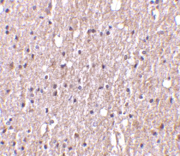
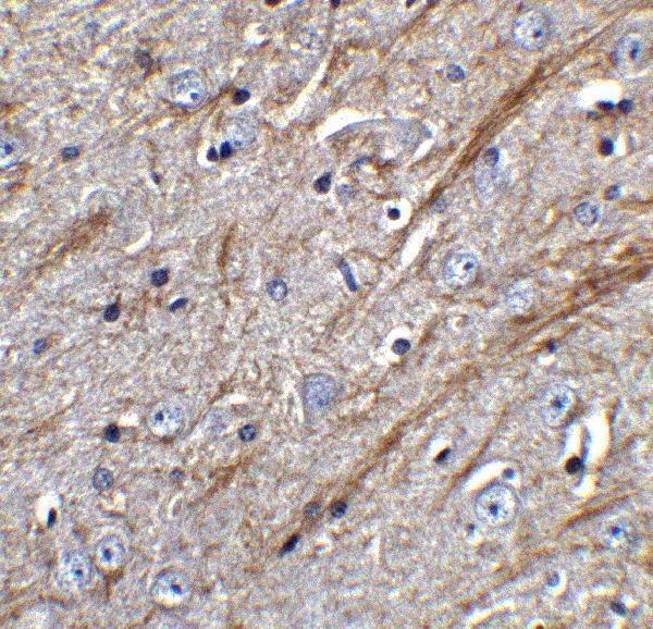
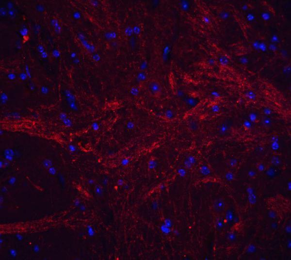
A02009-2 has IHC images from human and mouse paraffin-embedded brain tissue (IHC image captions) and an IF image from mouse brain tissue (IF image caption).
A02009-2 was demonstrated by IHC on human paraffin-embedded brain tissue at 10 μg/mL with formaldehyde fixation (human IHC image caption). The same SKU was demonstrated by IHC on mouse paraffin-embedded brain tissue at 5 μg/mL (mouse IHC image caption) and by IF on 4% paraformaldehyde-fixed mouse brain tissue (IF image caption).
Which to pick: Choose A02009-2 for tissue IHC: its application list includes IHC-P (catalog), and its images cover formaldehyde-fixed, paraffin-embedded human and mouse brain sections (IHC image captions). For IF, A02009-2 has a mouse brain image at 20 μg/mL (IF image caption); ICC validation is unreported (catalog). It is also the cross-species IHC choice here, with Human and Mouse reactivity (catalog) and IHC images for both species (IHC image captions); its host is rabbit, while clonality is unreported (catalog).