TOM1L1 / TOM1-like protein 1 · IHC design guide

Design Immunohistochemistry for TOM1L1

Plan TOM1L1 paraffin IHC around the cytoplasmic staining reported in most tissues (HPA tissue IHC). This guide covers fixation consistency, antibody titration, chromogenic detection and interpretation.

Evidence assembled Oct 2026 · For research use; verify linked source records and product datasheet before use
Immunohistochemistry protocol sheet for TOM1L1 (IHC for TOM1L1): expected localisation Predominantly cytoplasmic tissue staining (HPA tissue IHC), antibody A07598-2, validated IHC image, and IHC protocol steps
Printable TOM1L1 IHC protocol sheet — expected localisation Predominantly cytoplasmic tissue staining (HPA tissue IHC), antibody A07598-2, controls and protocol steps. Open the full TOM1L1 IHC guide →

TOM1L1 Immunohistochemistry Experimental Design Guide

Expected localisation, validated protocols, controls and antibodies — the at-a-glance facts below, then the full design guide.

Must know before staining
Expected localisation Predominantly cytoplasmic tissue staining (HPA tissue IHC)
Staining pattern Cytoplasmic staining in most tissues (HPA tissue IHC)
Antigen retrieval EDTA pH 8.0 HIER, heat-mediated (datasheet A07598-2)
Positive control ⓘ Adrenal gland+4 more · see all
Negative control ⓘ Skeletal muscle
Important caveats
Reasons your staining may differ from the expected pattern.
Fixation Matched tissue-IHC evidence does not establish the fixation claim. Validate the specimen-specific method before use. (selected-SKU IHC image A07598-2)
Caveat Staining awaits external verification (HPA tissue IHC)
Regulation Expression regulation unreported (UniProt)
Isoform / epitope 3 isoforms; shared epitope coverage unknown (UniProt)
Section 1

Recommended TOM1L1 IHC & IF Protocols

Start with the catalog antibody’s IHC-P protocol (datasheet A07598-2), then compare the published TOM1L1 tissue protocols (PMC4764922; PMC11568529; PMC9818872).

Recommended immunohistochemistry (IHC-P) protocol parameters
SampleParaffin-embedded human colon cancer tissue; fixative not specified (datasheet A07598-2)
FixationImage fixative and duration unreported (datasheet A07598-2); verify before use.
Sectioning4–5 µm sections on charged slides (standard)
DeparaffinisationXylene, graded ethanol series to water (standard)
Antigen retrievalHeat retrieval: EDTA pH 8.0 (datasheet A07598-2); 20 min, 95–100 °C (standard)
Peroxidase block3% H2O2, 10 min, room temperature (standard)
Blocking10% goat serum (datasheet A07598-2)
Primary antibodyRabbit anti-TOM1L1, 2-5 μg/ml (datasheet A07598-2)
Primary incubationOvernight at 4 °C (datasheet A07598-2)
DetectionHRP-conjugated secondary, DAB chromogen (datasheet A07598-2)
CounterstainHematoxylin, blue, dehydrate and mount (standard)
Expected resultTOM1L1-positive staining in glandular cells of adrenal gland (HPA tissue IHC: Medium). HPA tissue profile: Cytoplasmic expression in most tissues. No signal in the no-primary control.
💡Decision noteTry heat-mediated EDTA pH 8.0 retrieval first (datasheet A07598-2). For replication, follow the article’s reported retrieval conditions (PMC4764922; PMC9818872).
Section 2

What Is the Expected TOM1L1 Staining Pattern?

In paraffin-section IHC, expect mainly cytoplasmic TOM1L1 staining across many tissues (HPA: cytoplasmic expression in most tissues). Golgi and endosome associations may contribute to the pattern; only a small proportion is membrane-associated, and TOM1L1 has no transmembrane segment (UniProt O75674). HPA reports medium staining in several named cell populations, but rates its tissue IHC evidence Uncertain, pending external verification (HPA: tissue IHC).

What am I looking at on my slide?
Cytoplasmic chromogen in appendix or duodenal glandular cells at a medium level (HPA: tissue IHC).This matches an observed tissue and cell pattern (HPA: Medium in these glandular cells). Check the stained cells and compartment, not just the overall color of the section.
Cytoplasmic staining with a concentrated area near the nucleus or small intracellular puncta.This may fit the annotated Golgi and endosome locations (UniProt O75674). Those shapes are possible interpretations, not required IHC features or independently verified IF patterns.
Predominantly nuclear staining, with little corresponding cytoplasmic signal.This conflicts with the reported cytoplasmic tissue pattern and annotated locations (HPA: tissue IHC; UniProt O75674). Treat it as suspect and review controls before assigning it to TOM1L1.
Strong color in skeletal-muscle myocytes, or color that does not follow the expected cell boundaries.HPA reports TOM1L1 as not detected in those myocytes (HPA: skeletal muscle). Consider cross-reactivity or endogenous chromogenic activity; appearance alone cannot distinguish them.
Uniform haze across cells and spaces, or no signal in an expected positive cell population.Haze limits compartment scoring; absent signal conflicts with HPA's medium-stained examples (HPA: tissue IHC). Check assay controls and section quality before interpreting either result biologically.
💡Expected TOM1L1 appearanceA credible positive is predominantly cytoplasmic, approximately medium staining in an HPA-reported cell population such as appendix glandular cells (HPA: tissue IHC); isolated nuclear color or strong skeletal-muscle myocyte staining is suspect against the reported pattern (HPA: tissue IHC; UniProt O75674).
How each factor affects the staining
Cell and tissue selectionHPA reports medium staining in appendix, duodenal, endometrial and adrenal glandular cells, among others, and no detection in skeletal-muscle myocytes (HPA: tissue IHC). Select controls by cell population, not tissue name alone.
Intracellular location and topologyTOM1L1 is annotated in cytoplasm, Golgi stacks and endosome membranes; only a small fraction is membrane-associated and it lacks a transmembrane segment (UniProt O75674). A continuous cell-surface outline is therefore not the expected dominant readout.
Strength of IHC evidenceThe tissue profile is rated Uncertain because staining and RNA data have medium consistency, pending external verification; the listed antibody HPA022916 is also IHC Uncertain (HPA: tissue IHC; HPA: antibodies). Use concordant controls before making a firm localization claim.
Isoforms and antibody recognitionThree isoforms are listed (UniProt O75674). The supplied sources do not identify the antibody epitope or establish which isoforms its IHC signal detects, so apparent differences between specimens cannot be assigned to an isoform.
Phosphorylation and staining meaningSeveral modified residues and phosphorylation-dependent interactions are annotated (UniProt O75674). No phospho-specific IHC behavior is supplied; routine chromogenic intensity should not be read as a measure of TOM1L1 phosphorylation.
Why is my staining missing, weak or wrong?
SituationLikely causeNext action
No chromogen in an HPA-reported medium-stained glandular cell population.Possible assay failure or a missed cell population; the reported staining is evidence with Uncertain reliability (HPA: tissue IHC).Verify the relevant cells are present, review the run's positive control, then optimize the general IHC retrieval and antibody-dilution workflow if controls also fail.
Color spreads evenly over tissue and empty spaces.Nonspecific background or endogenous detection activity can obscure cytoplasmic staining (standard IHC practice).Inspect a primary-antibody omission control; review blocking, washes and chromogen development, then score only signal clearly above background.
Nuclei dominate the staining.A nuclear-dominant pattern conflicts with the reported cytoplasmic profile and annotated locations (HPA: tissue IHC; UniProt O75674).Compare a documented positive cell population and omission control on the same run; avoid calling isolated nuclear color TOM1L1 without further validation.
Skeletal-muscle myocytes stain strongly.HPA reports no detected staining in these myocytes (HPA: skeletal muscle); cross-reactivity or endogenous chromogenic activity is possible.Check whether color persists without primary antibody, then compare other control tissues. Do not treat the unexpected muscle signal alone as proof of expression.
A Golgi-like focus is absent despite clear cytoplasmic staining.The tissue IHC summary describes cytoplasmic expression, while Golgi and endosome locations are UniProt annotations (HPA: tissue IHC; UniProt O75674).Judge the IHC result against the reported cell-level cytoplasmic pattern; do not require distinct organelle puncta in a chromogenic section.
Can IF/ICC establish a matching TOM1L1 pattern?HPA supplies no main ICC-IF location or cell-line images for TOM1L1 (HPA: subcellular).Treat IF localization as unverified by this payload. Use the separate IF/ICC guide for that application; the UniProt location annotation alone does not validate an IF image.

Sample controls for TOM1L1 IHC & IF

🧪Run appendix first: glandular cells should stain (HPA: Appendix glandular cells, Medium). Run skeletal muscle as the negative tissue: myocytes are not detected (HPA: Skeletal muscle myocytes, Not detected). On the appendix slide, use neighboring cells without glandular staining as an internal background comparison; HPA does not identify a specific internal-negative cell type.
Positive control tissue: Adrenal gland (Glandular cells, HPA Medium)
Negative control tissue: Skeletal muscle (HPA Not detected)
ICC-IF cell lines (HPA subcellular resource): HPA carries no ICC-IF cell line for TOM1L1; derive a cell-line control from the positive tissue's cell type (Glandular cells) and confirm it by RNA or western blot first.
Technical controls: Include no-primary (secondary-only), host- and clonality-matched rabbit IgG isotype, and TOM1L1-knockout tissue if available; the selected primary is rabbit anti-TOM1L1 (caption: A07598-2). Quench endogenous peroxidase before HRP/DAB detection, especially in blood or inflammatory cells within appendix sections (caption: HRP/DAB detection; standard IHC practice).
⚠️Feasibility: A target-specific fixation window or fixation effect is unreported, and the selected-SKU paraffin-section caption does not state a fixative (caption: A07598-2). Heat retrieval in EDTA at pH 8.0 is documented for that IHC example, but whether retrieval is required has not been established (caption: A07598-2). The supplied evidence does not establish frozen sections or IF as easier, and no ICC-IF image-bearing cell lines are listed (HPA: subcellular record); account for endogenous peroxidase in appendix blood or inflammatory cells when interpreting DAB signal (standard IHC practice).

HPA tissue IHC evidence for TOM1L1

Comprehensive Human Protein Atlas IHC scoring per tissue (reliability: Uncertain — Medium consistency between antibody staining and RNA expression data. Pending external verification.). Rows are taken directly from the HPA tissue chart — click any row's HPA link to view the source.

Positive expression · recommended positive controls

TissueCell typeLevelEvidenceSource
Adrenal gland Glandular cells Medium Protein (IHC) HPA →
Appendix Glandular cells Medium Protein (IHC) HPA →
Breast Adipocytes Medium Protein (IHC) HPA →
Bronchus Respiratory epithelial cells Medium Protein (IHC) HPA →
Cerebellum Cells in granular layer Medium Protein (IHC) HPA →

Undetected expression · recommended negative controls

TissueCell typeLevelEvidenceSource
Skeletal muscle Myocytes Not detected Protein (IHC) HPA →
Section 3

Advanced TOM1L1 IHC Tips

Use compartment-aware controls and matched processing to troubleshoot TOM1L1 staining in paraffin-section chromogenic IHC (UniProt O75674; standard IHC practice).

Which retrieval conditions should I try first for TOM1L1 IHC?
Start with heat-mediated antigen retrieval in EDTA at pH 8.0 for paraffin-section TOM1L1 IHC (datasheet A07598-2). Keep heating and cooling conditions consistent across sections so differences in staining can be assessed alongside tissue morphology (standard IHC practice). The reported assay used 2 μg/ml primary antibody overnight at 4°C, providing a starting point when evaluating weak staining (datasheet A07598-2). If staining remains weak, compare retrieval conditions on adjacent sections while holding antibody concentration and detection constant (standard IHC practice). Excessive retrieval can damage morphology or increase nonspecific staining, so inspect both signal and section integrity (standard IHC practice).
How should I troubleshoot variable TOM1L1 staining after fixation?
Target-specific fixation sensitivity is unknown: the TOM1L1 caption identifies a paraffin-embedded section but does not state its fixative (datasheet A07598-2). Record fixative, fixation duration and tissue processing for each specimen, then compare sections processed alike before attributing staining differences to biology (standard IHC practice). Use the reported EDTA retrieval at pH 8.0 and 2 μg/ml primary antibody as consistent assay conditions during that comparison (datasheet A07598-2). Include a reference section in each staining run to help detect variation between runs (standard IHC practice). Do not infer a TOM1L1-specific fixation effect from tissue staining patterns or protein annotations (HPA: tissue IHC; UniProt O75674).
What staining pattern is plausible for TOM1L1 in tissue sections?
Assess TOM1L1 primarily as cytoplasmic staining, allowing a Golgi-associated or endosomal distribution where cellular detail is resolvable (HPA: cytoplasmic expression in most tissues; UniProt O75674 localisation). A small proportion is membrane-associated, and TOM1L1 has no transmembrane segment, so membrane staining alone needs careful validation (UniProt O75674 localisation and topology). Compare candidate signal with cell boundaries and morphology on the counterstained section rather than assigning individual puncta to organelles from chromogenic IHC alone (standard IHC practice). Keep retrieval at pH 8.0 consistent when comparing distributions across specimens (datasheet A07598-2). Diffuse nuclear-only staining is inconsistent with the supplied localisation annotations and warrants investigation (UniProt O75674 localisation).
Can this IHC assay distinguish TOM1L1 isoforms or phosphorylation states?
The record lists 3 TOM1L1 isoforms, but the supplied antibody caption does not map its epitope or establish isoform selectivity (UniProt O75674 isoforms; datasheet A07598-2). TOM1L1 has VHS and GAT domains at residues 22–154 and 200–288, respectively; domain annotation alone cannot identify what this antibody detects (UniProt O75674 domains; standard antibody interpretation). Several phosphorylation sites are annotated, including tyrosine 460, but routine staining does not establish phosphorylation specificity (UniProt O75674 modified residues; standard antibody interpretation). Compare isoform sequences with a verified immunogen or mapped epitope before making isoform claims (standard antibody validation practice). Use orthogonal validation before interpreting staining differences as changes in phosphorylation (standard antibody validation practice).
How can IF help check a questionable TOM1L1 IHC pattern?
Use IF as a complementary localisation check, while treating the supplied antibody evidence as paraffin-section chromogenic IHC evidence (datasheet A07598-2). Multiplex TOM1L1 with a validated marker for the expected cell type, selecting that marker from the tissue being examined and appropriate controls (standard IF practice). Choose spectrally separated fluorophores and inspect unstained tissue to place the TOM1L1 signal away from prominent tissue autofluorescence (standard IF practice). TOM1L1 is mainly cytoplasmic with some membrane association and no transmembrane segment; validate permeabilisation for access to the relevant epitope rather than assuming its orientation (UniProt O75674 localisation and topology; standard IF practice). Compare IF and IHC at the level of cells and compartments, accounting for their different detection methods (standard microscopy practice).
What should I check when TOM1L1 DAB staining looks widespread?
First inspect a section processed without primary antibody to assess detection-system staining, endogenous peroxidase and tissue pigment (standard IHC practice). Apply an appropriate peroxidase block before HRP detection, and review DAB development against the negative control (standard chromogenic IHC practice). The reported TOM1L1 assay used 10% goat serum blocking, 2 μg/ml primary antibody overnight at 4°C, and an HRP/DAB detection workflow (datasheet A07598-2). If background persists, titrate primary antibody and detection exposure on matched sections while keeping EDTA retrieval at pH 8.0 fixed (standard IHC practice; datasheet A07598-2). Evaluate whether remaining staining follows intact cytoplasm rather than folds, edges or damaged areas (HPA: cytoplasmic expression; standard IHC practice).
How should I score TOM1L1 staining across tissue samples? ⚠ ANSWER MARKED FOR VERIFICATION
Define the cell population and scoring region before measuring TOM1L1, then exclude folds, necrosis and poorly preserved areas (standard IHC quantification practice). For intact cells, report the percentage positive and staining intensity, or calculate an H-score using the same intensity thresholds across sections (standard IHC quantification practice). If counting positive cells or punctate profiles, express density per mm² of viable tissue and state the compartment assessed (standard image-analysis practice; UniProt O75674 localisation). Normalise comparisons to eligible cells or viable tissue area, and keep retrieval, antibody concentration and imaging settings consistent (standard IHC quantification practice). Document the reported 2 μg/ml primary concentration when following the catalog protocol (datasheet A07598-2).
How can I distinguish convincing TOM1L1 signal from IHC artefact?
A plausible positive pattern is cytoplasmic staining in intact cells, with possible Golgi or endosomal enrichment when morphology permits that distinction (HPA: tissue IHC; UniProt O75674 localisation). Check cell identity: HPA reports medium staining in colon endothelial cells, whereas the catalog image documents detection in a colon cancer tissue section without assigning a positive cell type (HPA: colon endothelial cells; datasheet A07598-2). Discount staining confined to section edges, folds or necrotic tissue, and compare it with a section lacking primary antibody (standard IHC practice). Investigate nuclear-only signal or uniform cell-surface outlines against the recorded localisation and absence of a transmembrane segment (UniProt O75674). Interpret apparent positives cautiously because HPA rates its tissue IHC reliability uncertain (HPA: reliability uncertain).
Boster reagents

Best TOM1L1 / TOM1-like protein 1 IHC Antibodies

A07598-2 has IHC and IF images from human paraffin sections (catalog image captions); its listed reactivity includes human, mouse and rat (catalog: reactivity).

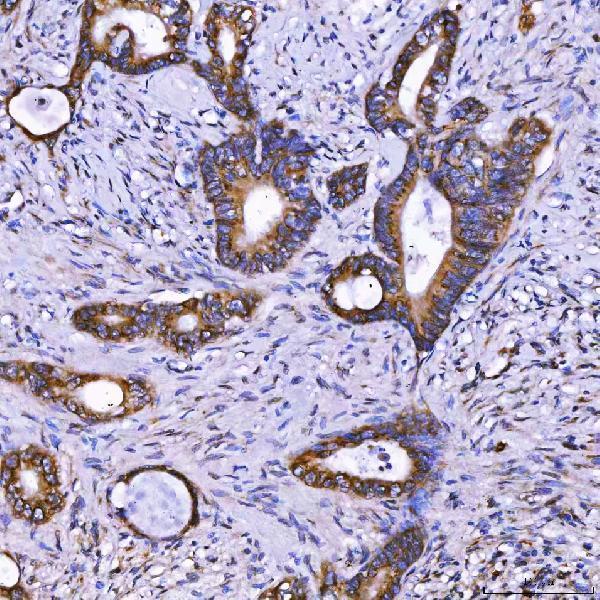
Real IHC data IHC analysis of TOM1L1 using anti-TOM1L1 antibody (A07598-2). TOM1L1 was detected in a paraffin-embedded section of human colon cancer tissue. Heat mediated antigen retrieval was performed in EDTA buffer (pH 8.0, epitope retrieval solution). The tissue section was blocked with 10% goat serum. The tissue section was then incubated with 2 μg/ml rabbit anti-TOM1L1 Antibody (A07598-2) overnight at 4°C. Peroxidase Conjugated Goat Anti-rabbit IgG was used as secondary antibody and incubated for 30 minutes at 37°C. The tissue section was developed using HRP Conjugated Rabbit IgG Super Vision Assay Kit (Catalog # SV0002) with DAB as the chromogen.
Anti-TOM1L1 Antibody ®
Cat # A07598-2

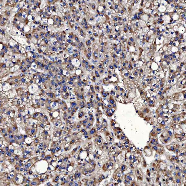
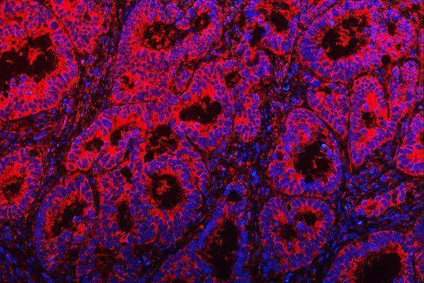
A07598-2 is listed for IHC and IF (catalog: applications). Its IHC images show human colon, liver, lung and thyroid cancer paraffin sections; its IF images show human rectal and ovarian cancer paraffin sections (catalog image captions).

Which to pick: For chromogenic tissue IHC, choose A07598-2: its IHC caption documents a human colon cancer paraffin section, EDTA retrieval at pH 8.0, and 2 μg/ml primary antibody with HRP/DAB detection (catalog: A07598-2 IHC caption); the fixative is unreported (catalog: A07598-2 IHC caption). For tissue IF, A07598-2 has paraffin section images at 5 μg/ml (catalog: A07598-2 IF captions); ICC validation is unreported (catalog: applications and image captions). For cross-species work, A07598-2 lists human, mouse and rat reactivity, while its documented IHC and IF images use human tissue (catalog: A07598-2 reactivity and image captions).

Each figure is that product's own IHC / IF validation image from its datasheet.

References

  1. UniProt Consortium. UniProt entry O75674 (TM1L1_HUMAN, TOM1-like protein 1).
  2. Human Protein Atlas. TOM1L1 tissue IHC expression (reliability: Uncertain).
  3. Human Protein Atlas. TOM1L1 subcellular location (ICC-IF): Highest expression in RPTEC/TERT1: 100.3 nTPM.
  4. Human Protein Atlas. TOM1L1 antibody validation summary (1 antibodies).
  5. TOM1L1 drives membrane delivery of MT1-MMP to promote ERBB2-induced breast cancer cell invasion. Nature communications 2016 — PMC4764922.
  6. Exploring the correlation between Tom1L1 and the efficacy of neoadjuvant chemotherapy for locally progressive mid-low rectal cancer. BMC cancer 2024 — PMC11568529.
  7. Identification and Validation of the Prognostic Panel in Clear Cell Renal Cell Carcinoma Based on Resting Mast Cells for Prediction of Distant Metastasis and Immunotherapy Response. Cells 2023 — PMC9818872.
  8. PubMed PMID:10329004 — UniProt-cited evidence.
  9. PubMed PMID:14702039 — UniProt-cited evidence.
  10. PubMed PMID:16625196 — UniProt-cited evidence.