This website uses cookies to ensure you get the best experience on our website.


- Table of Contents
Plan TOP2B paraffin IHC around its widespread nuclear tissue staining (HPA tissue IHC). Start the IHC-validated antibody at 1:100–1:300 (datasheet: A02750), using bone marrow hematopoietic cells as a positive tissue control (HPA tissue IHC).

Expected localisation, validated protocols, controls and antibodies — the at-a-glance facts below, then the full design guide.
| Expected localisation | Nuclear staining in tissue (HPA tissue IHC) | |
| Staining pattern | Widespread nuclear staining across tissue cell types (HPA tissue IHC) | |
| Antigen retrieval | Tris-EDTA pH 9.0 HIER, 95–98 °C, 20 min (rule: nuclear antigen) | |
| Positive control | Appendix+4 more · see all | |
| Negative control | None in HPA (detected in all 45 tissues); use no-primary + isotype controls |
| Fixation | Matched tissue-IHC evidence does not establish the fixation claim. Validate the specimen-specific method before use. (selected-SKU IHC image A02750) | |
| Caveat | Adipocytes may stain weakly despite the broad tissue pattern (HPA tissue IHC) | |
| Regulation | No intensity regulator reported (UniProt) | |
| Isoform / epitope | Two isoforms, Beta-1 and Beta-2; check epitope coverage (UniProt) |
The catalog antibody IHC-P protocol is accompanied by 2 published TOP2B protocols using paraffin sections (PMC13060179 methods; PMC13223395 methods).
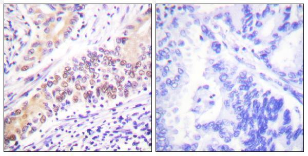
| Sample | Paraffin-embedded human lung carcinoma tissue; fixative not specified (datasheet A02750) |
| Fixation | Image fixative and duration unreported (datasheet A02750); verify before use. |
| Sectioning | 4–5 µm sections on charged slides (standard) |
| Deparaffinisation | Xylene, graded ethanol series to water (standard) |
| Antigen retrieval | Heat-induced epitope retrieval in Tris-EDTA buffer, pH 9.0, 20 min at 95–98 °C (standard rule: nuclear antigen) |
| Peroxidase block | 3% H2O2, 10 min, room temperature (standard) |
| Blocking | 10% normal serum of the secondary host, 30 min, room temperature (standard) |
| Primary antibody | Rabbit anti-TOP2B, 1:100 - 1:300 (datasheet A02750) |
| Primary incubation | Overnight at 4 °C (standard) |
| Detection | HRP-polymer secondary, DAB chromogen 5–10 min (standard) |
| Counterstain | Hematoxylin, blue, dehydrate and mount (standard) |
| Expected result | TOP2B-positive staining in glandular cells of appendix (HPA tissue IHC: High). HPA tissue profile: Ubiquitous nuclear expression. No signal in the no-primary control. |
TOP2B should stain nuclei across many cell types: HPA reports ubiquitous nuclear expression with supported IHC reliability and low tissue RNA specificity (HPA: tissue IHC). UniProt places TOP2B in the nucleoplasm and nucleolus and reports no transmembrane segment (UniProt Q02880: subcellular location; topology). Assess staining in the relevant cells rather than treating an entire tissue section as uniformly positive (HPA: tissue IHC).
| Clear nuclear staining in glandular or hematopoietic cells. | This fits reported high staining in appendix, cervix, duodenum and epididymis glandular cells, and bone marrow hematopoietic cells (HPA: tissue IHC). Judge whether the signal is centered on nuclei with visible tissue structure; intensity alone does not establish antibody specificity (general IHC practice). |
| Predominantly cytoplasmic or membranous staining with little nuclear signal. | This conflicts with the reported nuclear pattern and the absence of a transmembrane segment (HPA: tissue IHC; UniProt Q02880: topology). Consider nonspecific staining or a detection artefact before interpreting it as TOP2B. Recheck the staining pattern against a known-positive section (general IHC practice). |
| Strong signal confined to an unexpected cell population. | Compare the cells with the tissue-specific HPA entries before calling the pattern abnormal: TOP2B expression is broadly distributed, and HPA supplies no negative tissue list here (HPA: tissue IHC). If the signal remains discordant, investigate cross-reactivity or endogenous detection activity with appropriate controls (general IHC practice). |
| Diffuse color across nuclei, cytoplasm and extracellular spaces. | Diffuse background obscures the nuclear pattern reported for TOP2B (HPA: tissue IHC). Uneven reagent coverage, inadequate blocking or insufficient washing can produce background in chromogenic IHC; a no-primary control helps distinguish detection background from primary-antibody-associated signal (general IHC practice). |
| No nuclear staining in a section expected to be positive. | Absent signal in an HPA high-staining cell population warrants a technical check, but it does not establish that TOP2B is absent from the sample (HPA: tissue IHC; general IHC practice). Compare a known-positive section processed alongside it, then review retrieval, antibody dilution and detection steps (general IHC practice). |
| Cell and tissue context | HPA reports high staining in selected glandular, hematopoietic and glial populations, plus cells in the cerebellar molecular layer; adipocytes are listed as low (HPA: tissue IHC). Compare like cell populations when judging intensity, since these entries do not define a universal negative tissue (HPA: tissue IHC). |
| Antibody-specific validation | HPA lists IHC as Supported for HPA024120 and CAB004601; no IHC status is supplied for HPA050441 (HPA: antibody validation). These ratings support use of the reported patterns as references, but do not validate every catalog antibody or every staining condition (HPA: antibody validation; general IHC practice). |
| IF/ICC Q&A: where should fluorescence appear? | Mainly in the nucleoplasm; HPA calls this location enhanced in its ICC-IF record, while UniProt also lists the nucleolus (HPA: subcellular ICC-IF; UniProt Q02880: subcellular location). This localization answer does not specify an IF/ICC protocol. |
| Isoforms and epitope coverage | UniProt lists Beta-1 and Beta-2 isoforms, but the supplied record does not map an antibody epitope to either one (UniProt Q02880: isoforms). An unexpected pattern cannot be assigned to isoform recognition from these data alone; consult the chosen antibody's documented epitope before making that interpretation (general IHC practice). |
| Situation | Likely cause | Next action |
|---|---|---|
| Nuclei are weak in an HPA high-staining cell population. | The result conflicts with the reported high staining, but the payload provides no TOP2B-specific retrieval or fixation sensitivity data (HPA: tissue IHC). | Run a known-positive section in the same batch. Check the selected antibody's documented IHC-P retrieval, dilution and detection settings, then review section quality and reagent performance (general IHC practice). |
| Only cytoplasm or cell borders stain. | The compartment conflicts with nuclear localization and the reported lack of a transmembrane segment (HPA: tissue IHC; UniProt Q02880: topology). | Compare with a known-positive section and a no-primary control. Reassess antibody specificity and detection background before scoring the signal as TOP2B (general IHC practice). |
| Background covers the whole section. | A widespread deposit prevents reliable assessment of the expected nuclear pattern (HPA: tissue IHC). Background can arise during staining or chromogenic detection (general IHC practice). | Inspect no-primary and reagent controls; check washing, blocking and detection conditions, then repeat with the catalog antibody's documented IHC-P settings (general IHC practice). |
| A surprising cell type stains strongly. | HPA describes ubiquitous nuclear expression but reports intensity by cell population; a surprising signal is not automatically cross-reactivity (HPA: tissue IHC). | Identify the stained cells and compare them with HPA's matching tissue entry. If the pattern remains discordant, check no-primary controls and, where available, an independently validated antibody (HPA: tissue IHC; general IHC practice). |
| Adipocytes show strong, widespread staining. | HPA lists adipocytes as low, so strong staining merits scrutiny; the low entry is not proof that every adipocyte must be unstained (HPA: tissue IHC). | Confirm whether the deposit is nuclear, compare nearby cell populations, and inspect a no-primary control for background or endogenous detection activity (HPA: tissue IHC; general IHC practice). |
| Two antibodies produce different patterns. | The supplied HPA statuses differ by antibody and application; they do not establish equivalent IHC performance for all reagents (HPA: antibody validation). | Compare each antibody's documented IHC-P conditions and epitope information, then judge which pattern agrees with the HPA nuclear reference in matched cells (HPA: tissue IHC; general IHC practice). |


Comprehensive Human Protein Atlas IHC scoring per tissue (reliability: Supported — High consistency between antibody staining and RNA expression data.). Rows are taken directly from the HPA tissue chart — click any row's HPA link to view the source.
| Tissue | Cell type | Level | Evidence | Source |
|---|---|---|---|---|
| Appendix | Glandular cells | High | Protein (IHC) | HPA → |
| Bone marrow | Hematopoietic cells | High | Protein (IHC) | HPA → |
| Caudate | Glial cells | High | Protein (IHC) | HPA → |
| Cerebellum | Cells in molecular layer | High | Protein (IHC) | HPA → |
| Cerebral cortex | Glial cells | High | Protein (IHC) | HPA → |
| Tissue | Cell type | Level | Evidence | Source |
|---|---|---|---|---|
| None in HPA: TOP2B is detected in all 45 scored tissues. Use a no-primary (secondary-only) and an isotype control instead. | ||||
Troubleshoot TOP2B chromogenic IHC by assessing nuclear staining, section quality, and controls; IF/ICC considerations are included separately (UniProt Q02880 localisation; HPA tissue IHC).
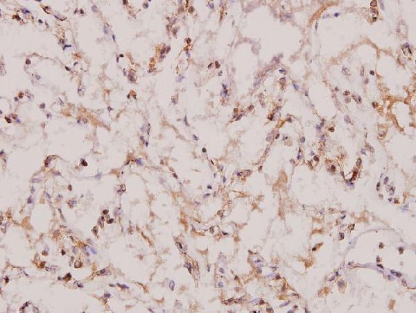
Two anti-TOP2B antibodies have IHC images from paraffin-embedded human lung carcinoma (each SKU’s image caption); A02750 also lists IF/ICC for human and mouse (catalog applications and reactivity), without an IF image (catalog images).
A02750 will render with an IHC image of paraffin-embedded human lung carcinoma, including a peptide-blocked comparison; it lists human and mouse reactivity and IF/ICC applications (A02750 image caption; catalog). A02750-1 will render with an IHC image of paraffin-embedded human lung carcinoma at 1:100; it lists human, mouse and rat reactivity (A02750-1 image caption; catalog).
Which to pick: For tissue IHC, either SKU has its own paraffin-section image; choose A02750 if the peptide-blocked comparison is useful (each SKU’s image caption). For IF/ICC, choose A02750 because it lists those applications and is polyclonal, although no IF image is supplied (A02750 catalog applications, dilution record and images). For rat samples, A02750-1 is the listed option; its IHC caption supports paraffin sections, but reports no fixative (A02750-1 catalog reactivity and image caption).