This website uses cookies to ensure you get the best experience on our website.


- Table of Contents
Use muscle tissues with high TPM2 staining as positive controls (HPA tissue IHC). This guide covers consistent fixation and chromogenic paraffin-section IHC, with attention to possible antibody cross-reactivity (standard IHC practice; HPA tissue IHC).

Expected localisation, validated protocols, controls and antibodies — the at-a-glance facts below, then the full design guide.
| Expected localisation | Cytoplasmic F-actin association expected; IHC site unverified (UniProt) | |
| Staining pattern | High in cardiac, skeletal and smooth muscle cells; site unreported (HPA tissue IHC) | |
| Antigen retrieval | EDTA pH 8.0 HIER, heat-mediated (datasheet A03082-1) | |
| Positive control | Heart muscle+4 more · see all | |
| Negative control | Adipose tissue+4 more · see all |
| Fixation | Keep paraffin-section fixation consistent across samples. (standard IHC practice; not target-specific) | |
| Caveat | Antibody may detect proteins from other genes (HPA tissue IHC) | |
| Regulation | Breast cancer-associated expression (UniProt) | |
| Isoform / epitope | 3 isoforms; check cytoplasmic epitope coverage (UniProt) |
The catalog antibody’s IHC-P protocol is accompanied by published TPM2 staining details from four PMC articles.
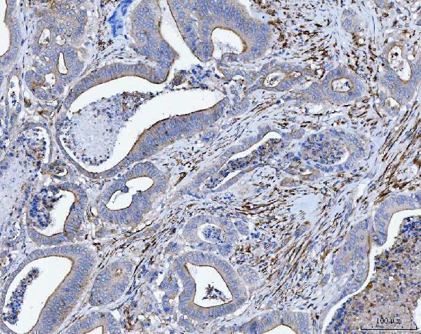
| Sample | Paraffin-embedded human colorectal adenocarcinoma tissue; fixative not specified (datasheet A03082-1) |
| Fixation | Image fixative and duration unreported (datasheet A03082-1); verify before use. |
| Sectioning | 4–5 µm sections on charged slides (standard) |
| Deparaffinisation | Xylene, graded ethanol series to water (standard) |
| Antigen retrieval | Heat retrieval: EDTA pH 8.0 (datasheet A03082-1); 20 min, 95–100 °C (standard) |
| Peroxidase block | 3% H2O2, 10 min, room temperature (standard) |
| Blocking | 10% goat serum (datasheet A03082-1) |
| Primary antibody | Rabbit anti-TPM2, 2-5 µg/ml (datasheet A03082-1) |
| Primary incubation | Overnight at 4 °C (datasheet A03082-1) |
| Detection | HRP-conjugated secondary, DAB chromogen (datasheet A03082-1) |
| Counterstain | Hematoxylin, blue, dehydrate and mount (standard) |
| Expected result | TPM2-positive staining in cardiomyocytes of heart muscle (HPA tissue IHC: High). HPA tissue profile: Expression in several tissues, most abundant in skeletal, heart and smooth muscle. No signal in the no-primary control. |
TPM2 should appear in the cytoplasm along actin-associated structures, especially in cardiomyocytes, skeletal myocytes, and smooth muscle cells (UniProt P07951: cytoskeleton and F-actin stress fibers; HPA: High in these cells). It has no transmembrane segment (UniProt P07951: topology). HPA rates its tissue IHC staining Approved but reports low agreement with RNA data and cautions that the antibodies may detect products of more than one gene (HPA: reliability description).
| Strong cytoplasmic staining in heart, skeletal, or smooth muscle, with little nuclear staining. | This fits the expected location and the strongest reported tissue patterns (UniProt P07951: cytoskeleton; HPA: High in cardiomyocytes, myocytes, and smooth muscle cells). Judge the pattern within identifiable cells; HPA's multi-gene caution limits how confidently staining can be assigned to TPM2 alone (HPA: reliability description). |
| Predominantly nuclear or crisp cell-surface staining, without a convincing cytoplasmic component. | Treat this as a compartment mismatch requiring investigation, since TPM2 associates with F-actin stress fibers and has no transmembrane segment (UniProt P07951: subcellular location and topology). Review morphology and the detection controls before scoring it as specific staining (general IHC practice). |
| Strong staining in an unexpected cell population, especially where HPA reports no detection. | For example, adipocytes and lymph-node non-germinal center cells are reported as Not detected (HPA: tissue IHC). Unexpected signal may reflect cross-reactivity or endogenous detection activity (general IHC practice); HPA also cautions that the antibodies may recognize products of more than one gene (HPA: reliability description). |
| Diffuse color across cells and surrounding tissue, with poor separation of compartments. | A widespread haze is difficult to interpret as TPM2 because the expected location is cytoskeletal (UniProt P07951: subcellular location). Check background with a primary-antibody omission control, then review blocking, washing, antibody concentration, and chromogen development as general IHC variables (general IHC practice). |
| No staining in a section expected to provide a strong positive control. | Heart, skeletal muscle, and smooth muscle contain cell populations scored High by HPA (HPA: tissue IHC). A blank result there calls for a check of tissue preservation, staining reagents, detection, and the catalog antibody's validated IHC-P conditions (general IHC practice); it does not by itself establish TPM2 absence. |
| Tissue and cell selection | Use heart, skeletal muscle, or smooth muscle for a high-signal reference (HPA: High). Leydig cells are also scored High, while breast glandular cells are scored Medium (HPA: tissue IHC). Score the named cell population rather than treating every cell in a tissue as equally positive (HPA: cell-level entries). |
| Specificity of the observed signal | HPA classifies tissue staining as Approved but reports low agreement with RNA and cautions that antibodies may detect products of more than one gene (HPA: reliability description). Its listed IHC antibodies, HPA047089 and HPA053624, are each Approved; neither is listed as Enhanced (HPA: antibody validation). |
| Isoforms and intracellular location | UniProt lists three TPM2 isoforms and describes an F-actin-associated cytoskeletal protein without a transmembrane segment (UniProt P07951: isoforms, location, topology). Isoform recognition cannot be inferred without antibody epitope information; avoid assigning a distinct staining pattern to any isoform from these sources alone. |
| Breast-tissue interpretation | UniProt reports TPM2 present in primary breast cancer tissue and absent from normal breast tissue, while HPA scores normal breast glandular cells Medium by IHC (UniProt P07951: tissue specificity; HPA: breast). Treat this source disagreement and HPA's multi-gene caution as limits on using breast staining alone to establish TPM2 specificity. |
| Situation | Likely cause | Next action |
|---|---|---|
| Known-positive muscle is blank or unexpectedly weak. | A control that HPA scores High has failed to show the expected signal (HPA: heart, skeletal muscle, smooth muscle). | Confirm the named cells are present; check the catalog antibody's IHC-P instructions, retrieval and dilution settings, reagent activity, and detection controls (general IHC practice). No TPM2-specific retrieval condition is supplied here. |
| Apparent nuclear or membrane-only staining dominates. | The pattern conflicts with TPM2's cytoskeletal location and lack of a transmembrane segment (UniProt P07951: location and topology). | Compare with a known-positive muscle section and an omission control; reassess cell boundaries and background before calling the compartment positive (HPA: High in muscle; general IHC practice). |
| Negative-reference cells show strong staining. | Adipocytes and lymph-node non-germinal center cells are reported Not detected; cross-reactivity or endogenous detection activity is possible (HPA: tissue IHC; general IHC practice). | Run a primary-antibody omission control, check endogenous detection activity, and interpret any residual signal cautiously given HPA's multi-gene warning (general IHC practice; HPA: reliability description). |
| Diffuse brown background obscures cell-level scoring. | General IHC causes include insufficient blocking or washing, excessive antibody concentration, or prolonged chromogen development (general IHC practice). | Use an omission control to locate background, then optimize those workflow variables against a known-positive muscle control (general IHC practice; HPA: High in muscle). |
| Breast staining appears inconsistent across the reference sources. | HPA reports Medium staining in normal breast glandular cells, whereas UniProt reports absence from normal breast tissue (HPA: breast; UniProt P07951: tissue specificity). | Record the observed cells and staining intensity, compare with muscle controls, and avoid using breast staining as sole evidence of specificity (HPA: reliability caution; general IHC practice). |
| IF/ICC: what pattern can be expected? | An actin-associated cytoplasmic pattern is plausible from UniProt, but HPA supplies no main subcellular location or ICC-IF images for this entry (UniProt P07951: F-actin stress fibers; HPA: subcellular record). | Treat that pattern as a hypothesis to validate with suitable IF/ICC controls; the HPA tissue IHC result alone does not establish an IF/ICC pattern (HPA: subcellular record; general IF practice). |
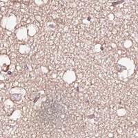


Comprehensive Human Protein Atlas IHC scoring per tissue (reliability: Approved — Low consistency between antibody staining and RNA expression data. Caution, targets protein from more than one gene. Pending external verification.). Rows are taken directly from the HPA tissue chart — click any row's HPA link to view the source.
| Tissue | Cell type | Level | Evidence | Source |
|---|---|---|---|---|
| Heart muscle | Cardiomyocytes | High | Protein (IHC) | HPA → |
| Skeletal muscle | Myocytes | High | Protein (IHC) | HPA → |
| Smooth muscle | Smooth muscle cells | High | Protein (IHC) | HPA → |
| Testis | Leydig cells | High | Protein (IHC) | HPA → |
| Adrenal gland | Glandular cells | Medium | Protein (IHC) | HPA → |
| Tissue | Cell type | Level | Evidence | Source |
|---|---|---|---|---|
| Adipose tissue | Adipocytes | Not detected | Protein (IHC) | HPA → |
| Lymph node | Non-germinal center cells | Not detected | Protein (IHC) | HPA → |
| Parathyroid gland | Glandular cells | Not detected | Protein (IHC) | HPA → |
| Small intestine | Glandular cells | Not detected | Protein (IHC) | HPA → |
| Spleen | Cells in red pulp | Not detected | Protein (IHC) | HPA → |
Troubleshoot TPM2 staining in paraffin sections by checking retrieval, controls, cellular localisation and scoring before interpreting chromogenic signal.
A03082-1 has real IHC and IF images from human paraffin sections (A03082-1 image captions); the catalog lists human, mouse and rat reactivity (catalog reactivity).
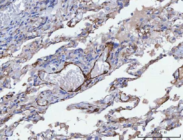
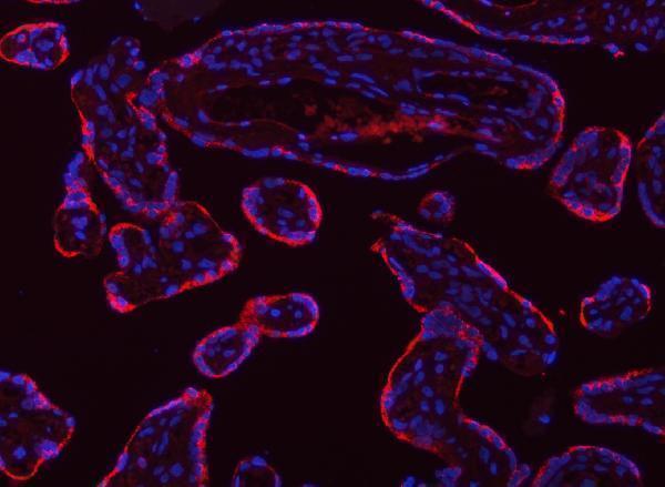
A03082-1 is listed for IHC and IF with human, mouse and rat reactivity (catalog applications and reactivity). Its captions show IHC in human colorectal adenocarcinoma, lung adenocarcinoma, placenta and spleen paraffin sections, and IF in human placenta paraffin sections (A03082-1 image captions).
Which to pick: For tissue IHC, choose A03082-1: its colorectal adenocarcinoma paraffin-section caption documents EDTA retrieval at pH 8.0 and 2 µg/ml primary antibody; the fixative is unreported (A03082-1 IHC caption). For IF, A03082-1 has a human placenta paraffin-section example at 5 μg/ml; no ICC example is shown (A03082-1 IF caption and catalog image captions). For cross-species work, A03082-1 lists mouse and rat reactivity, but the displayed IHC and IF examples are human (catalog reactivity; A03082-1 image captions).