Click image to see more details
-

-
-
-
-
+5
Product Info Summary
| SKU: | A03082-1 |
|---|---|
| Size: | 100 μg/vial |
| Reactive Species: | Human, Mouse, Rat |
| Host: | Rabbit |
| Application: | Flow Cytometry, IF, IHC, WB |
Customers Who Bought This Also Bought

Product info
Product Name
Anti-Tropomyosin 2/TPM2 Antibody Picoband®
SKU/Catalog Number
A03082-1
Size
100 μg/vial
Form
Lyophilized
Description
Boster Bio Anti-Tropomyosin 2/TPM2 Antibody Picoband® catalog # A03082-1. Tested in Flow Cytometry, IF, IHC, WB applications. This antibody reacts with Human, Mouse, Rat. The brand Picoband indicates this is a premium antibody that guarantees superior quality, high affinity, and strong signals with minimal background in Western blot applications. Only our best-performing antibodies are designated as Picoband, ensuring unmatched performance.
Storage & Handling
At -20°C for one year from date of receipt. After reconstitution, at 4°C for one month. It can also be aliquotted and stored frozen at -20°C for six months. Avoid repeated freezing and thawing.
Cite This Product
Anti-Tropomyosin 2/TPM2 Antibody Picoband® (Boster Biological Technology, Pleasanton CA, USA, Catalog # A03082-1)
Host
Rabbit
Contents
Each vial contains 4 mg Trehalose, 0.9 mg NaCl, 0.2 mg Na2HPO4.
Clonality
Polyclonal
Isotype
Rabbit IgG
Immunogen
A synthetic peptide corresponding to a sequence at the N-terminus of human Tropomyosin 2/TPM2, identical to the related mouse and rat sequences.
Cross-reactivity
No cross-reactivity with other proteins.
Reactive Species
A03082-1 is reactive to TPM2 in Human, Mouse, Rat
Observed Molecular Weight
38 kDa
Calculated molecular weight
32.9 kDa
Background of TPM2
β-Tropomyosin, also known as tropomyosin beta chain is a protein that in humans is encoded by the TPM2 gene. This gene encodes beta-tropomyosin, a member of the actin filament binding protein family, and mainly expressed in slow, type 1 muscle fibers. Mutations in this gene can alter the expression of other sarcomeric tropomyosin proteins, and cause cap disease, nemaline myopathy and distal arthrogryposis syndromes. Alternatively spliced transcript variants encoding different isoforms have been found for this gene.
Antibody Validation
Boster validates all antibodies on WB, IHC, ICC, Immunofluorescence, and ELISA with known positive control and negative samples to ensure specificity and high affinity, including thorough antibody incubations.
Application & Images
Applications
A03082-1 is guaranteed for Flow Cytometry, IF, IHC, WB Boster Guarantee
Assay Dilutions Recommendation
The recommendations below provide a starting point for assay optimization. The actual working concentration varies and should be decided by the user.
Western blot, 0.25-0.5 µg/ml, Human, Mouse, Rat
Immunohistochemistry(Paraffin-embedded Section), 2-5 µg/ml, Human
Immunofluorescence, 5 μg/ml, Human
Flow Cytometry (Fixed), 1-3 µg/1x106 cells, Human, Mouse, Rat
Positive Control
WB: human Hela whole cell, rat H9C2(2-1) whole cell, mouse skeletal muscle tissue
IHC: human colorectal adenocarcinoma tissue, human lung adenocarcinoma tissue, human placenta tissue, human spleen tissue
IF: human placenta tissue
FCM: SiHa cell, ANA-1 cell, RH35 cell
Validation Images & Assay Conditions
Click image to see more details
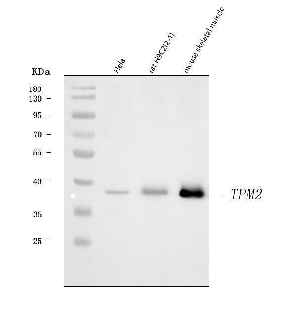
Western blot analysis of Tropomyosin 2/TPM2 using anti-Tropomyosin 2/TPM2 antibody (A03082-1).
Electrophoresis was performed on a 5-20% SDS-PAGE gel at 70V (Stacking gel) / 90V (Resolving gel) for 2-3 hours. The sample well of each lane was loaded with 30 ug of sample under reducing conditions.
Lane 1: human Hela whole cell lysates,
Lane 2: rat H9C2(2-1) whole cell lysates,
Lane 3: mouse skeletal muscle tissue lysates.
After electrophoresis, proteins were transferred to a nitrocellulose membrane at 150 mA for 50-90 minutes. Blocked the membrane with 5% non-fat milk/TBS for 1.5 hour at RT. The membrane was incubated with rabbit anti-Tropomyosin 2/TPM2 antigen affinity purified polyclonal antibody (Catalog # A03082-1) at 0.5 μg/mL overnight at 4°C, then washed with TBS-0.1%Tween 3 times with 5 minutes each and probed with a goat anti-rabbit IgG-HRP secondary antibody at a dilution of 1:5000 for 1.5 hour at RT. The signal is developed using an Enhanced Chemiluminescent detection (ECL) kit (Catalog # EK1002) with Tanon 5200 system. A specific band was detected for Tropomyosin 2/TPM2 at approximately 38 kDa. The expected band size for Tropomyosin 2/TPM2 is at 33 kDa.

Click image to see more details
IHC analysis of Tropomyosin 2/TPM2 using anti-Tropomyosin 2/TPM2 antibody (A03082-1).
Tropomyosin 2/TPM2 was detected in a paraffin-embedded section of human colorectal adenocarcinoma tissue. Heat mediated antigen retrieval was performed in EDTA buffer (pH 8.0, epitope retrieval solution). The tissue section was blocked with 10% goat serum. The tissue section was then incubated with 2 μg/ml rabbit anti-Tropomyosin 2/TPM2 Antibody (A03082-1) overnight at 4°C. Peroxidase Conjugated Goat Anti-rabbit IgG was used as secondary antibody and incubated for 30 minutes at 37°C. The tissue section was developed using HRP Conjugated Rabbit IgG Super Vision Assay Kit (Catalog # SV0002) with DAB as the chromogen.

Click image to see more details
IHC analysis of Tropomyosin 2/TPM2 using anti-Tropomyosin 2/TPM2 antibody (A03082-1).
Tropomyosin 2/TPM2 was detected in a paraffin-embedded section of human lung adenocarcinoma tissue. Heat mediated antigen retrieval was performed in EDTA buffer (pH 8.0, epitope retrieval solution). The tissue section was blocked with 10% goat serum. The tissue section was then incubated with 2 μg/ml rabbit anti-Tropomyosin 2/TPM2 Antibody (A03082-1) overnight at 4°C. Peroxidase Conjugated Goat Anti-rabbit IgG was used as secondary antibody and incubated for 30 minutes at 37°C. The tissue section was developed using HRP Conjugated Rabbit IgG Super Vision Assay Kit (Catalog # SV0002) with DAB as the chromogen.

Click image to see more details
IHC analysis of Tropomyosin 2/TPM2 using anti-Tropomyosin 2/TPM2 antibody (A03082-1).
Tropomyosin 2/TPM2 was detected in a paraffin-embedded section of human placenta tissue. Heat mediated antigen retrieval was performed in EDTA buffer (pH 8.0, epitope retrieval solution). The tissue section was blocked with 10% goat serum. The tissue section was then incubated with 2 μg/ml rabbit anti-Tropomyosin 2/TPM2 Antibody (A03082-1) overnight at 4°C. Peroxidase Conjugated Goat Anti-rabbit IgG was used as secondary antibody and incubated for 30 minutes at 37°C. The tissue section was developed using HRP Conjugated Rabbit IgG Super Vision Assay Kit (Catalog # SV0002) with DAB as the chromogen.

Click image to see more details
IHC analysis of Tropomyosin 2/TPM2 using anti-Tropomyosin 2/TPM2 antibody (A03082-1).
Tropomyosin 2/TPM2 was detected in a paraffin-embedded section of human spleen tissue. Heat mediated antigen retrieval was performed in EDTA buffer (pH 8.0, epitope retrieval solution). The tissue section was blocked with 10% goat serum. The tissue section was then incubated with 2 μg/ml rabbit anti-Tropomyosin 2/TPM2 Antibody (A03082-1) overnight at 4°C. Peroxidase Conjugated Goat Anti-rabbit IgG was used as secondary antibody and incubated for 30 minutes at 37°C. The tissue section was developed using HRP Conjugated Rabbit IgG Super Vision Assay Kit (Catalog # SV0002) with DAB as the chromogen.

Click image to see more details
Flow Cytometry analysis of SiHa cells using anti-Tropomyosin 2/TPM2 antibody (A03082-1).
Overlay histogram showing SiHa cells stained with A03082-1 (Blue line). To facilitate intracellular staining, cells were fixed with 4% paraformaldehyde and permeabilized with permeabilization buffer. The cells were blocked with 10% normal goat serum. And then incubated with rabbit anti-Tropomyosin 2/TPM2 Antibody (A03082-1, 1 μg/1x106 cells) for 30 min at 20°C. DyLight®488 conjugated goat anti-rabbit IgG (BA1127, 5-10 μg/1x106 cells) was used as secondary antibody for 30 minutes at 20°C. Isotype control antibody (Green line) was rabbit IgG (1 μg/1x106) used under the same conditions. Unlabelled sample without incubation with primary antibody and secondary antibody (Red line) was used as a blank control.

Click image to see more details
Flow Cytometry analysis of ANA-1 cells using anti-Tropomyosin 2/TPM2 antibody (A03082-1).
Overlay histogram showing ANA-1 cells stained with A03082-1 (Blue line). To facilitate intracellular staining, cells were fixed with 4% paraformaldehyde and permeabilized with permeabilization buffer. The cells were blocked with 10% normal goat serum. And then incubated with rabbit anti-Tropomyosin 2/TPM2 Antibody (A03082-1, 1 μg/1x106 cells) for 30 min at 20°C. DyLight®488 conjugated goat anti-rabbit IgG (BA1127, 5-10 μg/1x106 cells) was used as secondary antibody for 30 minutes at 20°C. Isotype control antibody (Green line) was rabbit IgG (1 μg/1x106) used under the same conditions. Unlabelled sample without incubation with primary antibody and secondary antibody (Red line) was used as a blank control.

Click image to see more details
Flow Cytometry analysis of RH35 cells using anti-Tropomyosin 2/TPM2 antibody (A03082-1).
Overlay histogram showing RH35 cells stained with A03082-1 (Blue line). To facilitate intracellular staining, cells were fixed with 4% paraformaldehyde and permeabilized with permeabilization buffer. The cells were blocked with 10% normal goat serum. And then incubated with rabbit anti-Tropomyosin 2/TPM2 Antibody (A03082-1, 1 μg/1x106 cells) for 30 min at 20°C. DyLight®488 conjugated goat anti-rabbit IgG (BA1127, 5-10 μg/1x106 cells) was used as secondary antibody for 30 minutes at 20°C. Isotype control antibody (Green line) was rabbit IgG (1 μg/1x106) used under the same conditions. Unlabelled sample without incubation with primary antibody and secondary antibody (Red line) was used as a blank control.

Click image to see more details
IF analysis of Tropomyosin 2/TPM2 using anti-Tropomyosin 2/TPM2 antibody (A03082-1).
Tropomyosin 2/TPM2 was detected in a paraffin-embedded section of human placenta tissue. Heat mediated antigen retrieval was performed in EDTA buffer (pH 8.0, epitope retrieval solution). The tissue section was blocked with 10% goat serum. The tissue section was then incubated with 5 μg/mL rabbit anti-Tropomyosin 2/TPM2 Antibody (A03082-1) overnight at 4°C. DyLight®550 Conjugated Goat Anti-Rabbit IgG (BA1135) was used as secondary antibody at 1:500 dilution and incubated for 30 minutes at 37°C. The section was counterstained with DAPI. Visualize using a fluorescence microscope and filter sets appropriate for the label used.
Specific Publications For Anti-Tropomyosin 2/TPM2 Antibody Picoband® (A03082-1)
Loading publications
Recommended Resources
Here are featured tools and databases that you might find useful.
- Boster's Pathways Library
- Protein Databases
- Bioscience Research Protocol Resources
- Data Processing & Analysis Software
- Photo Editing Software
- Scientific Literature Resources
- Research Paper Management Tools
- Molecular Biology Software
- Primer Design Tools
- Bioinformatics Tools
- Phylogenetic Tree Analysis
Customer Reviews
Have you used Anti-Tropomyosin 2/TPM2 Antibody Picoband®?
Share your experimental results or join a short interview to earn up to $1,000 in product credits or other rewards.
0 Reviews For Anti-Tropomyosin 2/TPM2 Antibody Picoband®
Customer Q&As
Have a question?
Find answers in Q&As, reviews.
Can't find your answer?
Submit your question
