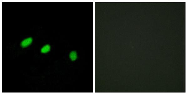
TRA2A / Transformer-2 protein homolog alpha · IHC design guide

Design Immunohistochemistry for TRA2A

Plan TRA2A paraffin IHC using the reported cytoplasmic staining in smooth muscle cells (HPA tissue IHC). Compare the observed cell type and compartment with controls, while accounting for the annotated nuclear location (UniProt) and uncertain tissue staining reliability (HPA tissue IHC).

Evidence assembled Oct 2026 · For research use; verify linked source records and product datasheet before use
Immunohistochemistry protocol sheet for TRA2A (IHC for TRA2A): expected localisation Cytoplasmic tissue staining (HPA tissue IHC); nuclear location (UniProt), antibody A03770, validated IHC image, and IHC protocol steps
Printable TRA2A IHC protocol sheet — expected localisation Cytoplasmic tissue staining (HPA tissue IHC); nuclear location (UniProt), antibody A03770, controls and protocol steps. Open the full TRA2A IHC guide →

TRA2A Immunohistochemistry Experimental Design Guide

Expected localisation, validated protocols, controls and antibodies — the at-a-glance facts below, then the full design guide.

Must know before staining
Expected localisation Cytoplasmic tissue staining (HPA tissue IHC); nuclear location (UniProt)
Staining pattern Cytoplasmic staining is most abundant in smooth muscle cells (HPA tissue IHC)
Antigen retrieval Tris-EDTA pH 9.0 HIER, 95–98 °C, 20 min (rule: nuclear antigen)
Positive control ⓘ Heart muscle+4 more · see all
Negative control ⓘ Adrenal gland+4 more · see all
Important caveats
Reasons your staining may differ from the expected pattern.
Fixation Matched tissue-IHC evidence does not establish the fixation claim. Validate the specimen-specific method before use. (selected-SKU IHC image A03770)
Caveat Tissue staining awaits external verification (HPA tissue IHC)
Regulation No expression regulator annotated (UniProt)
Isoform / epitope Four isoforms; epitope overlap is undetermined (UniProt)
Section 1

Recommended TRA2A IHC & IF Protocols

The catalog antibody’s IHC-P protocol is accompanied by one published TRA2A IHC protocol (PMC9275993).

Recommended immunohistochemistry (IHC-P) protocol parameters
SampleParaffin-embedded human brain tissue; fixative not specified (datasheet A03770)
FixationImage fixative and duration unreported (datasheet A03770); verify before use.
Sectioning4–5 µm sections on charged slides (standard)
DeparaffinisationXylene, graded ethanol series to water (standard)
Antigen retrievalHeat-induced epitope retrieval in Tris-EDTA buffer, pH 9.0, 20 min at 95–98 °C (standard rule: nuclear antigen)
Peroxidase block3% H2O2, 10 min, room temperature (standard)
Blocking10% normal serum of the secondary host, 30 min, room temperature (standard)
Primary antibodyRabbit anti-TRA2A, 1:100-1:300 (datasheet A03770)
Primary incubationOvernight at 4 °C (standard)
DetectionHRP-polymer secondary, DAB chromogen 5–10 min (standard)
CounterstainHematoxylin, blue, dehydrate and mount (standard)
Expected resultTRA2A-positive staining in cardiomyocytes of heart muscle (HPA tissue IHC: High). HPA tissue profile: Cytoplasmic expression, most abundant in smooth muscle cells. No signal in the no-primary control.
💡Decision noteStart with Tris-EDTA pH 9.0 at 95–98 °C for 20 min for nuclear TRA2A (page retrieval; UniProt Q13595); compare sodium citrate retrieval (PMC9275993).
Section 2

What Is the Expected TRA2A Staining Pattern?

For paraffin-section IHC, HPA reports predominantly cytoplasmic TRA2A staining, most abundant in smooth muscle cells; cardiomyocytes, kidney tubule cells and pancreatic exocrine cells also score High (HPA: tissue IHC). Interpret this cautiously: the tissue profile is Uncertain, and HPA054018 has Uncertain IHC validation (HPA: tissue IHC; HPA: antibody validation). UniProt places TRA2A in the nucleus and reports no transmembrane segment (UniProt Q13595: subcellular location and topology).

What am I looking at on my slide?
Strong cytoplasmic staining in smooth muscle cells, with staining in the expected cell populations of a comparison section.This matches the reported tissue IHC profile; cardiomyocytes, kidney tubule cells and pancreatic exocrine cells are also High (HPA: tissue IHC). It is a provisional positive pattern because the profile and HPA054018 IHC validation are Uncertain (HPA: tissue IHC; HPA: antibody validation).
Predominantly membrane-edge staining, with little signal in the expected cells or compartments.Treat a membrane-only pattern as suspect: TRA2A is annotated as nuclear and has no transmembrane segment (UniProt Q13595: location and topology). Check whether the signal follows section edges or persists when the primary antibody is omitted (general IHC practice).
Strong staining in cell populations scored Not detected, such as adrenal glandular cells or bronchial respiratory epithelial cells.Consider cross-reactivity or endogenous detection activity, especially if expected positive cells do not stain (HPA: tissue IHC; general IHC practice). A Not detected entry applies to the named cell population and does not make every cell in that tissue a negative control (HPA: tissue IHC).
A diffuse chromogen haze covers cells and surrounding tissue without clear cell boundaries.This cannot be scored as a TRA2A cell pattern; compare the no-primary control and inspect blocking, washing and detection background (general IHC practice). HPA's cytoplasmic tissue observation concerns stained cells, not uniform section haze (HPA: tissue IHC).
No convincing signal in smooth muscle cells on an otherwise intact section.The result conflicts with a High-scoring HPA cell population (HPA: tissue IHC). First check the run controls and detection steps, then review the catalog antibody's IHC-P instructions (general IHC practice). HPA's Uncertain reliability prevents calling this specimen TRA2A-negative from one failed stain (HPA: tissue IHC).
💡Expected TRA2A appearanceA provisional IHC positive shows discernible cytoplasmic staining strongest in smooth muscle cells, with High staining also reported in cardiomyocytes, kidney tubule cells and pancreatic exocrine cells; diffuse haze or strong signal in a named Not detected cell population warrants investigation (HPA: tissue IHC, Uncertain).
How each factor affects the staining
Tissue IHC evidence and antibody validationHPA calls the tissue profile Uncertain, pending external verification, and rates HPA054018 IHC Uncertain (HPA: tissue IHC; HPA: antibody validation). Treat agreement with the reported pattern as supporting evidence, not proof of antibody specificity.
Compartment differs by assayHPA tissue IHC describes cytoplasmic expression, while UniProt annotates a nuclear location (HPA: tissue IHC; UniProt Q13595: subcellular location). Keep assay and cell context attached to each observation; these sources do not resolve why the compartments differ.
Isoforms and processingUniProt lists 4 isoforms and a chain spanning residues 2–282, with no signal peptide or propeptide (UniProt Q13595: isoforms and processing). The payload gives no antibody epitope or isoform coverage, so an unexpected pattern cannot be assigned to an isoform.
IF/ICC Q: What localisation should be expected?A: HPA reports mainly nucleoli, with additional nucleoplasm and vesicles; nucleoli and nucleoplasm are Supported, and vesicles are Approved (HPA: subcellular ICC-IF). HPA054018 is ICC Supported (HPA: antibody validation). Apply that evidence to IF/ICC interpretation, not as an IHC-P protocol.
Why is my staining missing, weak or wrong?
SituationLikely causeNext action
The presumed positive section has no smooth muscle signal.The IHC run or detection may have failed; smooth muscle cells score High, although the tissue profile is Uncertain (HPA: tissue IHC; general IHC practice).Check that counterstain and run controls are acceptable, then review the catalog antibody's IHC-P instructions and detection reagents (general IHC practice). Repeat with a documented positive section before scoring the specimen negative.
The whole section is brown, including regions without identifiable positive cells.Diffuse background may reflect nonspecific detection or endogenous enzyme activity rather than cell-specific TRA2A signal (general IHC practice).Compare a no-primary control, inspect blocking and wash steps, and assess endogenous activity controls appropriate to the chromogen system (general IHC practice). Score only signal that resolves to cells.
Adrenal glandular cells or bronchial respiratory epithelial cells stain strongly.Those named populations are Not detected in HPA tissue IHC; cross-reactivity or detection background is possible, but the HPA profile is Uncertain (HPA: tissue IHC; general IHC practice).Confirm cell identity and compare the no-primary control with an HPA High cell population in the same run (HPA: tissue IHC; general IHC practice). Avoid interpreting one discrepant tissue as definitive specificity evidence.
Signal appears only at section edges or along membranes.Edge artefact or nonspecific deposition is plausible; UniProt annotates nuclear TRA2A without a transmembrane segment (UniProt Q13595: location and topology; general IHC practice).Inspect section quality and staining distribution, compare a no-primary control, and repeat on a well-preserved section if the pattern persists (general IHC practice).
Nuclear staining appears alongside the reported IHC cytoplasmic pattern.The sources disagree across assays: UniProt annotates nucleus, HPA tissue IHC describes cytoplasm, and HPA ICC-IF supports nucleoli and nucleoplasm (UniProt Q13595: location; HPA: tissue IHC; HPA: subcellular ICC-IF).Record nuclear and cytoplasmic staining separately by cell type. Compare positive and no-primary controls, and retain the IHC reliability caveat when interpreting either compartment (HPA: tissue IHC; general IHC practice).
Different specimens show different proportions of stained cells.HPA reports High, Medium, Low and Not detected levels in specified tissue cell populations, with Low tissue RNA specificity overall (HPA: tissue IHC). Those observations do not establish a universal intensity cutoff.Compare the same cell population across similarly processed sections, document intensity and distribution, and use a consistent run control (general IHC practice). Do not treat staining elsewhere in an HPA-listed tissue as the listed cell's result.

Sample controls for TRA2A IHC & IF

🧪Run kidney first and look for TRA2A staining in tubular cells (HPA: High in cells in tubules); use adrenal glandular cells as the negative tissue (HPA: Not detected in adrenal glandular cells). On the kidney slide, cells outside the stained tubules provide an internal background comparison, but should not be assumed TRA2A-negative without validation (HPA: High is assigned specifically to cells in tubules).
Positive control tissue: Heart muscle (Cardiomyocytes, HPA High)
Negative control tissue: Adrenal gland (HPA Not detected)
ICC-IF cell lines (HPA subcellular resource): HPA ICC-IF images show TRA2A in A-431, HeLa, U2OS, NIH 3T3, with annotated localisation: Nucleoli (supported) (HPA subcellular).
Technical controls: Include no-primary (secondary-only) and primary-host-matched isotype controls; match the antibody isotype for a monoclonal primary or use normal IgG from the host species for a polyclonal primary (standard IHC practice). Use TRA2A knockout material as a biological negative where available, and suppress endogenous peroxidase activity in kidney sections before chromogenic detection (standard IHC practice).
⚠️Feasibility: No target-specific fixation window or retrieval dependency is reported in the supplied evidence; optimize antigen retrieval empirically for paraffin sections (standard IHC practice). The selected A03770 paraffin-section brain caption does not report a fixative, so it does not establish fixation (A03770 tissue-IHC caption). ICC-IF images are available in A-431, HeLa, U2OS and NIH 3T3 cells (HPA: subcellular), but the supplied evidence does not establish that IF or frozen sections are easier; kidney sections warrant an endogenous peroxidase control for chromogenic detection (standard IHC practice). The selected A03770 tissue-IHC caption documents paraffin sections, but does not specify the fixative (selected-SKU IHC image A03770).

HPA tissue IHC evidence for TRA2A

Comprehensive Human Protein Atlas IHC scoring per tissue (reliability: Uncertain — Pending external verification.). Rows are taken directly from the HPA tissue chart — click any row's HPA link to view the source.

Positive expression · recommended positive controls

TissueCell typeLevelEvidenceSource
Heart muscle Cardiomyocytes High Protein (IHC) HPA →
Kidney Cells in tubules High Protein (IHC) HPA →
Pancreas Exocrine glandular cells High Protein (IHC) HPA →
Smooth muscle Smooth muscle cells High Protein (IHC) HPA →
Bone marrow Hematopoietic cells Medium Protein (IHC) HPA →

Undetected expression · recommended negative controls

TissueCell typeLevelEvidenceSource
Adrenal gland Glandular cells Not detected Protein (IHC) HPA →
Appendix Glandular cells Not detected Protein (IHC) HPA →
Breast Adipocytes Not detected Protein (IHC) HPA →
Bronchus Respiratory epithelial cells Not detected Protein (IHC) HPA →
Caudate Glial cells Not detected Protein (IHC) HPA →
Section 3

Advanced TRA2A IHC Tips

Troubleshoot TRA2A staining in paraffin sections by checking retrieval, compartment, controls, and cell specific scoring before interpreting chromogenic signal.

What retrieval should I try first when TRA2A staining is weak?
Start with Tris-EDTA pH 9.0 heat retrieval at 95–98 °C for 20 min (page retrieval setting: nuclear antigen). Let sections cool in retrieval buffer, then compare a test section with one processed without retrieval while keeping antibody incubation and chromogen development identical (standard IHC practice). Score nuclear signal separately from cytoplasmic signal because UniProt places TRA2A in the nucleus, whereas the HPA tissue IHC profile describes cytoplasmic expression (UniProt Q13595: subcellular location; HPA tissue IHC: profile). If signal remains weak, vary retrieval duration around the starting condition and check tissue preservation before changing antibody concentration (standard IHC practice).
Could fixation explain weak or uneven TRA2A staining?
Target specific sensitivity of TRA2A to fixation is unknown from the supplied evidence; the A03770 paraffin section caption does not report a fixative (A03770 tissue IHC caption). Record the fixative, fixation interval, section thickness, and time in storage for each block before comparing staining across specimens (standard IHC practice). On adjacent sections, hold retrieval at Tris-EDTA pH 9.0, 95–98 °C, 20 min while testing the affected block beside a well preserved reference block (page retrieval setting; standard IHC practice). If morphology and staining both vary across a section, review processing records and repeat with consistently processed material; neither HPA tissue patterns nor TRA2A topology establishes a fixation effect (standard IHC practice; HPA tissue IHC: profile; UniProt Q13595: topology).
How should I assess nuclear versus cytoplasmic TRA2A staining?
Mark nuclei with a light hematoxylin counterstain and assess nuclear and cytoplasmic chromogen in separate fields (standard chromogenic IHC practice). UniProt places TRA2A in the nucleus, and HPA subcellular data support nucleolar and nucleoplasmic localization (UniProt Q13595: subcellular location; HPA subcellular: supported locations). HPA tissue IHC instead describes predominantly cytoplasmic expression, with reliability rated Uncertain pending external verification (HPA tissue IHC: profile and reliability). Document each compartment separately, including the fraction of stained cells, and compare peptide blocked staining where the A03770 control is available (standard IHC practice; A03770 tissue IHC caption).
Could isoform coverage or epitope accessibility change the staining pattern?
TRA2A has 4 annotated isoforms and an RNA recognition motif spanning residues 119–197 (UniProt Q13595: isoforms and domains). Check the catalog antibody's stated immunogen or epitope against each isoform sequence before treating staining differences as expression differences (standard antibody validation practice). The supplied A03770 image includes staining blocked with its synthesized peptide, but the caption does not locate that peptide within TRA2A (A03770 tissue IHC caption). If epitope coverage remains unclear, compare the same tissue regions with an independently validated antibody to a documented region, and report compartment specific results without assigning an isoform from chromogenic staining alone (standard IHC validation practice).
How can IF help check a disputed TRA2A IHC pattern?
Use IF as a separate localization check, pairing TRA2A with a validated marker for the cell type being scored in the IHC section (standard multiplex IF practice). Choose spectrally separated fluorophores and place the weaker expected signal in a channel with low tissue autofluorescence; include single stain and unstained controls (standard IF practice). For an intracellular TRA2A epitope, use controlled permeabilization, such as 0.1% Triton X-100 for 5–10 min, and verify that the marker signal survives that treatment (UniProt Q13595: no transmembrane segment; standard IF practice). Compare nucleolar and nucleoplasmic signal with the chromogenic compartment scores, while treating the HPA tissue IHC cytoplasmic profile as uncertain (HPA subcellular: supported locations; HPA tissue IHC: reliability).
What should I check when diffuse brown staining obscures TRA2A?
Run no primary and isotype controls where appropriate, and inspect whether brown deposit follows tissue edges, damaged areas, or cell boundaries (standard chromogenic IHC practice). Block endogenous peroxidase, for example with 3% hydrogen peroxide for 10 min, before peroxidase detection, and compare a section with a shorter DAB development time (standard IHC practice). Reduce background through appropriate protein blocking, thorough washes, and antibody titration using the catalog instructions rather than assuming a dilution (standard IHC practice). Assess any remaining signal by compartment: UniProt annotates nuclear TRA2A, while the HPA tissue IHC cytoplasmic profile has Uncertain reliability (UniProt Q13595: subcellular location; HPA tissue IHC: profile and reliability).
How should I quantify TRA2A across sections with mixed staining? ⚠ ANSWER MARKED FOR VERIFICATION
Define cell populations and nuclear versus cytoplasmic compartments before scoring, then sample comparable viable regions across sections (standard IHC scoring practice). For each compartment, record percentage positive and intensity on a 0–3 scale; an H-score sums each intensity multiplied by its percentage, giving a 0–300 range (standard IHC scoring practice). Normalize positive cell counts to the number of eligible cells, or report positive cell density per mm² of viable tissue, while holding DAB development and imaging settings constant (standard IHC practice). Report nuclear and cytoplasmic results separately because UniProt annotates nuclear localization and the HPA tissue IHC cytoplasmic pattern is rated uncertain (UniProt Q13595: subcellular location; HPA tissue IHC: profile and reliability).
When is a TRA2A positive result convincing rather than artefactual?
A convincing result is reproducible in intact cells, follows a defined compartment, and exceeds the matched no primary control (standard IHC interpretation practice). Nuclear staining is consistent with UniProt, and nucleolar or nucleoplasmic staining has supporting HPA subcellular evidence (UniProt Q13595: subcellular location; HPA subcellular: supported locations). Check the identity of stained cells against the section morphology: HPA reports high staining in smooth muscle cells, but rates its tissue IHC profile Uncertain (HPA tissue IHC: positive cells and reliability). Treat staining confined to edges, necrotic areas, or sites of endogenous enzyme activity as suspect, and compare with the peptide blocked A03770 image only within its documented paraffin tissue context (standard IHC practice; A03770 tissue IHC caption).
Boster reagents

Best TRA2A / Transformer-2 protein homolog alpha IHC Antibodies

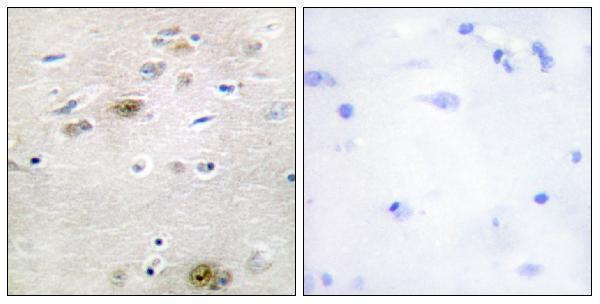
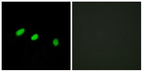
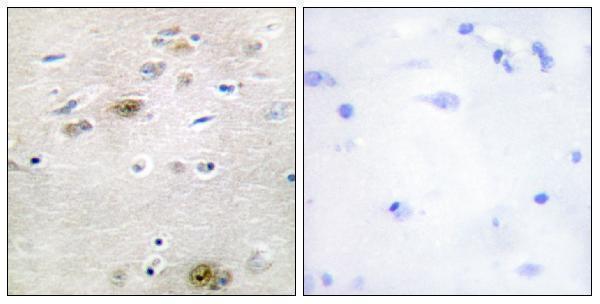
Anti-TRA2A antibodies A03770, A03770-1 and A30486 have IHC images from paraffin-embedded human brain tissue (catalog IHC image captions); A03770 and A30486 also have IF images from HeLa cells (catalog IF image captions).

Real IHC data Immunohistochemistry analysis of paraffin-embedded human brain tissue, using TRA-2 alpha Antibody. The picture on the right is blocked with the synthesized peptide.
Anti-Tra-2 alpha Antibody
Cat # A03770
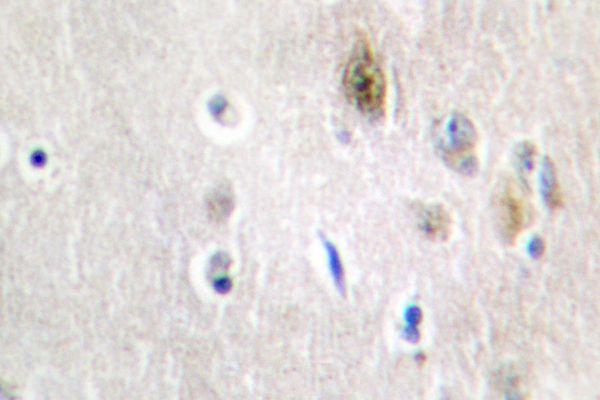
Real IHC data Immunohistochemistry (IHC) analyzes of Tra-2α (G241) pAb in paraffin-embedded human brain tissue.
Anti-Tra-2 Alpha (G241) TRA2A Antibody
Cat # A03770-1
Real IHC data Immunohistochemistry analysis of paraffin-embedded human brain tissue, using TRA-2 alpha Antibody. The picture on the right is blocked with the synthesized peptide.
Anti-TRA-2 alpha Antibody
Cat # A30486

A03770 and A30486 each have an IHC image from paraffin-embedded human brain tissue and an IF image from HeLa cells (A03770 and A30486 image captions); both list Human and Mouse reactivity (catalog reactivity). A03770-1 has an IHC image from paraffin-embedded human brain tissue, lists IF among its applications, and lists Human, Mouse and Rat reactivity (A03770-1 image caption; catalog applications and reactivity).

Which to pick: For tissue IHC, all three have images from paraffin-embedded human brain tissue (each SKU’s IHC image caption); start within the listed IHC dilutions of 1:100–1:300 for A03770 or A30486, or 1:50–1:200 for A03770-1 (catalog dilutions). For IF/ICC, choose A30486 when ICC is required because it lists both ICC and IF and has a HeLa IF image; A03770 also has a HeLa IF image but lists IF without ICC (catalog applications; A30486 and A03770 IF image captions). For Rat samples, A03770-1 is the only listed Rat-reactive option (catalog reactivity); its IHC image shows human brain tissue only, and the fixative is unreported in all three IHC image captions (each SKU’s IHC image caption).

Each figure is that product's own IHC / IF validation image from its datasheet.

References

  1. UniProt Consortium. UniProt entry Q13595 (TRA2A_HUMAN, Transformer-2 protein homolog alpha).
  2. Human Protein Atlas. TRA2A tissue IHC expression (reliability: Uncertain).
  3. Human Protein Atlas. TRA2A subcellular location (ICC-IF): Mainly localized to the nucleoli. In addition localized to the nucleoplasm and vesicles..
  4. Human Protein Atlas. TRA2A antibody validation summary (1 antibodies).
  5. An Integrative Study of Protein-RNA Condensates Identifies Scaffolding RNAs and Reveals Players in Fragile X-Associated Tremor/Ataxia Syndrome. Cell reports 2018 — PMC6315285.
  6. Transformer 2 alpha homolog is a downstream gene of hypoxia-inducible factor 1 subunit alpha and is involved in the progression of pancreatic cancer. Bioengineered 2022 — PMC9275993.
  7. Splicing factor TRA2B is required for neural progenitor survival. The Journal of comparative neurology 2014 — PMC3855887.
  8. PubMed PMID:8799144 — UniProt-cited evidence.
  9. PubMed PMID:14702039 — UniProt-cited evidence.
  10. PubMed PMID:12853948 — UniProt-cited evidence.