TRAF6 / TNF receptor-associated factor 6 · IHC design guide

Design Immunohistochemistry for TRAF6

Plan TRAF6 IHC in paraffin sections using the cytoplasmic tissue pattern reported by HPA (HPA tissue IHC). Compare staining across matched sections with consistent fixation and appropriate controls (standard IHC practice).

Evidence assembled Oct 2026 · For research use; verify linked source records and product datasheet before use
Immunohistochemistry protocol sheet for TRAF6 (IHC for TRAF6): expected localisation Cytoplasmic in most tissues (HPA tissue IHC), antibody M00185-1, validated IHC image, and IHC protocol steps
Printable TRAF6 IHC protocol sheet — expected localisation Cytoplasmic in most tissues (HPA tissue IHC), antibody M00185-1, controls and protocol steps. Open the full TRAF6 IHC guide →

TRAF6 Immunohistochemistry Experimental Design Guide

Expected localisation, validated protocols, controls and antibodies — the at-a-glance facts below, then the full design guide.

Must know before staining
Expected localisation Cytoplasmic in most tissues (HPA tissue IHC)
Staining pattern Cytoplasmic staining in glandular and other cells (HPA tissue IHC)
Antigen retrieval Tris-EDTA pH 9.0 HIER, 95–98 °C, 20 min (rule: nuclear antigen)
Positive control ⓘ Adrenal gland+4 more · see all
Negative control ⓘ None in HPA (detected in all 45 tissues); use no-primary + isotype controls
Important caveats
Reasons your staining may differ from the expected pattern.
Fixation Keep fixation conditions consistent across sections. (standard IHC practice; not target-specific)
Caveat Staining and RNA expression show medium consistency (HPA tissue IHC)
Regulation Expression regulation not specified (UniProt)
Isoform / epitope One 1–522 chain; no isoforms or processing listed (UniProt)
Section 1

Recommended TRAF6 IHC & IF Protocols

Compare the catalog antibody’s IHC-P protocol with published TRAF6 workflows for glioblastoma, rat intestine, and liver tissue (PMC9685220; PMC4005622; PMC5041287).

Recommended immunohistochemistry (IHC-P) protocol parameters
SampleParaffin-embedded Rat ovary tissue; fixative not specified (datasheet M00185-1)
FixationImage fixative and duration unreported (datasheet M00185-1); verify before use.
Sectioning4–5 µm sections on charged slides (standard)
DeparaffinisationXylene, graded ethanol series to water (standard)
Antigen retrievalHeat-induced epitope retrieval in Tris-EDTA buffer, pH 9.0, 20 min at 95–98 °C (standard rule: nuclear antigen)
Peroxidase block3% H2O2, 10 min, room temperature (standard)
Blocking10% normal serum of the secondary host, 30 min, room temperature (standard)
Primary antibodyRabbit monoclonal (clone BOF-20) anti-TRAF6, 1:50-1:200 (datasheet M00185-1)
Primary incubationOvernight at 4 °C (standard)
DetectionHRP-polymer secondary, DAB chromogen 5–10 min (standard)
CounterstainHematoxylin, blue, dehydrate and mount (standard)
Expected resultTRAF6-positive staining in glandular cells of adrenal gland (HPA tissue IHC: Medium). HPA tissue profile: Cytoplasmic expression in most tissues. No signal in the no-primary control.
💡Decision noteStart with Tris-EDTA pH 9.0 retrieval at 95–98 °C for 20 min (page retrieval rule); the glioblastoma protocol also used pH 9 retrieval (PMC9685220).
Section 2

What Is the Expected TRAF6 Staining Pattern?

TRAF6 is mainly cytoplasmic in most tissues, with medium staining reported in glandular, hematopoietic, neuronal and endothelial cells (HPA tissue IHC). Nuclear and cell-cortex pools are also reported, and TRAF6 has no transmembrane segment (UniProt Q9Y4K3). HPA rates tissue IHC reliability Approved, with medium consistency between staining and RNA expression (HPA).

What am I looking at on my slide?
Cytoplasmic stain in listed glandular, marrow, neuronal or endothelial cells (HPA: Medium).Matches the reported tissue pattern (HPA: cytoplasmic in most tissues).
Crisp plasma-membrane-only stain (UniProt Q9Y4K3: no transmembrane segment).Unexpected; assess staining artifact and antibody specificity (UniProt Q9Y4K3; general IHC practice).
Strong signal confined to lung macrophages (HPA: Low).Assess cross-reactivity or endogenous detection activity (HPA; general IHC practice).
Diffuse stain across cells and empty regions (general IHC practice).Suggests background from blocking or detection; review controls (general IHC practice).
No stain in bone-marrow hematopoietic cells (HPA: Medium).Check the positive control and detection steps (HPA; general IHC practice).
💡Expected TRAF6 appearanceA convincing positive shows cytoplasmic stain in HPA-listed medium-staining cells; diffuse haze or membrane-only stain is suspect (HPA tissue IHC; UniProt Q9Y4K3 topology; general IHC practice).
How each factor affects the staining
Subcellular pools (UniProt Q9Y4K3)Cytoplasm, cell cortex and nucleus are reported; nuclear stain can be real (UniProt Q9Y4K3).
Topology (UniProt Q9Y4K3)No transmembrane segment; isolated surface staining warrants review (UniProt Q9Y4K3).
IHC evidence (HPA)Three listed antibodies are IHC Approved; RNA–stain consistency is medium (HPA).
IF/ICC expectation? (HPA)HPA reports mitochondria and nucleoli, both uncertain; assess IF separately (HPA subcellular).
Why is my staining missing, weak or wrong?
SituationLikely causeNext action
No stain in marrow hematopoietic cells (HPA: Medium).Possible weak detection or section-processing failure (general IHC practice).Check positive control, retrieval and detection steps (general IHC practice).
Diffuse slide-wide color (general IHC practice).Possible incomplete blocking or excess antibody (general IHC practice).Review no-primary control; titrate antibody and improve washes (general IHC practice).
Membrane-only stain (UniProt Q9Y4K3: no transmembrane segment).Possible nonspecific binding (general IHC practice).Compare an independent IHC Approved antibody and controls (HPA; general IHC practice).
Strong stain in lung macrophages (HPA: Low).Possible endogenous peroxidase or cross-reactivity (general IHC practice).Use a peroxidase block and no-primary control (general IHC practice).
Nuclear-only stain (HPA: mainly cytoplasmic; UniProt: nucleus reported).May reflect a nuclear pool or nonspecific signal (UniProt Q9Y4K3; general IHC practice).Compare cytoplasmic staining and an independent IHC Approved antibody (HPA; general IHC practice).

Sample controls for TRAF6 IHC & IF

🧪Run bone marrow first: hematopoietic cells should stain at the Medium level (HPA: bone marrow, hematopoietic cells). HPA detects TRAF6 in all 45 scored tissues, so there is no supported negative tissue; use no-primary and isotype controls, and treat any nonreactive cells on the positive slide as local background comparators rather than a validated TRAF6-negative population (HPA: no negative rows; standard IHC practice).
Positive control tissue: Adrenal gland (Glandular cells, HPA Medium)
Negative control tissue: None in HPA: TRAF6 is detected in all 45 scored tissues. Use a no-primary (secondary-only) and an isotype control instead.
ICC-IF cell lines (HPA subcellular resource): HPA ICC-IF images show TRAF6 in A-431, U-251MG, U2OS, with annotated localisation: Mitochondria (uncertain) (HPA subcellular).
Technical controls: Include a no-primary, secondary-only slide and an isotype control matched to the primary antibody’s host species, clonality and concentration; use a validated TRAF6-knockout specimen or immunizing-peptide competition, if available, as a biological specificity control (standard IHC practice). Quench endogenous peroxidase in bone marrow before chromogenic detection because hematopoietic cells can contribute peroxidase background (HPA: bone marrow, hematopoietic cells; standard IHC practice).
⚠️Feasibility: A target-specific fixation window or fixation effect is unreported; the selected M00185-1 rat ovary paraffin-section caption gives a 1:50 dilution but does not report the fixative (selected tissue-IHC caption). Retrieval dependence is unreported, so optimize antigen retrieval on the paraffin sections rather than inferring it from HPA staining (HPA: positive rows; standard IHC practice). No matched evidence establishes that frozen sections or IF are easier for TRAF6; for the selected bone marrow control, endogenous peroxidase is a practical chromogenic artefact to manage (application: IHC-P; HPA: bone marrow, hematopoietic cells; standard IHC practice).

HPA tissue IHC evidence for TRAF6

Comprehensive Human Protein Atlas IHC scoring per tissue (reliability: Approved — Medium consistency between antibody staining and RNA expression data.). Rows are taken directly from the HPA tissue chart — click any row's HPA link to view the source.

Positive expression · recommended positive controls

TissueCell typeLevelEvidenceSource
Adrenal gland Glandular cells Medium Protein (IHC) HPA →
Appendix Glandular cells Medium Protein (IHC) HPA →
Bone marrow Hematopoietic cells Medium Protein (IHC) HPA →
Breast Glandular cells Medium Protein (IHC) HPA →
Caudate Neuronal cells Medium Protein (IHC) HPA →

Undetected expression · recommended negative controls

TissueCell typeLevelEvidenceSource
None in HPA: TRAF6 is detected in all 45 scored tissues. Use a no-primary (secondary-only) and an isotype control instead.
Section 3

Advanced TRAF6 IHC Tips

Use compartment, cell type and matched controls to interpret TRAF6 staining in paraffin sections (UniProt Q9Y4K3; HPA tissue IHC).

How should I troubleshoot weak TRAF6 staining after antigen retrieval?
Start with Tris-EDTA at pH 9.0, heated to 95–98 °C for 20 min (page retrieval setting). If staining remains weak, compare a citrate buffer at pH 6.0 on adjacent sections while holding antibody concentration, detection and imaging conditions constant (general IHC practice). Examine a known expressing cell population in the same run before increasing retrieval intensity; TRAF6 has a broad cytoplasmic tissue profile, so a blank slide alone cannot identify the failed step (HPA tissue IHC; general IHC practice). Record signal and tissue damage together, because stronger heating can improve epitope exposure while reducing section integrity (general IHC practice).
Could fixation explain weak or uneven TRAF6 staining in paraffin sections?
The selected 1:50 rat ovary image specifies paraffin embedding but does not report its fixative, so target-specific fixation sensitivity is unknown (catalog antibody M00185-1 caption). Do not infer fixation tolerance from TRAF6 localisation, its lack of a transmembrane segment, or HPA tissue staining (UniProt Q9Y4K3; HPA tissue IHC). For a fixation comparison, process matched specimens through the same embedding, retrieval and chromogenic detection steps, changing only the fixation condition where feasible (general IHC practice). Include a morphology check and a control section in each run, then report the actual fixative and fixation duration with the staining result (general IHC practice).
Where should convincing TRAF6 staining appear in tissue sections?
Expect predominantly cytoplasmic staining across many tissues, while interpreting cell identity alongside compartment because the HPA tissue profile is broad (HPA tissue IHC). UniProt also lists cell cortex, nucleus and lipid droplet localisation, including nuclear TRAF6 in some lymphocyte settings, so nuclear signal requires context rather than automatic rejection (UniProt Q9Y4K3). HPA subcellular images suggest mitochondrial and nucleolar localisation, but both assignments are marked uncertain and should not set a mandatory IHC pattern (HPA subcellular). Compare adjacent cells, the counterstain and a matched negative control before calling isolated puncta or nuclear deposits TRAF6 positive (general IHC practice).
How do epitope uncertainty and TRAF6 processing affect antibody interpretation?
UniProt lists one TRAF6 chain spanning residues 1–522, no annotated isoforms, no transmembrane segment and no listed glycosylation sites or modified residues in this record (UniProt Q9Y4K3). Its MATH domain spans residues 350–499, but the supplied catalog caption does not identify this antibody's epitope, so domain membership cannot establish binding specificity (UniProt Q9Y4K3; catalog antibody M00185-1 caption). Check the antibody's documented immunogen or epitope before attributing a staining difference to epitope masking (general IHC practice). If that information remains unavailable, compare retrieval conditions and a second independently characterised antibody on matched sections, documenting concordant cell and compartment patterns (general IHC practice).
How can IF help assess a TRAF6 pattern seen by chromogenic IHC?
Use IF as a separate comparison, pairing TRAF6 with a marker of the cell population being scored in IHC and checking whether both signals occupy the same cells (general IF practice). Choose a fluorophore in a channel with low measured tissue autofluorescence, then inspect single-channel images and unstained controls before interpreting puncta or overlap (general IF practice). TRAF6 has no transmembrane segment and is reported in cytoplasm and nucleus; permeabilisation should therefore permit access to intracellular epitopes, with its strength adjusted to preserve morphology (UniProt Q9Y4K3; general IF practice). Because the antibody epitope is unspecified, establish IF fixation and permeabilisation conditions independently of the paraffin-section caption (catalog antibody M00185-1 caption; general IF practice).
What should I change when TRAF6 DAB staining has high background?
First inspect a no-primary control and the tissue's intrinsic pigment, then compare their distribution with the TRAF6 slide under identical illumination (general IHC practice). Quench endogenous peroxidase before HRP detection, use an appropriate protein block and wash consistently between incubations; these are general chromogenic workflow controls, not TRAF6-specific validation (general IHC practice). The selected paraffin-section caption reports an antibody dilution of 1:50, which is a documented image condition rather than a universal optimum (catalog antibody M00185-1 caption). Titrate around that documented condition on matched sections and stop DAB development at a defined time, recording both background and cellular signal (general IHC practice).
How should I score TRAF6 staining across heterogeneous tissue regions? ⚠ ANSWER MARKED FOR VERIFICATION
Define the cell population and compartment before scoring, since the HPA profile is broadly cytoplasmic and includes different cell types across tissues (HPA tissue IHC). For cell-based comparisons, record the percentage of positive cells and staining intensity, then calculate an H-score on the same defined cell population using a consistent 0–3 intensity scale (general IHC practice). If counting discrete positive cells, report density per mm² of viable, analysable tissue and state how the region was selected (general IHC practice). Normalise group comparisons to the same cell type or tissue area, acquisition settings and batch controls, excluding necrotic or folded regions (general IHC practice).
How can I distinguish genuine TRAF6 staining from section artefacts?
A plausible result identifies staining within intact cells and fits the predominantly cytoplasmic tissue profile, while allowing context-dependent nuclear localisation described for TRAF6 (HPA tissue IHC; UniProt Q9Y4K3). Check cell identity carefully: HPA reports low staining in ovarian stroma cells, so a strong rat ovary signal should be assigned to a specific cell population before comparison with that profile (HPA tissue IHC; catalog antibody M00185-1 caption). Discount deposits concentrated at section edges, folds or necrotic areas unless they persist in well-preserved regions and controls (general IHC practice). Compare no-primary and peroxidase-block controls when diffuse brown signal could reflect endogenous enzyme activity, and avoid treating isolated nuclear or mitochondrial-looking puncta as proof of specificity (general IHC practice; HPA subcellular).
Boster reagents

Best TRAF6 / TNF receptor-associated factor 6 IHC Antibodies

The catalog lists human, mouse, and rat reactivity (catalog reactivity); the images show TRAF6 staining in human and rat paraffin sections and K562 cells (catalog image captions).

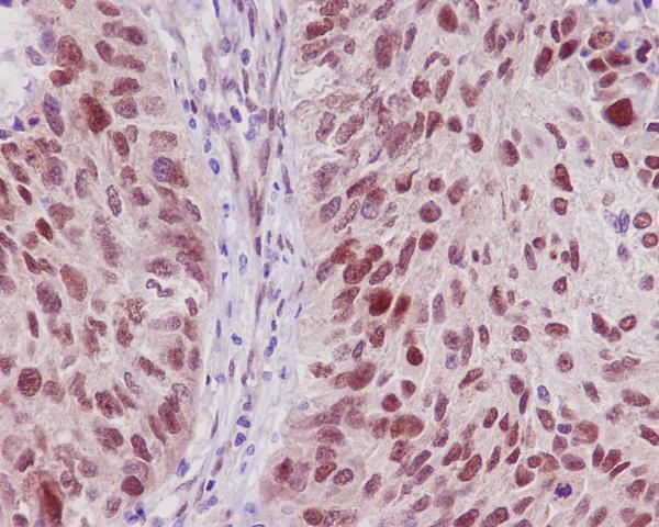
Real IHC data Immunohistochemical analysis of paraffin-embedded Rat ovary, using the Antibody at 1:50 dilution.
Anti-TRAF6 Rabbit Monoclonal Antibody
Cat # M00185-1
Real IHC data Immunohistochemical analysis of paraffin-embedded human lung carcinoma, using TRAF6 Antibody.
Anti-TRAF6 Rabbit Monoclonal Antibody
Cat # M00185
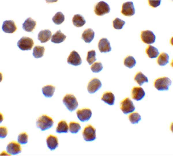
Real IF data Immunocytochemistry of TRAF6 in K562 cells with TRAF6 antibody at 0.5 μg/mL.
Anti-TRAF6 Antibody
Cat # A00185-1

M00185-1 shows rat ovary paraffin-section IHC at 1:50, while M00185 shows human lung carcinoma paraffin-section IHC (catalog IHC captions). A00185-1 shows K562 cell ICC at 0.5 μg/mL and IF at 20 μg/mL (catalog image captions).

Which to pick: For tissue IHC, choose rabbit monoclonal M00185-1 for rat paraffin sections or rabbit monoclonal M00185 for human paraffin sections, matching each SKU’s own image (catalog host, clone, applications and IHC captions); the fixative is unreported in both captions. For IF/ICC, A00185-1 has K562 cell images for both methods, while M00185 lists IF/ICC without an IF image (catalog applications and image captions). For IHC across human, mouse, and rat, both M00185-1 and M00185 list all three species, but their IHC images document rat and human samples, respectively (catalog reactivity and IHC captions).

Each figure is that product's own IHC / IF validation image from its datasheet.

References

  1. UniProt Consortium. UniProt entry Q9Y4K3 (TRAF6_HUMAN, TNF receptor-associated factor 6).
  2. Human Protein Atlas. TRAF6 tissue IHC expression (reliability: Approved).
  3. Human Protein Atlas. TRAF6 subcellular location (ICC-IF): Mainly localized to the mitochondria. In addition localized to the nucleoli..
  4. Human Protein Atlas. TRAF6 antibody validation summary (3 antibodies).
  5. Comparing tumor microRNA profiles of patients with long‑ and short‑term‑surviving glioblastoma. Molecular medicine reports 2023 — PMC9685220.
  6. TRAF6 promotes chemoresistance to paclitaxel of triple negative breast cancer via regulating PKM2-mediated glycolysis. Cancer medicine 2023 — PMC10587986.
  7. Glutamine attenuates the inhibitory effect of methotrexate on TLR signaling during intestinal chemotherapy-induced mucositis in a rat. Nutrition & metabolism 2014 — PMC4005622.
  8. Relationship between TRAF6 and deterioration of HCC: an immunohistochemical and in vitro study. Cancer cell international 2016 — PMC5041287.
  9. PubMed PMID:8837778 — UniProt-cited evidence.
  10. PubMed PMID:14702039 — UniProt-cited evidence.
  11. PubMed PMID:16554811 — UniProt-cited evidence.