Click image to see more details
-

-
-
-
-
+7
Product Info Summary
| SKU: | M00185 |
|---|---|
| Size: | 100 μl |
| Reactive Species: | Human, Mouse, Rat |
| Host: | Rabbit |
| Application: | IF, IHC, ICC, WB |
Customers Who Bought This Also Bought

Product info
Product Name
Anti-TRAF6 Rabbit Monoclonal Antibody
SKU/Catalog Number
M00185
BM4061 is an alternative SKU for this antibody, used in previous lots.
Size
100 μl
Form
Liquid
Description
Boster Bio Anti-TRAF6 Rabbit Monoclonal Antibody catalog # M00185. Tested in WB, IHC, ICC/IF applications. This antibody reacts with Human, Mouse, Rat.
Storage & Handling
Store at -20°C for one year. For short term storage and frequent use, store at 4°C for up to one month. Avoid repeated freeze-thaw cycles.
Cite This Product
Anti-TRAF6 Rabbit Monoclonal Antibody (Boster Biological Technology, Pleasanton CA, USA, Catalog # M00185)
Host
Rabbit
Contents
Rabbit IgG in stabilizing components, phosphate buffered saline, pH 7.4, 150mM NaCl, 0.02% sodium azide and 50% glycerol.
*This antibody is supplied in a stabilized formulation.
Compatibility with conjugation reactions depends on the chemistry of the conjugation method used.
For conjugation methods that are not compatible with the stabilizing components present in this formulation, a carrier-free antibody format is required.
Clonality
Monoclonal
Clone Number
BOG-20
Isotype
Rabbit IgG
Immunogen
A synthesized peptide derived from human TRAF6
Reactive Species
M00185 is reactive to TRAF6 in Human, Mouse, Rat
Observed Molecular Weight
58 kDa
Calculated molecular weight
59.6 kDa
Antibody Validation
Boster validates all antibodies on WB, IHC, ICC, Immunofluorescence, and ELISA with known positive control and negative samples to ensure specificity and high affinity, including thorough antibody incubations.
Application & Images
Applications
M00185 is guaranteed for IF, IHC, ICC, WB Boster Guarantee
Recommend Dilution
WB 1:500-2000
IHC 1:50-200
ICC/IF 1:50-200
Tested application
Suggested blocking solution with 5% non-fat milk or BSA; (*)Recommended protein loading: 20-40 µg per lane
Use TE buffer pH 9.0 for antigen retrieval; (*) citrate buffer pH 6.0 is an alternative.
Validation Images & Assay Conditions
Click image to see more details
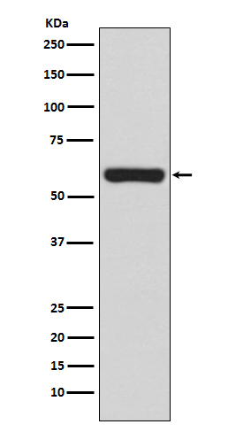
Western blot analysis of TRAF6 expression in Jurkat cell lysate.

Click image to see more details
The regulatory effects of MOIG on NF-κB and JAK2/STAT3 pathway of TNF-α-stimulated FLSs cells. FLSs were incubated with TNF-α for 12 h and subsequently treated with MOIG for 24 h, the proteins were extracted to analyze expression of associated proteins by Western blot. ( A–C ) The expression of p-JAK2, JAK2, p-STAT3, STAT3. ( D–G ) The expression of TRAF6, p-p65, p65 and IκB-α, respectively. The data represents the mean ± SD (n = 3). The experiments were repeated for three times. # p < 0.05, ## p < 0.01, ### p < 0.001 vs. normal ctrl group; * p < 0.05, ** p < 0.01, *** p < 0.001 vs. TNF-α model group.
Index in PubMed under a CC BY license. PMID: 39444614

Click image to see more details
calpain2a interacts with TRAF6 and inhibits the NF-κB Signaling Pathway. a List of TRAF6 interactome based on Label-free quantification intensity (section). b , c MKC cells seeded in 6 cm 2 dishes. After 24 h, cell lysates were immunoprecipitated (IP) with anti-TRAF6 or anti-calpain2a affinity gels. Then the immunoprecipitates and cell lysates were analyzed by IB with the anti-TRAF6 and anti-calpain2a Abs, respectively. d , e HEK 293 cells seeded in 6 cm 2 dishes were transfected with the plasmids calpain2a-Flag and TRAF6-Myc (2 μg each). After 24 h, cell lysates were immunoprecipitated (IP) with anti-Myc d or anti-Flag e affinity gels. Then the immunoprecipitates and cell lysates were analyzed by IB with the anti-Myc and anti-Flag Abs, respectively. f The same amino acids among human calpain2a (Hu-calpain2), mouse calpain2a (Mu-calpain2), zebrafish calpain2a (ZF-calpain2a) and miiuy croaker calpain2a (M-calpain2a) are highlighted with black background. g – i EPC cells were transfected with calpain2a-Flag or empty vector together with the NF-κB, IL-1β, and IL-8 luciferase reporters. At 24 h post-transfection, cells were untreated (Mock) or treated with LPS for 6 h. The luciferase activity value was achieved against the Renilla luciferase activity ( n = 3 per group). Western blot analysis was used to measure the expression of transiently transfected calpain2a-Flag. The expression of Tubulin was used as a loading control. Data were analyzed by two-way ANOVA ( g , h , i ). ** p < 0.01. All experiments were performed in at least three independent experiments.
Index in PubMed under a CC BY license. PMID: 37002312

Click image to see more details
The interaction of calpain2a with TRAF6. a , b Schematic diagrams of domain organization in miiuy croaker calpain2a, TRAF6 and mutants used in this study. “+“ represents the interaction with TRAF6 or calpain2a, “-“ means no interaction. c HEK293 cells were transfected with mock, calpain2a-Flag wild type (WT), and calpain2a-Flag truncated mutants, or TRAF6-HA as indicated. At 24 h post-transfection, transfected cells were extracted and cell lysates were subjected to immunoprecipitation with anti-HA antibody followed by IB using anti-HA or anti-Flag antibody. d – f HEK293 cells were transfected with mock, TRAF6-HA wild type (WT), and TRAF6-HA truncated mutants, or calpain2a-Flag as indicated. At 24 h post-transfection, transfected cells were extracted and cell lysates were subjected to immunoprecipitation with anti-HA antibody d , f or anti-Flag antibody e followed by IB using anti-HA or anti-Flag antibody. g The interaction site of TRAF6 and calpain2a. h EPC cells were transfected with TRAF6-HA, calpain2a-Flag or calpain2a-Flag truncated mutants together with NF-κB and IL-1β luciferase reporters. After 36 h post-transfection, the luciferase activity value was achieved against the renilla luciferase activity ( n = 3 per group). i , j EPC cells were transfected with mock, calpain2a-mCherry, and TRAF6-GFP. DAPI-stained nuclei are shown in blue. calpain2a was detected with red fluorescence, and TRAF6 was detected with green fluorescence. Scale bar, 10 μm. All experiments were performed in at least three independent experiments. Data were analyzed by one-way ANOVA ( h ). ** p < 0.01.
Index in PubMed under a CC BY license. PMID: 37002312

Click image to see more details
calpain2a attenuates autoubiquitination of TRAF6. a HEK293 cells were transfected with TRAF6-Myc, TRAF6-HA, or empty vector. After 24 h post-transfection, the cells were lysed and IP analyses with HA antibody. b HEK293 cells were transfected with TRAF6-Myc, TRAF6-HA, empty vector, or calpain2a-Flag. After 24 h post-transfection, the cells were lysed and IP analyses with HA antibody. c HEK293 cells were transfected with TRAF6-Myc, TRAF6-HA, empty vector, or different concentrations of calpain2a-ΔCysPc-Flag. After 24 h post-transfection, the cells were lysed and IP analyses with HA antibody. d , e HEK293 cells were cotransfected with TRAF6-Myc, TRAF6-HA and WT-ubiquitin-His or K63O-ubiquitin-His (in which only lysine 63 is kept) together with calpain2a-Flag, calpain2a-ΔCysPc-Flag or empty vector. After 24 h post-transfection, the cells were lysed and purified with Ni-NTA agarose. The immunoprecipitates and input immunoblot analysis with anti-Myc, anti-Flag, anti-HA, and anti-Tubulin Abs. All experiments were performed in at least three independent experiments.
Index in PubMed under a CC BY license. PMID: 37002312

Click image to see more details
calpain2a inhibits TRAF6 protein expression. a The time gradient experiment of empty vectors or calpain2a-Myc plasmids together with TRAF6-Flag was conducted in EPC cells. The cell lysates were subjected to IB with anti-Myc, anti-Flag, and anti–Tubulin Abs. RNA was extracted from cells and reverse transcribed, then TRAF6 and actin were amplified by PCR primers. b EPC cells were seeded in 12-well plates overnight and co-transfected with TRAF6-Flag and calpain2a-Myc (0.3, 0.6, or 0.9 μg) for 48 h. The expression of TRAF6-Flag and calpain2a-Myc proteins were detected by Western blotting. RNA was extracted from cells and reverse transcribed, then TRAF6 and actin were amplified by PCR primers. c calpain2a-Myc or empty vectors were co-transfected with TRAF6-Flag into EPC cells. At 36 h post-transfection, the transfected cells were treated with cycloheximide (CHX) for 2 or 4 h. d MKC cells were transfected with calpain2a-Flag or empty vectors. At 48 h post-transfection, the cell lysates were subjected to IB with anti-TRAF6, anti-Flag, and anti-Tubulin Abs. e , f EPC cells were transfected with the indicated plasmids in the presence or absence of MG132, 3-MA, NH 4 CL, E-64 (50 or 75 μM), or PMSF for 6 h before immunoblot analysis was performed. g MKC cells were transfected with calpain2a-Flag in the presence or absence of E-64 (50 or 75 μM) for 6 h before immunoblot analysis was performed. h calpain2a-Flag and calpain2a-ΔCysPc-Flag were co-transfected with TRAF6-HA into EPC cells. At 48 h post-transfection, the cell lysates were subjected to IB with indicated Abs. i calpain2a-Flag and calpain2a-ΔCysPc-Flag were co-transfected into MKC cells. At 48 h post-transfection, the cell lysates were subjected to IB with anti-TRAF6, anti-Flag, and anti-Tubulin Abs. j IL-1β , IL-8 mRNA in MKC stably transduced with calpain2a-Flag and calpain2a-ΔCysPc-Flag and treated with saline (0) or challenged with LPS for 4 h ( n = 3 per group). Relative mRNA level was normalized to the expression of the gene encoding β-actin in each sample. All experiments were performed in at least three independent experiments. Data were analyzed by two-way ANOVA ( j ). ** p < 0.01.
Index in PubMed under a CC BY license. PMID: 37002312

Click image to see more details
calpain2a inhibits TRAF6 ubiquitin-ligase activity. a Ubiquitination of endogenous TRAF6 in MKC cells transduced with calpain2a-Flag and calpain2a-ΔCysPc-Flag and unchallenged (−) or challenged with LPS (+), assessed by immunoblot analysis with anti-ubiquitin after immunoprecipitation with anti-TRAF6 and input immunoblot analysis with indicated Abs. b , c HEK293 cells were cotransfected with TRAF6-HA and WT-ubiquitin-His or K63O-ubiquitin-His (in which only lysine 63 is kept) together with calpain2a-Flag, calpain2a-ΔCysPc-Flag or empty vector. After 24 h post-transfection, the cells were lysed and purified with Ni-NTA agarose. d , e HEK293 cells were cotransfected with TRAF6-HA and WT-ubiquitin-His or K63O-ubiquitin-His (in which only lysine 63 is kept) together with calpain2a-Flag (0.5 μg, 1 μg), calpain2a-ΔCysPc-Flag (0.5 μg, 1 μg) or empty vector. After 24 h post-transfection, the cells were lysed and purified with Ni-NTA agarose. f Ubiquitination of overexpressed TRAF6 in MKC cells transduced with calpain2a-ΔCysPc-Flag, assessed by immunoblot analysis with anti-ubiquitin after immunoprecipitation with anti-Myc and input immunoblot analysis with indicated Abs. g , h HEK293 cells were cotransfected with TAK1-Myc, TRAF6-HA and WT-ubiquitin-His or K63O-ubiquitin-His (in which only lysine 63 is kept) together with calpain2a-Flag, calpain2a-ΔCysPc-Flag or empty vector. After 24 h post-transfection, the cells were lysed and purified with Ni-NTA agarose. All the immunoprecipitates and input immunoblot analysis with anti-Myc, anti-Flag, anti-HA, and anti-Tubulin Abs. All experiments were performed in at least three independent experiments.
Index in PubMed under a CC BY license. PMID: 37002312

Click image to see more details
calpain2a inhibits TRAF6-mediated ubiquitination of IRF7 and IRF3. a , b HEK293 cells were cotransfected with IRF3-Myc, TRAF6-HA and WT-ubiquitin-His or K63O-ubiquitin-His (in which only lysine 63 is kept) together with calpain2a-Flag, calpain2a-ΔCysPc-Flag or empty vector. After 24 h post-transfection, the cells were lysed and purified with Ni-NTA agarose. c , d HEK293 cells were cotransfected with IRF7-Myc, TRAF6-HA and WT-ubiquitin-His or K63O-ubiquitin-His (in which only lysine 63 is kept) together with calpain2a-Flag, calpain2a-ΔCysPc-Flag or empty vector. After 24 h post-transfection, the cells were lysed and purified with Ni-NTA agarose. All the immunoprecipitates and input with anti-Myc, anti-Flag, anti-HA, and anti-Tubulin Abs. All experiments were performed in at least three independent experiments. e Model detailing the roles of calpain2a in TRAF6-mediated signaling pathways. Upon LPS and virus, TRAF6 could be activated, homo-oligomerization and auto-ubiquitination. BECN1, ECSIT, IRF7 and IRF3 are ubiquitinated by TRAF6 and trigger the activation of downstream signals. calpain2a as a negative regulator targets TRAF6 to inhibit TRAF6-mediated signaling pathways.
Index in PubMed under a CC BY license. PMID: 37002312

Click image to see more details
calpain2a inhibits TRAF6-mediated ubiquitination of ECSIT and BECN1. a HEK293 cells were transfected with ECSIT-Myc, TRAF6-Flag, or empty vector. After 24 h post-transfection, the cells were lysed and IP analyses with Flag antibody. b HEK293 cells were transfected with TAK1-Flag, ECSIT-Myc, or empty vector. After 24 h post-transfection, the cells were lysed and IP analyses with Myc antibody. c HEK293 cells were transfected with TRAF6-Myc, ECSIT-HA, empty vector, or different concentrations of calpain2a-ΔCysPc-Flag. After 24 h post-transfection, the cells were lysed and IP analyses with Myc antibody. d HEK293 cells were cotransfected with ECSIT-Myc, TRAF6-HA, WT-ubiquitin-His together with calpain2a-Flag, calpain2a-ΔCysPc-Flag or empty vector. After 24 h post-transfection, the cells were lysed and purified with Ni-NTA agarose. e HEK293 cells were transfected with BECN1-Flag, TRAF6-Myc, or empty vector. After 24 h post-transfection, the cells were lysed and IP analyses with Myc antibody. f HEK293 cells were cotransfected with BECN1-HA, TRAF6-Myc, WT-ubiquitin-His together with calpain2a-Flag, calpain2a-ΔCysPc-Flag or empty vector. After 24 h post-transfection, the cells were lysed and purified with Ni-NTA agarose. The immunoprecipitates and input with anti-Myc, anti-Flag, anti-HA, and anti-Tubulin Abs. All experiments were performed in at least three independent experiments.
Index in PubMed under a CC BY license. PMID: 37002312

Click image to see more details
Effects of JTG on expression of associated proteins and NF-κB pathway of osteoclast induced from BMMs with RANKL and LPS. BMMs were incubated with RANKL and JTG for 48 h, the proteins were extracted to analyze associated proteins of osteoclast by Western blot. A : a Western blot imagines for expression of NFATc1, c-Fos, Cathepsin K and MMP9. A : b – e The quantification analysis of NFATc1, c-Fos, Cathepsin K and MMP9 based on the results of A : a by ECL detection system, respectively. B : a The images of Western blot for TRAF6, P-P65, P65 and IκBα. B : b – d The quantification analysis of TRAF6, P-P65/P65 and IκBα based on the results of B : a by using an ECL detection system, respectively. Each point represents the mean ± SD (n = 3). The experiments were repeated for three times. * P < 0.05, ** P < 0.01 compared with control group
Index in PubMed under a CC BY license. PMID: 36195960

Click image to see more details
Immunohistochemical analysis of paraffin-embedded human lung carcinoma, using TRAF6 Antibody.
Specific Publications For Anti-TRAF6 Rabbit Monoclonal Antibody (M00185)
Loading publications
Recommended Resources
Here are featured tools and databases that you might find useful.
- Boster's Pathways Library
- Protein Databases
- Bioscience Research Protocol Resources
- Data Processing & Analysis Software
- Photo Editing Software
- Scientific Literature Resources
- Research Paper Management Tools
- Molecular Biology Software
- Primer Design Tools
- Bioinformatics Tools
- Phylogenetic Tree Analysis
Customer Reviews
Have you used Anti-TRAF6 Rabbit Monoclonal Antibody?
Share your experimental results or join a short interview to earn up to $1,000 in product credits or other rewards.
0 Reviews For Anti-TRAF6 Rabbit Monoclonal Antibody
Customer Q&As
Have a question?
Find answers in Q&As, reviews.
Can't find your answer?
Submit your question
3 Customer Q&As for Anti-TRAF6 Rabbit Monoclonal Antibody
Question
Would M00185 anti-TRAF6 Rabbit Monoclonal antibody work on parafin embedded sections? If so, which fixation method do you recommend we use (PFA, paraformaldehyde, other)?
Verified Customer
Verified customer
Asked: 2019-06-06
Answer
As indicated on the product datasheet, M00185 anti-TRAF6 Rabbit Monoclonal antibody as been tested on IF. It is best to use PFA for fixation because it has better tissue penetration ability. PFA needs to be prepared fresh before use. Long term stored PFA turns into formalin, as the PFA molecules congregate and become formalin.
Boster Scientific Support
Answered: 2019-06-06
Question
I see that the anti-TRAF6 Rabbit Monoclonal antibody M00185 works with IF, what is the protocol used to produce the result images on the product page?
Verified Customer
Verified customer
Asked: 2017-07-05
Answer
You can find protocols for IF on the "support/technical resources" section of our navigation menu. If you have any further questions, please send an email to support@bosterbio.com
Boster Scientific Support
Answered: 2017-07-05
Question
Does anti-TRAF6 Rabbit Monoclonal antibody M00185 work for IF with secondary oocyte?
N. Evans
Verified customer
Asked: 2016-03-10
Answer
According to the expression profile of secondary oocyte, TRAF6 is highly expressed in secondary oocyte. So, it is likely that anti-TRAF6 Rabbit Monoclonal antibody M00185 will work for IF with secondary oocyte.
Boster Scientific Support
Answered: 2016-03-10
