This website uses cookies to ensure you get the best experience on our website.


- Table of Contents
Plan TREX2 IHC-P around nuclear staining in several tissues (HPA tissue IHC). Compare a medium-staining tissue such as tonsil with a documented not-detected tissue such as adipose tissue, and interpret results cautiously because tissue IHC reliability is uncertain (HPA tissue IHC).

Expected localisation, validated protocols, controls and antibodies — the at-a-glance facts below, then the full design guide.
| Expected localisation | Nuclear staining in several tissues (HPA tissue IHC) | |
| Staining pattern | Nuclear staining in glandular and germinal center cells (HPA tissue IHC) | |
| Antigen retrieval | Tris-EDTA pH 9.0 HIER, 95–98 °C, 20 min (rule: nuclear antigen) | |
| Positive control | Adrenal gland+4 more · see all | |
| Negative control | Adipose tissue+4 more · see all |
| Fixation | Keep fixation consistent across sections. (standard IHC practice; not target-specific) | |
| Caveat | Tissue IHC staining has uncertain reliability (HPA tissue IHC) | |
| Regulation | Staining-related regulation is unreported (UniProt) | |
| Isoform / epitope | 2 isoforms; epitope impact is undetermined (UniProt) |
The catalog antibody’s IHC-P protocol is paired with published TREX2 protocols for laryngeal cancer sections (PMC6499986) and HNSCC tissue microarrays (PMC4673170).
| Sample | Tissue sections; selected-image fixative not specified (standard IHC workflow) |
| Fixation | Image fixative and duration unreported (datasheet A08960); verify before use. |
| Sectioning | 4–5 µm sections on charged slides (standard) |
| Deparaffinisation | Xylene, graded ethanol series to water (standard) |
| Antigen retrieval | Heat-induced epitope retrieval in Tris-EDTA buffer, pH 9.0, 20 min at 95–98 °C (standard rule: nuclear antigen) |
| Peroxidase block | 3% H2O2, 10 min, room temperature (standard) |
| Blocking | 10% normal serum of the secondary host, 30 min, room temperature (standard) |
| Primary antibody | Rabbit anti-TREX2, 2.5 μg/mL (datasheet A08960) |
| Primary incubation | Overnight at 4 °C (standard) |
| Detection | HRP-polymer secondary, DAB chromogen 5–10 min (standard) |
| Counterstain | Hematoxylin, blue, dehydrate and mount (standard) |
| Expected result | TREX2-positive staining in glandular cells of adrenal gland (HPA tissue IHC: Medium). HPA tissue profile: Nuclear expression in several tissues. No signal in the no-primary control. |
TREX2 is a nuclear protein with no transmembrane segment (UniProt Q9BQ50: location and topology). In paraffin section IHC, expect nuclear staining in cell populations reported at medium intensity, including skin fibroblasts, testis Leydig cells, and lymph node germinal center cells (HPA: tissue IHC). Treat the pattern as a guide for comparison: the available tissue IHC antibody is rated Uncertain (HPA: antibody IHC validation).
| Distinct nuclear chromogen in skin fibroblasts, testis Leydig cells, or lymph node germinal center cells. | This fits TREX2's nuclear location and the reported medium staining in those cell populations (UniProt Q9BQ50: location; HPA: tissue IHC). Judge the named cells separately from surrounding tissue. A matching pattern is supportive, but the antibody's IHC rating is Uncertain, so morphology alone does not establish specificity (HPA: antibody IHC validation). |
| Predominantly cytoplasmic, membranous, or extracellular staining with little nuclear signal. | This conflicts with the annotated nuclear location and absence of a transmembrane segment (UniProt Q9BQ50: location and topology). Check the counterstain to locate nuclei, then review background and detection controls as general IHC practice. Do not score a compartment that disagrees with the expected pattern as convincing TREX2 without independent validation. |
| Strong staining in adipocytes or liver cholangiocytes, especially when expected nuclei are weak. | Those cell populations are reported as not detected in HPA tissue IHC (HPA: adipose tissue and liver). Unexpected staining warrants a cross-reactivity or endogenous detection activity check under general IHC practice. HPA's Uncertain IHC rating limits how confidently any single positive or negative field can be interpreted (HPA: antibody IHC validation). |
| Diffuse chromogen across nuclei and surrounding tissue, with poorly defined cell boundaries. | This is hard to reconcile with a cell-specific nuclear pattern (UniProt Q9BQ50: location; HPA: nuclear expression in several tissues). General IHC causes to assess include inadequate blocking, excess detection reagent, or incomplete washing. Compare a no-primary control and nearby cell populations before assigning any diffuse color to TREX2. |
| No visible signal in a section containing skin fibroblasts or testis Leydig cells. | These are reported at medium intensity, making them useful comparison populations, though they are not guaranteed positives in every preparation (HPA: tissue IHC; HPA: IHC reliability Uncertain). First verify that the relevant cells are present and the detection run worked. Review the antibody's validated IHC-P conditions before calling the tissue negative. |
| Compartment and topology | TREX2 is annotated in the nucleus, with no transmembrane segment or signal peptide (UniProt Q9BQ50: location, topology and processing). Nuclear localization is therefore the primary reading criterion; these annotations do not explain an isolated membrane or extracellular pattern. |
| Cell population and tissue | HPA reports medium staining in several named cell populations, while adipocytes and liver cholangiocytes are not detected (HPA: tissue IHC). Compare the specified cells, not whole-section darkness. A not-detected entry describes that observation; it is not proof that every cell or preparation from the tissue must be negative. |
| IHC evidence strength | HPA describes nuclear expression in several tissues, but rates tissue IHC reliability and the listed antibody's IHC validation as Uncertain (HPA: tissue IHC; HPA: antibody IHC validation). Use the distribution as an expectation that needs controls, rather than a definitive specificity test. |
| Isoforms and processing | UniProt lists 2 isoforms and a chain spanning residues 1–236, with no signal peptide or propeptide (UniProt Q9BQ50: isoforms and processing). The payload gives no antibody epitope or isoform coverage, so it cannot predict an isoform-specific stain or justify a processing-based explanation for missing signal. |
| RNA versus protein readout | HPA calls RNA tissue enhanced in blood vessel and skin, while its protein IHC observations specify particular stained cell populations (HPA: RNA specificity and tissue IHC). Use the protein observations for slide interpretation; RNA enrichment alone does not establish strong chromogenic staining in every cell of either tissue. |
| Situation | Likely cause | Next action |
|---|---|---|
| Expected nuclei are blank in a reported medium-staining population. | The relevant cells may be absent from the field, or the IHC run may have failed; HPA medium staining is observational rather than guaranteed (HPA: tissue IHC; HPA: reliability Uncertain). | Confirm cell identity on the counterstain, check a working run control, and review the catalog antibody's IHC-P protocol as general IHC practice. The supplied sources do not establish TREX2-specific fixation or retrieval sensitivity. |
| Color is strongest outside nuclei. | The distribution conflicts with TREX2's nuclear annotation (UniProt Q9BQ50: location); nonspecific staining or detection background are general IHC possibilities. | Compare nuclear counterstain alignment and a no-primary control. Score only convincing cell-associated nuclear signal while investigating the discordant compartment under general IHC practice. |
| Unexpected cells stain as strongly as the comparison population. | Cross-reactivity or endogenous detection activity may contribute under general IHC practice; HPA reports some populations as not detected and rates IHC Uncertain (HPA: tissue IHC and validation). | Check tissue morphology and detection controls, including the relevant endogenous activity control for the chromogen system. Reassess the claim against the specific HPA cell population, not the tissue name alone. |
| The whole section shows diffuse color. | General IHC background can obscure the nuclear, cell-specific distribution expected from UniProt and HPA (UniProt Q9BQ50: location; HPA: tissue IHC). | Inspect a no-primary control and review blocking, reagent concentration, washing, and chromogen development as general IHC practice; repeat scoring only where nuclei and cell boundaries remain clear. |
| A reported not-detected population has no staining. | That finding agrees with the listed HPA observation for the specific cell population, such as adipocytes or liver cholangiocytes (HPA: tissue IHC). | Record the cell population and run control. Do not treat a blank field alone as proof of antibody specificity or successful staining; HPA rates the IHC evidence Uncertain (HPA: antibody IHC validation). |
| IF/ICC: should a fluorescent nuclear pattern be expected? | UniProt annotates TREX2 as nuclear, but HPA supplies no main ICC-IF location or cell-line images for it (UniProt Q9BQ50: location; HPA: subcellular record). | Treat nuclear fluorescence as a hypothesis, not an HPA-validated ICC-IF result. Assess IF/ICC staining with its own controls and guide; the tissue IHC observations do not establish an IF/ICC pattern. |
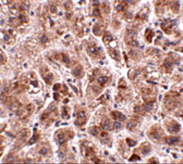
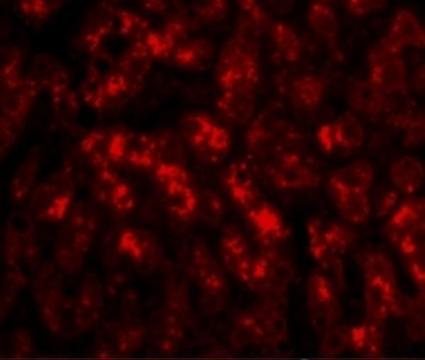


Comprehensive Human Protein Atlas IHC scoring per tissue (reliability: Uncertain — External characterization data supports antibody staining.). Rows are taken directly from the HPA tissue chart — click any row's HPA link to view the source.
| Tissue | Cell type | Level | Evidence | Source |
|---|---|---|---|---|
| Adrenal gland | Glandular cells | Medium | Protein (IHC) | HPA → |
| Endometrium | Glandular cells | Medium | Protein (IHC) | HPA → |
| Esophagus | Squamous epithelial cells | Medium | Protein (IHC) | HPA → |
| Lymph node | Germinal center cells | Medium | Protein (IHC) | HPA → |
| Skin | Fibroblasts | Medium | Protein (IHC) | HPA → |
| Tissue | Cell type | Level | Evidence | Source |
|---|---|---|---|---|
| Adipose tissue | Adipocytes | Not detected | Protein (IHC) | HPA → |
| Caudate | Glial cells | Not detected | Protein (IHC) | HPA → |
| Cerebellum | Cells in granular layer | Not detected | Protein (IHC) | HPA → |
| Epididymis | Glandular cells | Not detected | Protein (IHC) | HPA → |
| Fallopian tube | Glandular cells | Not detected | Protein (IHC) | HPA → |
Troubleshoot TREX2 staining in paraffin sections by checking nuclear localisation, cell identity, assay controls, and the limits of the available tissue evidence.
A08960 has IHC data from human liver tissue and IF data from human liver cells; its listed reactivity is human, mouse, and rat (catalog image captions; catalog reactivity).
A08960 will render with an IHC image of human liver tissue at 2.5 μg/mL (IHC image caption). The same SKU lists IF and shows human liver cells at 20 μg/mL (catalog applications; IF image caption).
Which to pick: Choose A08960 for paraffin-section tissue IHC based on its IHC-P listing and human liver tissue image; the fixative is unreported (catalog applications; IHC image caption). Choose A08960 for IF based on its human liver cell image; ICC-specific validation is unreported (catalog applications; IF image caption). It is the listed option for human, mouse, and rat reactivity, although both images show human samples and clonality is unreported (catalog reactivity; IHC/IF image captions; catalog clone field).