TRIM21 / E3 ubiquitin-protein ligase TRIM21 · IHC design guide

Design Immunohistochemistry for TRIM21

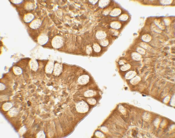
Plan TRIM21 paraffin-section IHC around cytoplasmic staining, with bone marrow hematopoietic cells as a positive control (HPA tissue IHC). The A02079 IHC-P validation provides a starting antibody concentration of 5 μg/mL for chromogenic detection (datasheet A02079: IHC-P).

Evidence assembled Oct 2026 · For research use; verify linked source records and product datasheet before use
Immunohistochemistry protocol sheet for TRIM21 (IHC for TRIM21): expected localisation Cytoplasmic staining in most tissues (HPA tissue IHC), antibody A02079, validated IHC image, and IHC protocol steps
Printable TRIM21 IHC protocol sheet — expected localisation Cytoplasmic staining in most tissues (HPA tissue IHC), antibody A02079, controls and protocol steps. Open the full TRIM21 IHC guide →

TRIM21 Immunohistochemistry Experimental Design Guide

Expected localisation, validated protocols, controls and antibodies — the at-a-glance facts below, then the full design guide.

Must know before staining
Expected localisation Cytoplasmic staining in most tissues (HPA tissue IHC)
Staining pattern Cytoplasmic signal in most tissues (HPA tissue IHC)
Antigen retrieval Citrate pH 6 HIER, heat-mediated (datasheet A02079)
Positive control ⓘ Bone marrow+4 more · see all
Negative control ⓘ Adrenal gland+3 more · see all
Important caveats
Reasons your staining may differ from the expected pattern.
Fixation Formaldehyde-fixed paraffin sections (datasheet A02079: IHC-P) (selected-SKU IHC image A02079)
Caveat Marrow peroxidase can mimic chromogenic signal (HPA tissue IHC; standard IHC practice)
Regulation Up-regulated by IFN/JAK/STAT signaling (UniProt)
Isoform / epitope 2 isoforms; antibody epitope coverage is unreported (UniProt; datasheet A02079)
Section 1

Recommended TRIM21 IHC & IF Protocols

The catalog antibody’s IHC-P protocol is followed by published TRIM21 staining parameters from three PMC articles (PMC13526992; PMC13338432; PMC10048917).

Recommended immunohistochemistry (IHC-P) protocol parameters
SampleFormaldehyde-fixed, paraffin-embedded human small intestine tissue (datasheet A02079)
FixationImage formalin-fixed; duration unreported (datasheet A02079); verify before use.
Sectioning4–5 µm sections on charged slides (standard)
DeparaffinisationXylene, graded ethanol series to water (standard)
Antigen retrievalHeat retrieval: Citrate pH 6 (datasheet A02079); 20 min, 95–100 °C (standard)
Peroxidase block3% H2O2, 10 min, room temperature (standard)
Blocking10% serum (datasheet A02079)
Primary antibodyRabbit anti-TRIM21, 5 μg/mL (datasheet A02079)
Primary incubationOvernight at 4 °C (standard)
DetectionHRP-polymer secondary, DAB chromogen 5–10 min (standard)
CounterstainHematoxylin, blue, dehydrate and mount (standard)
Expected resultTRIM21-positive staining in hematopoietic cells of bone marrow (HPA tissue IHC: High). HPA tissue profile: Cytoplasmic expression in most tissues. No signal in the no-primary control.
💡Decision noteStart with heat-mediated citrate retrieval at pH 6 for the catalog antibody (datasheet: A02079); the published protocols specify citrate or heat retrieval without a pH (PMC13526992; PMC10048917).
Section 2

What Is the Expected TRIM21 Staining Pattern?

In paraffin IHC, TRIM21 is usually cytoplasmic, with high staining in bone marrow hematopoietic cells and several glandular epithelia (HPA: cytoplasmic in most tissues; High). Nuclear localization is also documented (UniProt P19474: nucleus). TRIM21 has no transmembrane segment (UniProt P19474 topology). HPA rates tissue IHC Enhanced, with medium consistency between staining and RNA expression (HPA: reliability).

What am I looking at on my slide?
Cytoplasmic stain in marrow hematopoietic cells (HPA: High)Expected pattern (HPA: cytoplasmic expression in most tissues).
Membrane-only or extracellular stain (UniProt P19474 topology)Unexpected for a protein without a transmembrane segment; assess artefact (UniProt P19474; IHC practice).
Strong adrenal glandular stain (HPA: Not detected)Check for cross-reactivity or endogenous detection activity (IHC practice).
Uniform haze across cells and stroma (IHC practice)May reflect inadequate blocking or washing (IHC practice).
No marrow hematopoietic signal (HPA: High)Unexpected; check assay sensitivity with a positive control (HPA: High; IHC practice).
💡Expected TRIM21 appearanceHigh cytoplasmic staining in bone marrow hematopoietic cells supports a positive result (HPA: High; cytoplasmic); membrane-only staining or stromal haze is suspect (UniProt P19474 topology; IHC practice).
How each factor affects the staining
Tissue choice (HPA IHC)Marrow hematopoietic cells are High; adrenal glandular cells are Not detected (HPA IHC).
IF/ICC: where is TRIM21 seen?The approved location is nucleoplasm (HPA ICC-IF); UniProt P19474 also lists cytoplasm.
Antibody validation (HPA)CAB004566 is IHC Enhanced; HPA005673 is IHC Approved (HPA antibodies).
Subcellular context (UniProt P19474)TRIM21 is listed in cytoplasm, nucleus, P-bodies and stress granules (UniProt P19474).
Why is my staining missing, weak or wrong?
SituationLikely causeNext action
No marrow cell staining (HPA: High)Assay sensitivity may be inadequate (IHC practice).Check the positive control and the validated IHC protocol (IHC practice).
Membrane-only staining (UniProt P19474 topology)Off-target staining is possible (IHC practice).Compare with a second IHC-validated antibody and controls (IHC practice).
Strong adrenal glandular staining (HPA: Not detected)Cross-reactivity or endogenous detection activity is possible (IHC practice).Run a no-primary control and compare tissue patterns (IHC practice).
Diffuse chromogen haze (IHC practice)Blocking or washing may be inadequate (IHC practice).Review blocking, washes and detection conditions (IHC practice).
Nuclear staining in IHC (UniProt P19474: nucleus)Nuclear localization is documented (UniProt P19474; HPA ICC-IF: nucleoplasm).Assess cell type and cytoplasmic pattern before scoring (HPA IHC; IHC practice).
Signal in a no-primary control (IHC practice)Endogenous detection activity may contribute (IHC practice).Check the detection-system controls and blocking step (IHC practice).

Sample controls for TRIM21 IHC & IF

🧪Run duodenum first and score its glandular cells, where TRIM21 staining is High (HPA: High in duodenal glandular cells). Use adrenal gland as the negative tissue (HPA: Not detected in adrenal glandular cells); on the duodenum slide, expect non-glandular cells to show little or no signal relative to the glandular cells, and assess them as a background check rather than an established negative population.
Positive control tissue: Bone marrow (Hematopoietic cells, HPA High)
Negative control tissue: Adrenal gland (HPA Not detected)
ICC-IF cell lines (HPA subcellular resource): HPA ICC-IF images show TRIM21 in HUVEC/TERT2, U-251MG, U2OS, with annotated localisation: Nucleoplasm (approved) (HPA subcellular).
Technical controls: Include a no-primary, secondary-only control and a concentration-matched rabbit IgG isotype control matched to the primary antibody’s clonality (selected-SKU caption: goat anti-rabbit secondary); a TRIM21-knockout sample provides a biological specificity control. Quench endogenous peroxidase in duodenum, and block endogenous biotin if using avidin–biotin detection (standard IHC practice).
⚠️Feasibility: A TRIM21-specific fixation window or fixation effect is unreported, and the exact selected-SKU tissue-IHC caption does not establish the fixative (selected-SKU caption: fixative unreported). The paraffin-section example uses heat retrieval in citrate buffer at pH 6, so begin there and verify retrieval for the chosen specimen (selected-SKU caption: citrate buffer, pH 6). Frozen-section feasibility and whether IF/ICC is easier are unreported; HPA documents nucleoplasmic IF signal, while endogenous intestinal peroxidase or biotin can complicate chromogenic IHC (HPA: Nucleoplasm approved; standard IHC practice).

HPA tissue IHC evidence for TRIM21

Comprehensive Human Protein Atlas IHC scoring per tissue (reliability: Enhanced — Medium consistency between antibody staining and RNA expression data.). Rows are taken directly from the HPA tissue chart — click any row's HPA link to view the source.

Positive expression · recommended positive controls

TissueCell typeLevelEvidenceSource
Bone marrow Hematopoietic cells High Protein (IHC) HPA →
Breast Glandular cells High Protein (IHC) HPA →
Bronchus Respiratory epithelial cells High Protein (IHC) HPA →
Colon Glandular cells High Protein (IHC) HPA →
Duodenum Glandular cells High Protein (IHC) HPA →

Undetected expression · recommended negative controls

TissueCell typeLevelEvidenceSource
Adrenal gland Glandular cells Not detected Protein (IHC) HPA →
Cerebellum Cells in granular layer Not detected Protein (IHC) HPA →
Soft tissue Peripheral nerve Not detected Protein (IHC) HPA →
Spleen Cells in red pulp Not detected Protein (IHC) HPA →
Section 3

Advanced TRIM21 IHC Tips

Troubleshoot TRIM21 staining in paraffin sections by checking retrieval, cell identity and compartment before interpreting changes in chromogenic signal.

How should I retrieve TRIM21 in paraffin sections when staining is weak?
Use heat-mediated citrate retrieval at pH 6 for TRIM21 paraffin-section IHC (datasheet A02079). Keep heating, cooling and section handling consistent across comparison slides, because retrieval differences can change staining intensity (standard IHC practice). The selected tissue-IHC image used citrate pH 6, followed by 5 µg/mL primary antibody overnight at 4°C (caption A02079). If staining remains weak, compare a small retrieval time series while holding antibody concentration and detection constant (standard IHC practice). Include a concurrently processed positive control so stronger background is not mistaken for improved TRIM21 detection (standard IHC practice).
Could fixation explain weak or patchy TRIM21 staining?
TRIM21-specific sensitivity to fixation is unknown from the supplied evidence; do not infer it from tissue staining patterns or protein features (supplied evidence; HPA tissue IHC; UniProt P19474). For paraffin-section IHC, compare sections with documented, matched fixation and processing histories before changing antibody conditions (standard IHC practice). Uneven staining within a section calls for inspection of tissue preservation, section attachment and reagent coverage (standard IHC practice). Retain citrate retrieval at pH 6 and the same detection settings during that comparison (datasheet A02079; standard IHC practice). Record any fixation differences as possible technical variables, without assigning a TRIM21-specific fixation effect (standard IHC practice).
Should TRIM21 stain cytoplasm, nuclei or discrete puncta?
Expect substantial cytoplasmic staining in tissue sections, while judging compartment patterns against cell morphology (HPA tissue IHC: cytoplasmic expression in most tissues; standard IHC practice). Nuclear TRIM21 is biologically plausible because UniProt lists the nucleus and reports nuclear entry after nitric oxide exposure (UniProt P19474; PubMed:18361920). HPA subcellular imaging identifies the nucleoplasm as its approved location, so nuclear signal alone is not grounds for rejection (HPA subcellular). Cytoplasmic dots may reflect processing bodies, stress granules or autophagy-associated structures, but morphology alone cannot assign their identity (UniProt P19474; standard IHC practice). Compare each compartment with control staining and local tissue background before scoring it (standard IHC practice).
Can this antibody distinguish TRIM21 isoforms or explain compartment differences?
TRIM21 has 2 listed isoforms, but the supplied antibody evidence does not map the recognized epitope or establish isoform discrimination (UniProt P19474; supplied evidence). Its B30.2/SPRY domain spans residues 268–465, and a phosphoserine is reported at residue 266; neither fact identifies this antibody's binding site (UniProt P19474). Do not label a staining pattern isoform-specific or phosphorylation-dependent without epitope and orthogonal validation (standard IHC practice). Compare stained compartments across matched sections using the same citrate pH 6 retrieval and primary conditions (datasheet A02079; caption A02079; standard IHC practice). If compartment assignment matters, document antibody specificity and score nuclear and cytoplasmic signal separately (standard IHC practice).
How can IF help check the cellular source of TRIM21 IHC signal?
On the separate IF/ICC guide, multiplex TRIM21 with a marker for the cell population implicated by the IHC morphology, then compare signals within individual cells (standard IF practice). Choose fluorophores after checking the specimen's autofluorescence and use single-label controls to assess channel bleed-through (standard IF practice). TRIM21 has no transmembrane segment and is reported in cytoplasmic and nuclear compartments, so permeabilisation should allow intracellular epitope access when the antibody's epitope is intracellular (UniProt P19474; standard IF practice). The supplied A02079 tissue image establishes paraffin-section IHC conditions, not IF performance (caption A02079). Interpret IF puncta cautiously because their identity requires a compartment marker or another independent check (UniProt P19474; standard IF practice).
What should I check when TRIM21 staining is diffuse or background is high?
First inspect a no-primary control for detection-system signal and compare it with the stained section under the same development conditions (standard chromogenic IHC practice). Block endogenous peroxidase when using HRP detection, and distinguish residual enzyme activity from antibody-dependent staining (standard chromogenic IHC practice). The selected image used 10% serum for 1 h at room temperature, 5 µg/mL primary overnight at 4°C, and HRP secondary at 1:250 (caption A02079). If background persists, titrate primary antibody and shorten chromogen development while preserving the control comparison (standard IHC practice). Diffuse cytoplasmic signal can be plausible for TRIM21, so reject it only after evaluating controls and tissue morphology (HPA tissue IHC; standard IHC practice).
How should I quantify TRIM21 across differently stained sections? ⚠ ANSWER MARKED FOR VERIFICATION
Define the cell population and compartment before measuring TRIM21, then apply identical inclusion rules across sections (standard IHC practice). An H-score combines the percentages of cells at each intensity level; report it separately for cytoplasm and nucleus if both are assessed (standard IHC practice; UniProt P19474). Alternatively, report percentage of positive cells or positive-cell density per mm² within a prespecified tissue area (standard IHC practice). Normalize cell counts to the eligible cells or analyzed tissue area, and keep retrieval, staining and image settings matched (standard IHC practice; datasheet A02079). State the positivity threshold and exclude folds, damaged edges and necrotic areas consistently (standard IHC practice).
How do I distinguish true TRIM21 staining from section artefacts?
Prioritize signal in intact cells with interpretable morphology and compare its compartment with reported cytoplasmic and nuclear TRIM21 locations (HPA tissue IHC; HPA subcellular; UniProt P19474; standard IHC practice). Cytoplasmic signal in many tissues is plausible, but cell-type expectations vary: HPA reports high staining in bone-marrow hematopoietic cells and no detection in adrenal glandular cells (HPA tissue IHC). Treat staining concentrated at section edges, folds or necrotic regions as suspect until adjacent intact tissue and controls support it (standard IHC practice). Use a no-primary control to check endogenous enzyme or detection background in the chromogenic workflow (standard IHC practice). Resolve unexpected nuclear or punctate patterns with compartment-aware controls before assigning biological meaning (UniProt P19474; standard IHC practice).
Boster reagents

Best TRIM21 / E3 ubiquitin-protein ligase TRIM21 IHC Antibodies

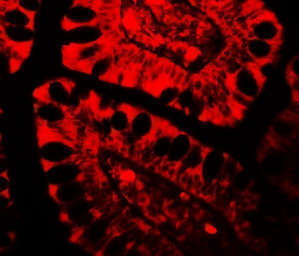
A02079 has IHC data from human small intestine paraffin sections and IF data from human tissue (catalog image captions); A02079-2 has IF/ICC data from human cells (catalog IF caption).

Real IHC data Immunohistochemistry Validation of TRIM21 in Human Small Intestine Tissue Immunohistochemical analysis of paraffin-embedded human small intestine tissue using anti-TRIM21 antibody (A02079) at 5 μg/ml. Tissue was fixed with formaldehyde and blocked with 10% serum for 1 h at RT; antigen retrieval was by heat mediation with a citrate buffer (pH6). Samples were incubated with primary antibody overnight at 4˚C. A goat anti-rabbit IgG H&L (HRP) at 1/250 was used as secondary. Counter stained with Hematoxylin.
Anti-TRIM21 Antibody
Cat # A02079
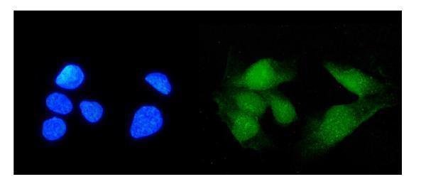
Real IF data IF analysis of TRIM21/SS-A using anti-TRIM21/SS-A antibody (A02079-2). TRIM21/SS-A was detected in immunocytochemical section of U20S cells. Enzyme antigen retrieval was performed using IHC enzyme antigen retrieval reagent (AR0022) for 15 mins. The cells were blocked with 10% goat serum. And then incubated with 2μg/mL rabbit anti-TRIM21/SS-A Antibody (A02079-2) overnight at 4°C. DyLight®488 Conjugated Goat Anti-Rabbit IgG (BA1127) was used as secondary antibody at 1:100 dilution and incubated for 30 minutes at 37°C. The section was counterstained with DAPI. Visualize using a fluorescence microscope and filter sets appropriate for the label used.
Anti-TRIM21/SS-A Antibody ®
Cat # A02079-2

A02079 is listed for human IHC-P and IF, with images from human small intestine paraffin sections and human small intestine tissue (catalog applications, reactivity, and image captions). A02079-2 is listed for IF and ICC, with an image from U20S cells; its listed reactivity is human, mouse, and rat (catalog applications, reactivity, and IF caption).

Which to pick: For tissue IHC, choose A02079: its own IHC image shows formaldehyde-fixed human small intestine paraffin sections, with citrate pH 6 heat retrieval and primary antibody at 5 μg/mL (A02079 IHC caption). For IF/ICC, A02079-2 has a human cell image at 2 μg/mL; A02079 also has a human tissue IF image at 20 μg/mL (catalog IF captions). For mouse or rat work, A02079-2 lists those species as reactive, but its supplied IF/ICC image is from human cells and it has no listed IHC-P application (catalog reactivity, applications, and IF caption).

Each figure is that product's own IHC / IF validation image from its datasheet.

References

  1. UniProt Consortium. UniProt entry P19474 (RO52_HUMAN, E3 ubiquitin-protein ligase TRIM21).
  2. Human Protein Atlas. TRIM21 tissue IHC expression (reliability: Enhanced).
  3. Human Protein Atlas. TRIM21 subcellular location (ICC-IF): Localized to the nucleoplasm..
  4. Human Protein Atlas. TRIM21 antibody validation summary (2 antibodies).
  5. Targeting AQP5-mediated arginine deprivation in gastric cancer stem cells restores NK cell anti-tumor immunity. Cell reports. Medicine 2025 — PMC12490235.
  6. TRIM21 suppresses uterine corpus endometrial carcinoma progression by promoting K48 -linked ubiquitination and degradation of SOAT1. Frontiers in immunology 2026 — PMC13526992.
  7. Pro-inflammatory cytokine IFN-γ protects against renal fibrosis by promoting E3 ubiquitin ligase Trim21-mediated Loxl2 degradation in tubular epithelial cells. Cell death & disease 2026 — PMC13338432.
  8. TRIM21 Expression as a Prognostic Biomarker for Progression-Free Survival in HNSCC. International journal of molecular sciences 2023 — PMC10048917.
  9. PubMed PMID:1985094 — UniProt-cited evidence.
  10. PubMed PMID:1985112 — UniProt-cited evidence.
  11. PubMed PMID:7713506 — UniProt-cited evidence.