This website uses cookies to ensure you get the best experience on our website.


- Table of Contents
Plan chromogenic TRIM24 IHC on paraffin sections using 2 μg/mL catalog antibody, as shown in its tissue captions (datasheet M03258-2). Compare staining with high thyroid glandular cell signal and undetected adipocyte signal, and score nuclear and cytoplasmic staining separately (HPA tissue IHC).

Expected localisation, validated protocols, controls and antibodies — the at-a-glance facts below, then the full design guide.
| Expected localisation | Nuclear, with cytoplasmic staining in several tissues (HPA tissue IHC) | |
| Staining pattern | Cells across tissues: nuclear, sometimes cytoplasmic (HPA tissue IHC) | |
| Antigen retrieval | EDTA pH 8.0 HIER, heat-mediated (datasheet M03258-2) | |
| Positive control | Thyroid gland+4 more · see all | |
| Negative control | Adipose tissue+4 more · see all |
| Fixation | Matched tissue-IHC evidence does not establish the fixation claim. Validate the specimen-specific method before use. (selected-SKU IHC image M03258-2) | |
| Caveat | Antibody staining and RNA show medium consistency (HPA tissue IHC) | |
| Regulation | Early DNA damage lowers TRIM24 (UniProt) | |
| Isoform / epitope | 2 isoforms, Long and Short; check epitope coverage (UniProt) |
Compare the catalog antibody’s IHC-P protocol (datasheet: M03258-2) with three published TRIM24 IHC protocols (PMC5076458; PMC8805319; PMC3661592).
| Sample | Paraffin-embedded human adenocarcinoma of the colon tissue; fixative not specified (datasheet M03258-2) |
| Fixation | Image fixative and duration unreported (datasheet M03258-2); verify before use. |
| Sectioning | 4–5 µm sections on charged slides (standard) |
| Deparaffinisation | Xylene, graded ethanol series to water (standard) |
| Antigen retrieval | Heat retrieval: EDTA pH 8.0 (datasheet M03258-2); 20 min, 95–100 °C (standard) |
| Peroxidase block | 3% H2O2, 10 min, room temperature (standard) |
| Blocking | 10% goat serum (datasheet M03258-2) |
| Primary antibody | Mouse monoclonal (clone 4G6C2) anti-TRIM24, 2 μg/ml (datasheet M03258-2) |
| Primary incubation | Overnight at 4 °C (datasheet M03258-2) |
| Detection | HRP-conjugated secondary, DAB chromogen (datasheet M03258-2) |
| Counterstain | Hematoxylin, blue, dehydrate and mount (standard) |
| Expected result | TRIM24-positive staining in glandular cells of thyroid gland (HPA tissue IHC: High). HPA tissue profile: General nuclear expression with additional cytoplasmic expression in several tissues. No signal in the no-primary control. |
TRIM24 is predominantly nuclear and has no transmembrane segment (UniProt O15164: subcellular location; topology). In tissue IHC, expect nuclear staining in thyroid glandular cells at high intensity and in several other epithelial and trophoblastic cell populations at medium intensity (HPA: tissue IHC). HPA describes general nuclear expression with additional cytoplasmic staining in several tissues; its tissue IHC reliability is Approved, with medium consistency between staining and RNA expression (HPA: tissue IHC).
| Distinct nuclear staining in thyroid glandular cells, with weaker staining in some other listed positive cell populations. | This fits the reported distribution: thyroid glandular cells are High; colon and duodenum glandular cells, esophageal squamous epithelial cells, ovarian follicle cells, and placental trophoblastic cells are Medium (HPA: tissue IHC). Compare cells within the same section before treating every difference in intensity as a technical failure (general IHC practice). |
| Predominantly cytoplasmic staining with little or no nuclear signal in an expected positive cell population. | Investigate the compartment call and possible staining artefact: predominant nuclear localisation is expected (UniProt O15164: subcellular location), although HPA reports additional cytoplasmic expression in several tissues (HPA: tissue IHC). Cytoplasmic signal alone cannot establish a false positive, because UniProt also records cytoplasmic localisation and context dependent movement to mitochondria (UniProt O15164: subcellular location). |
| Strong staining in a cell population listed as not detected, such as adipocytes or heart muscle cardiomyocytes. | Treat the unexpected result as a specificity question, including possible antibody cross-reactivity or endogenous detection activity (general IHC practice). HPA reports TRIM24 as Not detected in those particular cell populations (HPA: tissue IHC); that reference observation does not prove every specimen of that tissue must be negative. |
| Diffuse color across nuclei, cytoplasm, stroma, or empty areas without clear cell boundaries. | A uniform haze cannot be scored as the expected cell and compartment pattern (general IHC practice; HPA: general nuclear expression). Consider nonspecific binding, incomplete blocking, excess detection reagent, or endogenous detection activity as general IHC causes. Judge the signal against a matched negative detection control and a known positive tissue section (general IHC practice). |
| No discernible nuclear signal in thyroid glandular cells on a section expected to serve as a positive reference. | HPA reports High staining in thyroid glandular cells (HPA: tissue IHC), so absence of signal calls for a workflow check. Confirm that glandular cells are present and assess retrieval, antibody incubation, detection reagents, and counterstain with appropriate controls (general IHC practice). HPA's Approved rating has medium consistency with RNA data, so interpret a negative specimen cautiously (HPA: tissue IHC). |
| Reference tissue and cell population | Choose the actual cell population named in the reference: thyroid glandular cells are High, whereas adipocytes and heart muscle cardiomyocytes are Not detected (HPA: tissue IHC). HPA also reports low tissue RNA specificity, so its staining list should guide comparison without becoming an absolute rule for every specimen (HPA: tissue IHC). |
| Reported IHC antibody validation | HPA043495 is listed as IHC Approved; HPA061717 has no IHC validation status in the supplied antibody record (HPA: antibody validation). The overall tissue IHC profile is Approved with medium consistency between staining and RNA expression (HPA: tissue IHC). Record which antibody produced the slide before comparing it with that reference pattern (general IHC practice). |
| Compartment and topology | Nuclear staining has the strongest routine expectation: UniProt calls TRIM24 predominantly nuclear, and HPA reports general nuclear tissue expression (UniProt O15164: subcellular location; HPA: tissue IHC). The protein has no transmembrane segment (UniProt O15164: topology). Additional cytoplasmic staining is documented, so assess its cell context alongside nuclear signal (HPA: tissue IHC). |
| Isoforms and epitope coverage | UniProt lists 2 isoforms, Long and Short (UniProt O15164: isoforms). The payload gives no antibody epitope or isoform coverage, so isoform specific staining cannot be predicted from it. If comparing antibodies, obtain their documented epitope information before attributing a difference in tissue staining to an isoform (general IHC practice). |
| IF/ICC Q: what localisation should an independent image show? | A: HPA's ICC-IF summary places TRIM24 in the nucleoplasm, with an enhanced main location; images are listed for A-431, U-251MG, U2OS, and Hep-G2 (HPA: subcellular ICC-IF). This supports a nuclear localisation check but does not supply an IHC staining intensity for those cell lines (HPA: subcellular ICC-IF). |
| Situation | Likely cause | Next action |
|---|---|---|
| Thyroid positive reference is blank. | The expected glandular cells may be absent from the evaluated area, or an IHC workflow step may have failed (general IHC practice); thyroid glandular cells are High in the HPA reference (HPA: tissue IHC). | First identify intact glandular cells, then check retrieval, primary antibody incubation, chromogenic detection, and the positive control run (general IHC practice). Do not infer target specific fixation sensitivity from this result; none is supplied. |
| Signal is mainly cytoplasmic in a reference positive tissue. | The section may have background or a compartment assignment problem (general IHC practice). Additional cytoplasmic staining occurs in several tissues, but TRIM24 is predominantly nuclear (HPA: tissue IHC; UniProt O15164: subcellular location). | Inspect nuclear boundaries and compare the staining with a negative detection control and a known positive section (general IHC practice). Record the cytoplasmic component separately; do not score cytoplasmic color alone as the expected nuclear result. |
| A listed negative population stains strongly. | Cross-reactivity or endogenous detection activity is possible (general IHC practice). HPA reports adipocytes and heart muscle cardiomyocytes as Not detected (HPA: tissue IHC). | Check a control omitting the primary antibody for detection related signal, then compare another documented reference cell population and the antibody's IHC validation status (general IHC practice; HPA: antibody validation). |
| The whole section shows diffuse brown haze. | Nonspecific binding, inadequate blocking, or excessive detection signal are general IHC possibilities (general IHC practice). A haze lacks the predominantly nuclear pattern reported for TRIM24 (UniProt O15164: subcellular location; HPA: tissue IHC). | Review blocking and detection controls, then adjust reagent conditions using the antibody's documented IHC instructions (general IHC practice). Score only staining that can be assigned to cells and compartments. |
| A faint section is being called negative. | Some named populations are Low, including lung alveolar cells and salivary gland glandular cells (HPA: tissue IHC). Weak target signal can be hard to separate from counterstain or background (general IHC practice). | Compare the same cell population with a positive reference and a negative detection control, and inspect nuclei before assigning an intensity category (general IHC practice). Avoid using a low staining population as the sole assay check (HPA: tissue IHC). |
| Results differ between two antibodies or between IHC and IF/ICC. | Antibody validation and assay context differ: HPA043495 is IHC Approved, while the supplied record gives HPA061717 no IHC status; HPA's ICC-IF localisation is nucleoplasmic (HPA: antibody validation; HPA: subcellular ICC-IF). | Keep each antibody and assay result identified, compare nuclear localisation first, and review documented epitope coverage before proposing an isoform explanation (general IHC practice; UniProt O15164: 2 isoforms). |


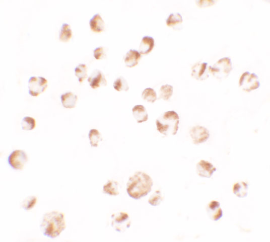


Comprehensive Human Protein Atlas IHC scoring per tissue (reliability: Approved — Medium consistency between antibody staining and RNA expression data.). Rows are taken directly from the HPA tissue chart — click any row's HPA link to view the source.
| Tissue | Cell type | Level | Evidence | Source |
|---|---|---|---|---|
| Thyroid gland | Glandular cells | High | Protein (IHC) | HPA → |
| Colon | Glandular cells | Medium | Protein (IHC) | HPA → |
| Duodenum | Glandular cells | Medium | Protein (IHC) | HPA → |
| Esophagus | Squamous epithelial cells | Medium | Protein (IHC) | HPA → |
| Fallopian tube | Ciliated cells (ciliary rootlets) | Medium | Protein (IHC) | HPA → |
| Tissue | Cell type | Level | Evidence | Source |
|---|---|---|---|---|
| Adipose tissue | Adipocytes | Not detected | Protein (IHC) | HPA → |
| Adrenal gland | Glandular cells | Not detected | Protein (IHC) | HPA → |
| Cerebellum | Cells in granular layer | Not detected | Protein (IHC) | HPA → |
| Cerebral cortex | Endothelial cells | Not detected | Protein (IHC) | HPA → |
| Cervix | Glandular cells | Not detected | Protein (IHC) | HPA → |
Troubleshoot TRIM24 staining in paraffin sections by checking retrieval, nuclear localisation and cell type before comparing staining intensity across samples.
The catalog shows real TRIM24 IHC data from human paraffin sections (M03258-2 IHC captions) and IF/ICC data from MCF-7 and EL4 cells (M03258-2 and A03258 IF captions).
M03258-2 has IHC images from human paraffin sections of colon adenocarcinoma, thyroid cancer, ovarian serous tumor, and breast cancer, plus IF in MCF-7 cells (M03258-2 image captions). A03258 has ICC and IF images in EL4 cells, with mouse ICC/IF validation stated in its catalog entry (A03258 image captions and validation text).
Which to pick: Choose M03258-2 for tissue IHC: its own caption documents a human paraffin section with EDTA retrieval, while the fixative is unreported (M03258-2 IHC caption). For IF/ICC, choose M03258-2 for MCF-7 cells or A03258 for EL4 cells (respective IF captions). A03258 lists human, mouse, and rat reactivity for cross-species planning, but its pictured IF/ICC validation is in mouse EL4 cells (A03258 catalog and validation text).