TRIP12 / E3 ubiquitin-protein ligase TRIP12 · IHC design guide

Design Immunohistochemistry for TRIP12

Plan TRIP12 IHC on paraffin sections using its nuclear and nucleolar tissue pattern as a guide (HPA tissue IHC). This guide covers fixation, staining controls and interpretation, including cell populations with no detectable signal (HPA tissue IHC).

Evidence assembled Oct 2026 · For research use; verify linked source records and product datasheet before use
Immunohistochemistry protocol sheet for TRIP12 (IHC for TRIP12): expected localisation Nuclear and nucleolar (HPA tissue IHC), antibody A05449, validated IHC image, and IHC protocol steps
Printable TRIP12 IHC protocol sheet — expected localisation Nuclear and nucleolar (HPA tissue IHC), antibody A05449, controls and protocol steps. Open the full TRIP12 IHC guide →

TRIP12 Immunohistochemistry Experimental Design Guide

Expected localisation, validated protocols, controls and antibodies — the at-a-glance facts below, then the full design guide.

Must know before staining
Expected localisation Nuclear and nucleolar (HPA tissue IHC)
Staining pattern General nuclear and nucleolar staining; most abundant in testis (HPA tissue IHC)
Antigen retrieval Tris-EDTA pH 9.0 HIER, 95–98 °C, 20 min (rule: nuclear antigen)
Positive control ⓘ Appendix+4 more · see all
Negative control ⓘ Adipose tissue+3 more · see all
Important caveats
Reasons your staining may differ from the expected pattern.
Fixation Keep fixation conditions consistent across sections. (standard IHC practice; not target-specific)
Caveat Adipocytes and cholangiocytes may lack detectable signal (HPA tissue IHC)
Regulation Most abundant in testis (HPA tissue IHC)
Isoform / epitope Five isoforms; check antibody epitope coverage (UniProt)
Section 1

Recommended TRIP12 IHC & IF Protocols

The catalog antibody protocol is accompanied by published TRIP12 chromogenic IHC methods for human gastric tissue (PMC6138561) and human post-mortem brain (PMC8639668).

Recommended immunohistochemistry (IHC-P) protocol parameters
SampleTissue sections; selected-image fixative not specified (standard IHC workflow)
FixationImage fixative and duration unreported (datasheet A05449); verify before use.
Sectioning4–5 µm sections on charged slides (standard)
DeparaffinisationXylene, graded ethanol series to water (standard)
Antigen retrievalHeat-induced epitope retrieval in Tris-EDTA buffer, pH 9.0, 20 min at 95–98 °C (standard rule: nuclear antigen)
Peroxidase block3% H2O2, 10 min, room temperature (standard)
Blocking10% normal serum of the secondary host, 30 min, room temperature (standard)
Primary antibodyRabbit anti-TRIP12, 5 μg/mL (datasheet A05449)
Primary incubationOvernight at 4 °C (standard)
DetectionHRP-polymer secondary, DAB chromogen 5–10 min (standard)
CounterstainHematoxylin, blue, dehydrate and mount (standard)
Expected resultTRIP12-positive staining in glandular cells of appendix (HPA tissue IHC: Medium). HPA tissue profile: General nuclear and nucleolar expression, most abundant in testis. No signal in the no-primary control.
💡Decision noteStart with Tris-EDTA pH 9.0 retrieval at 95–98 °C for 20 min (page antigen retrieval rule); TRIP12 is nucleoplasmic (UniProt Q14669).
Section 2

What Is the Expected TRIP12 Staining Pattern?

TRIP12 is a nuclear, nucleoplasmic protein without a transmembrane segment (UniProt Q14669: location and topology). In paraffin-section IHC, expect mainly nuclear and sometimes nucleolar staining across multiple cell types; HPA describes general nuclear and nucleolar expression, most abundant in testis (HPA: tissue IHC). Its tissue IHC profile has Enhanced reliability, reflecting consistency between antibody staining and RNA expression (HPA: tissue IHC).

What am I looking at on my slide?
Clear nuclear staining in glandular cells of colon or appendix, or respiratory epithelium of bronchus.These are plausible positive results: HPA reports Medium staining in each named cell population (HPA: tissue IHC). Assess signal in the recorded cell type, since an entire section need not stain uniformly (general IHC practice).
Predominantly cytoplasmic or membranous signal, with little convincing nuclear staining.This conflicts with TRIP12’s nuclear, nucleoplasmic location (UniProt Q14669: location) and HPA’s nuclear and nucleolar tissue profile (HPA: tissue IHC). Treat it as a possible artefact; check controls and detection background before assigning it to TRIP12 (general IHC practice).
Strong staining in a cell population reported as Not detected, such as adipocytes in adipose tissue.HPA reports TRIP12 as Not detected in adipocytes (HPA: tissue IHC). Unexpected signal warrants a cross-reactivity or endogenous-detection check, especially if it is extranuclear (general IHC practice); one HPA observation alone does not prove that every adipocyte must be negative.
Diffuse colour across tissue, stroma and nuclei, without clear cellular boundaries.This is hard to score as specific nuclear TRIP12 staining (UniProt Q14669: location; general IHC practice). Review a no-primary control, blocking and chromogen development for nonspecific background or endogenous detection activity (general IHC practice).
No nuclear signal in a purported positive-control section.Check that the control includes a cell type HPA actually scored Medium, such as colon glandular cells or cerebral-cortex neurons (HPA: tissue IHC). If it does, review tissue preservation, retrieval, antibody preparation and detection controls before calling the sample negative (general IHC practice).
💡Expected TRIP12 appearanceCall a convincing positive result when the relevant cells show discernible nuclear, sometimes nucleolar, chromogenic staining, consistent with HPA’s Medium examples; predominantly cytoplasmic, membranous or indiscriminately diffuse colour is suspect (HPA: tissue IHC; UniProt Q14669: location; general IHC practice).
How each factor affects the staining
Compartment used for scoringScore nuclei first: UniProt places TRIP12 in the nucleoplasm, while HPA describes nuclear and nucleolar tissue staining (UniProt Q14669: location; HPA: tissue IHC). A nucleolar accent can fit the tissue profile, but is not required in every positive cell (HPA: tissue IHC).
Cell population and tissue choiceHPA reports Medium staining in several epithelial and neuronal populations, Low staining in specified populations, and Not detected in others (HPA: tissue IHC). Match interpretation to the named cell population; tissue-wide labels can obscure these differences (general IHC practice).
Antibody validationHPA036835 and HPA045893 each have Enhanced IHC validation (HPA: antibody records). The tissue profile also carries Enhanced reliability (HPA: tissue IHC). These support comparison with the reported pattern but do not identify the epitope or establish performance for every specimen.
Topology, processing and isoformsUniProt lists no transmembrane segment or signal peptide and lists five isoforms (UniProt Q14669: topology, processing and isoforms). Interpret membrane-only staining cautiously; epitope coverage across isoforms cannot be inferred from these records without an epitope assignment.
IF/ICC Q&A: where should signal appear?In IF/ICC, HPA reports approved localisation to nuclear speckles and images in RT-4, Rh30 and U2OS cells (HPA: subcellular record). This answers the location question for that assay; speckle detail need not be resolved in chromogenic paraffin-section IHC (general microscopy practice).
Why is my staining missing, weak or wrong?
SituationLikely causeNext action
Positive-control nuclei are blank.The selected section may lack a reported positive cell population, or the staining run may have failed (HPA: tissue IHC; general IHC practice).Use a supplied Medium example, such as colon glandular cells, and verify the detection run with appropriate controls; then review retrieval and antibody preparation (HPA: tissue IHC; general IHC practice).
Colour is chiefly cytoplasmic or outlines cell membranes.The distribution disagrees with the expected nuclear location (UniProt Q14669: location; HPA: tissue IHC).Compare the same run with a known positive section and no-primary control; assess whether background or misplaced signal is being mistaken for nuclear staining (general IHC practice).
A reported Not detected cell type stains strongly.Cross-reactivity or endogenous detection activity is possible; HPA lists adipocytes, cholangiocytes, chondrocytes and splenic red-pulp cells as Not detected in the specified tissues (HPA: tissue IHC; general IHC practice).Confirm the precise cell identity and compartment, then inspect no-primary and detection controls before interpreting the unexpected signal (general IHC practice).
All compartments show hazy brown background.Nonspecific staining, insufficient blocking or excess chromogen development may obscure a nuclear pattern (general IHC practice).Review the no-primary control and background-reduction steps, and judge positives only where nuclear signal separates clearly from surrounding tissue (general IHC practice).
Results differ between fields or tissue blocks.HPA records cell-specific Medium, Low and Not detected levels; variation in represented cells can change the apparent result (HPA: tissue IHC).Compare like cell populations and score localisation and intensity separately, using a reported positive population on the same run for context (HPA: tissue IHC; general IHC practice).
IF/ICC shows speckles, while IHC looks broadly nuclear.HPA describes nuclear speckles in ICC-IF and general nuclear and nucleolar staining in tissue IHC; the assays present different spatial detail (HPA: subcellular record; HPA: tissue IHC; general microscopy practice).Interpret each image against its own assay reference and confirm nuclear localisation; do not require resolved speckles in the chromogenic section (HPA: subcellular record; HPA: tissue IHC; general microscopy practice).

Sample controls for TRIP12 IHC & IF

🧪Run colon first and look for TRIP12 staining in glandular cells (HPA: Medium in colon glandular cells). Use adipose tissue adipocytes as the negative comparison (HPA: Not detected in adipocytes); cells without specific staining on the colon slide should show counterstain alone, although HPA does not identify a particular internal-negative colon cell type (HPA: colon glandular-cell row).
Positive control tissue: Appendix (Glandular cells, HPA Medium)
Negative control tissue: Adipose tissue (HPA Not detected)
ICC-IF cell lines (HPA subcellular resource): HPA ICC-IF images show TRIP12 in RT-4, Rh30, U2OS, with annotated localisation: Nuclear speckles (approved) (HPA subcellular).
Technical controls: Include a no-primary, secondary-only control; a host- and isotype-matched control antibody; and, if available, a TRIP12-knockout sample as a biological negative (standard IHC practice). Block endogenous peroxidase in colon sections, and block endogenous biotin if using avidin–biotin detection (standard chromogenic IHC practice).
⚠️Feasibility: A target-specific fixation window or fixation effect is unreported, and the selected A05449 rat-colon caption does not state its fixative (selected-SKU tissue-IHC caption). Retrieval dependency is unreported; optimize antigen retrieval on paraffin sections and assess specific nuclear staining (UniProt Q14669: nucleoplasm; standard IHC practice). The supplied evidence does not establish whether frozen sections or IF are easier; exclude luminal debris and edge staining when scoring colon glands (standard IHC practice).

HPA tissue IHC evidence for TRIP12

Comprehensive Human Protein Atlas IHC scoring per tissue (reliability: Enhanced — High consistency between antibody staining and RNA expression data.). Rows are taken directly from the HPA tissue chart — click any row's HPA link to view the source.

Positive expression · recommended positive controls

TissueCell typeLevelEvidenceSource
Appendix Glandular cells Medium Protein (IHC) HPA →
Bronchus Respiratory epithelial cells Medium Protein (IHC) HPA →
Caudate Neuronal cells Medium Protein (IHC) HPA →
Cerebral cortex Neuronal cells Medium Protein (IHC) HPA →
Colon Glandular cells Medium Protein (IHC) HPA →

Undetected expression · recommended negative controls

TissueCell typeLevelEvidenceSource
Adipose tissue Adipocytes Not detected Protein (IHC) HPA →
Liver Cholangiocytes Not detected Protein (IHC) HPA →
Soft tissue Chondrocytes Not detected Protein (IHC) HPA →
Spleen Cells in red pulp Not detected Protein (IHC) HPA →
Section 3

Advanced TRIP12 IHC Tips

Troubleshoot TRIP12 staining in paraffin section chromogenic IHC using nuclear localisation, documented tissue patterns and controls matched to each run.

Which retrieval conditions should I try first for weak TRIP12 staining?
Start with Tris-EDTA pH 9.0 heat-induced epitope retrieval at 95–98 °C for 20 min (page retrieval setting: nuclear antigen). Keep section thickness, cooling time and antibody incubation consistent while comparing retrieval runs, so changes in nuclear staining can be attributed to retrieval (standard IHC practice). Score intact nuclei in colon glandular cells as a reference population; their staining is reported as medium (HPA: colon glandular cells, medium). If signal remains weak, compare a milder retrieval condition on adjacent sections while watching for tissue lifting and diffuse background (standard IHC practice). Record the alternative buffer and pH as a tested fallback, not as a documented TRIP12 condition (page retrieval setting).
Could fixation explain weak or uneven nuclear TRIP12 staining?
Target-specific sensitivity of TRIP12 to fixation is unknown from the supplied evidence; the rat colon image caption does not state a fixative (A05449 caption: fixative not stated). For paraffin sections, document fixation method and duration before comparing specimens, because fixation and processing can alter epitope access (standard IHC practice). Compare similarly processed control and test sections in the same staining run, using intact nuclei and tissue morphology to judge whether weak signal tracks processing differences (standard IHC practice). If staining varies across a section, inspect folds, drying and poorly preserved areas before changing retrieval or antibody concentration (standard IHC practice). Do not assign the variation to a TRIP12-specific fixation effect without direct validation (supplied fixation evidence: none).
What subcellular pattern supports specific TRIP12 staining in tissue?
Prioritise staining within intact nuclei because TRIP12 is assigned to the nucleoplasm (UniProt Q14669: subcellular location). A punctate nuclear pattern can be compatible with its approved nuclear speckle localisation, although chromogenic IHC may not resolve individual speckles (HPA: nuclear speckles; standard IHC practice). HPA also describes general nuclear and nucleolar tissue expression, so assess the whole nuclear compartment rather than requiring one sharply defined pattern (HPA: tissue IHC profile). Compare nuclei with the counterstain and check whether cytoplasmic colour persists outside nuclear boundaries (standard IHC practice). Predominantly membrane-associated staining is unexpected for a protein with no transmembrane segment and warrants a control review (UniProt Q14669: topology).
How should isoforms and epitope uncertainty affect antibody selection?
TRIP12 has 5 annotated isoforms, so determine whether the antibody’s stated immunogen lies in a region shared by the isoforms relevant to the experiment (UniProt Q14669: isoforms; standard IHC practice). The supplied product caption gives no epitope position, so it cannot establish which isoforms the catalog antibody detects (A05449 caption: epitope not stated). TRIP12 contains a WWE domain at residues 797–911 and a HECT domain at 1960–2067; map any disclosed immunogen against these regions (UniProt Q14669: domains). It also has annotated modified residues, including phosphoserines at 12, 119, 127 and 142, but their effect on this antibody’s staining is untested (UniProt Q14669: modified residues; supplied antibody evidence).
How can I adapt the localisation check to multiplex IF?
For secondary IF/ICC work, pair TRIP12 with a validated marker for the expected cell population, such as a glandular cell marker when examining colon epithelium (HPA: colon glandular cells, medium; standard IF practice). Choose fluorophores after inspecting unstained tissue for autofluorescence, placing the weaker signal in a cleaner spectral channel and checking single-stain controls for bleed-through (standard IF practice). TRIP12 is nucleoplasmic and has no transmembrane segment, so permeabilisation must allow antibody access through the cell and nuclear membranes (UniProt Q14669: subcellular location and topology; standard IF practice). Compare the TRIP12 channel with a nuclear counterstain; nuclear speckles are a reported IF localisation, not a required appearance in chromogenic tissue IHC (HPA: nuclear speckles; standard imaging practice).
What should I change when TRIP12 chromogenic staining is diffuse?
First separate colour confined to nuclei from diffuse colour in stroma, cytoplasm or slide edges; TRIP12 is assigned to the nucleoplasm (UniProt Q14669: subcellular location; standard IHC practice). Run a no-primary control to assess secondary reagent, chromogen and endogenous enzyme contributions, and use a peroxidase block when the detection system requires one (standard chromogenic IHC practice). Adjust blocking, wash stringency and antibody concentration one variable at a time, then check whether nuclear contrast improves without losing signal in reference tissue (standard IHC practice). The reported rat colon image used 5 µg/ml, but that caption does not establish an optimal concentration for another processing or detection workflow (A05449 caption).
How should I quantify TRIP12 across differently cellular tissue regions? ⚠ ANSWER MARKED FOR VERIFICATION
Define the scored cell population and nuclear compartment before analysis, because TRIP12 has reported nuclear expression and tissue staining varies by cell type (UniProt Q14669: subcellular location; HPA: tissue IHC profile). For a per-cell result, report the percentage of positive nuclei and an H-score using prespecified intensity categories; keep thresholds fixed across the comparison (standard IHC scoring practice). Normalise counts to the number of eligible intact nuclei, or report positive nuclear density per mm² of analysed tissue when cell density is itself relevant (standard image analysis practice). Exclude folds, necrosis and section edges with the same rules for every specimen, and report the number of regions and cells scored (standard IHC practice).
How do I distinguish a true TRIP12 signal from artefact?
A credible result shows staining associated with intact nuclei in an appropriate cell population, consistent with TRIP12’s nucleoplasmic annotation and reported tissue patterns (UniProt Q14669: subcellular location; HPA: tissue IHC profile). Colon glandular cells have reported medium staining, whereas adipocytes are reported as not detected; use those patterns as context rather than absolute controls across different specimens (HPA: colon glandular cells, medium; adipocytes, not detected). Treat predominant membrane colour, strong edge-only staining and colour in necrotic areas as reasons to inspect specificity and processing (UniProt Q14669: no transmembrane segment; standard IHC practice). Check a no-primary control for endogenous peroxidase or detection background before calling diffuse DAB deposition TRIP12 positive (standard chromogenic IHC practice).
Boster reagents

Best TRIP12 / E3 ubiquitin-protein ligase TRIP12 IHC Antibodies

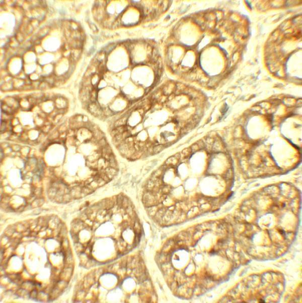
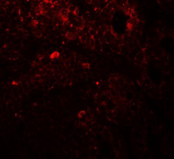
A05449 has IHC and IF images from rat colon and an ICC image from HeLa cells (catalog image captions); the catalog lists human, mouse and rat reactivity (catalog reactivity).

Real IHC data Immunohistochemistry of TRIP12 in rat colon tissue with TRIP12 antibody at 5 μg/ml.
Anti-TRIP12 Antibody
Cat # A05449

A05449 is listed for IHC-P, IF and ICC (catalog applications). Its images show rat-colon IHC at 5 μg/mL, rat-colon IF at 20 μg/mL and HeLa ICC at 2 μg/mL (catalog image captions).

Which to pick: For tissue IHC, choose A05449: IHC-P is listed, and its IHC image shows rat colon at 5 μg/mL (catalog applications; A05449 IHC caption). The IHC caption does not report section processing or fixative (A05449 IHC caption). For IF/ICC or cross-species work, A05449 lists both applications and human, mouse and rat reactivity; its images show rat-colon IF and HeLa ICC, while clonality is unreported (catalog applications and reactivity; A05449 IF/ICC captions; catalog clone field).

Each figure is that product's own IHC / IF validation image from its datasheet.

References

  1. UniProt Consortium. UniProt entry Q14669 (TRIPC_HUMAN, E3 ubiquitin-protein ligase TRIP12).
  2. Human Protein Atlas. TRIP12 tissue IHC expression (reliability: Enhanced).
  3. Human Protein Atlas. TRIP12 subcellular location (ICC-IF): Localized to the nuclear speckles..
  4. Human Protein Atlas. TRIP12 antibody validation summary (2 antibodies).
  5. Helicobacter pylori pathogen regulates p14ARF tumor suppressor and autophagy in gastric epithelial cells. Oncogene 2018 — PMC6138561.
  6. TRIP12 ubiquitination of glucocerebrosidase contributes to neurodegeneration in Parkinson's disease. Neuron 2021 — PMC8639668.
  7. The TRIP12 E3 ligase induces SWI/SNF component BRG1-β-catenin interaction to promote Wnt signaling. Nature communications 2025 — PMC12141612.
  8. USP7 overexpression prevents the progression of clear cell renal cell carcinoma by enhancing pyroptosis via TRIP12 deubiquitination. Cancer biology & therapy 2025 — PMC12439559.
  9. PubMed PMID:20208519 — UniProt-cited evidence.
  10. PubMed PMID:7584044 — UniProt-cited evidence.
  11. PubMed PMID:15815621 — UniProt-cited evidence.