This website uses cookies to ensure you get the best experience on our website.


- Table of Contents
Real validated TRPC5 Western blot protocols, expected-band and isoform facts, troubleshooting for weak or shifted signal, and recommended anti-TRPC5 WB antibodies. Everything you need to plan the experiment before you commit precious samples.
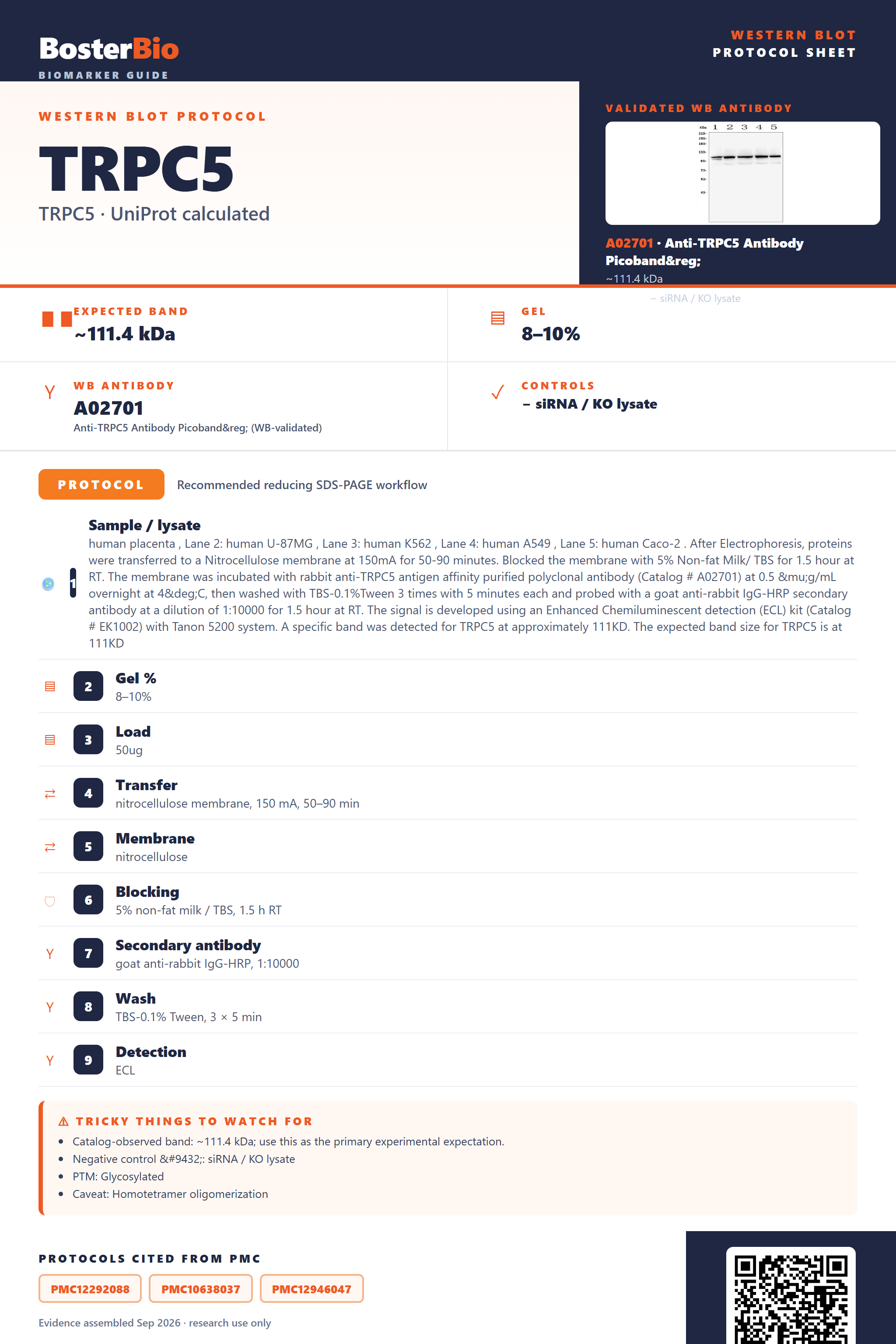
Expected bands, validated protocols, controls and antibodies — the at-a-glance facts below, then the full design guide.
| Expected band | ~111.4 kDa | |
| Observed band | ~111 kDa | |
| Gel | 8–10% | |
| Negative control | siRNA / KO lysate |
| PTM | Glycosylated | |
| Caveat | Homotetramer oligomerization | |
| Regulation | LPS-suppressed | |
| Isoform | 1 isoform(s) |
Literature-validated Western blot parameters for TRPC5 — gel percentage, transfer, blocking, antibody incubation and detection, extracted from published methods.
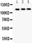
| Sample / lysate | human placenta , Lane 2: human U-87MG , Lane 3: human K562 , Lane 4: human A549 , Lane 5: human Caco-2 . After Electrophoresis, proteins were transferred to a Nitrocellulose membrane at 150mA for 50-90 minutes. Blocked the membrane with 5% Non-fat Milk/ TBS for 1.5 hour at RT. The membrane was incubated with rabbit anti-TRPC5 antigen affinity purified polyclonal antibody (Catalog # A02701) at 0.5 μg/mL overnight at 4°C, then washed with TBS-0.1%Tween 3 times with 5 minutes each and probed with a goat anti-rabbit IgG-HRP secondary antibody at a dilution of 1:10000 for 1.5 hour at RT. The signal is developed using an Enhanced Chemiluminescent detection (ECL) kit (Catalog # EK1002) with Tanon 5200 system. A specific band was detected for TRPC5 at approximately 111KD. The expected band size for TRPC5 is at 111KD |
| Gel % | 8–10% |
| Load | 50ug |
| Transfer | nitrocellulose membrane, 150 mA, 50–90 min |
| Membrane | nitrocellulose |
| Blocking | 5% non-fat milk / TBS, 1.5 h RT |
| Secondary antibody | goat anti-rabbit IgG-HRP, 1:10000 |
| Wash | TBS-0.1% Tween, 3 × 5 min |
| Detection | ECL |
| Exposure / imaging | Tanon 5200 |
| Observed band | 111 kDa |
TRPC5 has a predicted mass of 111.4 kDa and runs at the same ~111 kDa on reducing SDS-PAGE, with only a minor shift from its single N461 glycosylation site.
| single sharp band at ~111 kDa | matches the calculated mass of the 973-residue TRPC5 polypeptide with minimal net shift from modification |
| band running slightly above the unmodified 111.4 kDa backbone or showing a faint upper shoulder | reflects the single N-linked glycosylation site at Asn461 adding modest mass |
| higher-molecular-weight species (multiples of ~111 kDa) under non-reducing or incompletely denaturing conditions | reflects the native homotetrameric channel assembly (ANK-repeat mediated) not fully dissociated by SDS/heat |
| no extra bands attributable to alternate transcripts | only one annotated isoform exists for TRPC5, so multi-band patterns are unlikely to come from splicing |
| very faint or absent signal in some whole-cell lysates despite loading | TRPC5 is a multi-pass cell-membrane channel protein that can be under-solubilized by mild lysis conditions |
| Predicted mass (111.4 kDa, 973 aa) | sets the baseline expected migration position on SDS-PAGE |
| N-linked glycosylation at Asn461 | adds modest mass, can nudge the band slightly above the unmodified backbone or add slight heterogeneity |
| Homotetrameric channel assembly (non-covalent, via ANK repeats) | can leave higher-order oligomeric species on the gel if denaturation is incomplete, though full reducing/denaturing treatment collapses it to the ~111 kDa monomer |
| Intrachain disulfide bond (Cys553-Cys558) | stabilizes a local loop within the monomer rather than linking separate subunits, so it does not itself generate a dimer-sized band |
| Recombinant/truncated antigen standards | smaller recombinant fragments used in some antibody QC lanes run well below the native full-length size and should not be used to judge expected native band position |
| Situation | Likely cause | Next action |
|---|---|---|
| Band higher than expected | incomplete denaturation of the non-covalent homotetrameric channel assembly | boil samples longer in fresh reducing SDS sample buffer with DTT or beta-mercaptoethanol before loading |
| Band lower than expected | comparison against a truncated or non-native recombinant standard rather than full-length native TRPC5 | confirm the antibody immunogen region and benchmark lysate bands against a full-length native positive control, not a recombinant fragment |
| Broad smear instead of sharp band | heterogeneous glycosylation at the single N461 site or aggregation of this multi-pass membrane protein | run a PNGase F deglycosylation control and optimize lysis/solubilization to limit aggregation |
| Weak or no signal | poor solubilization of a multi-pass membrane protein by standard lysis buffers | use a detergent-based lysis buffer suited to membrane proteins and increase total protein loaded |
| Multiple bands | partial proteolytic degradation of the large 973-residue membrane protein during extraction | add fresh protease inhibitors and keep samples cold throughout lysis and processing |

Comprehensive Human Protein Atlas IHC scoring per tissue. Rows are taken directly from the HPA tissue chart — click any row's HPA link to view the source.
| Tissue | Cell type | Level | Evidence | Source |
|---|
| Tissue | Cell type | Level | Evidence | Source |
|---|
Deeper troubleshooting and optimisation questions for TRPC5, answered from its protein features.
BosterBio's TRPC5 antibodies are among the best-performing WB antibodies on the market — well cited, thoroughly validated, and orthogonally cross-validated against negative tissues and complementary methods.
These TRPC5 antibodies are top-performing Boster reagents, extensively cited and rigorously validated by Western blot, including orthogonal confirmation against recombinant protein controls and predicted molecular weight matching, giving researchers confident, reproducible detection across sample types.
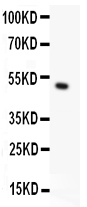
Which to pick: Both A02701 and PB9271 include real Western blot validation images. Choose PB9271 for recombinant-protein-confirmed specificity with matching predicted/observed 50KD band; choose A02701 for validated detection in electrophoresed sample lysates using standard SDS-PAGE conditions.