This website uses cookies to ensure you get the best experience on our website.


- Table of Contents
Plan chromogenic TSG101 IHC around the cytoplasmic and membranous tissue pattern (HPA tissue IHC). This guide identifies high-staining cell populations for controls and flags cell-cycle-dependent nuclear localisation when interpreting unexpected staining (HPA tissue IHC; UniProt).

Expected localisation, validated protocols, controls and antibodies — the at-a-glance facts below, then the full design guide.
| Expected localisation | Cytoplasmic and membranous tissue staining (HPA tissue IHC) | |
| Staining pattern | High glandular staining; generally cytoplasmic and membranous (HPA tissue IHC) | |
| Antigen retrieval | Tris-EDTA pH 9.0 HIER, 95–98 °C, 20 min (rule: nuclear antigen) | |
| Positive control | Appendix+4 more · see all | |
| Negative control | Adipose tissue |
| Fixation | Keep paraffin-section fixation consistent across samples (standard IHC practice; not target-specific) | |
| Caveat | Cell-cycle-dependent nuclear localisation may alter the pattern (UniProt) | |
| Regulation | Intensity regulation not specified (UniProt) | |
| Isoform / epitope | 2 isoforms; confirm antibody epitope coverage (UniProt) |
Compare the catalog antibody’s IHC-P protocol with published cervical and renal tissue IHC procedures (PMC3997686; PMC6332891).
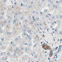
| Sample | Paraffin-embedded Rat liver tissue; fixative not specified (datasheet M01233) |
| Fixation | Image fixative and duration unreported (datasheet M01233); verify before use. |
| Sectioning | 4–5 µm sections on charged slides (standard) |
| Deparaffinisation | Xylene, graded ethanol series to water (standard) |
| Antigen retrieval | Heat-induced epitope retrieval in Tris-EDTA buffer, pH 9.0, 20 min at 95–98 °C (standard rule: nuclear antigen) |
| Peroxidase block | 3% H2O2, 10 min, room temperature (standard) |
| Blocking | 10% normal serum of the secondary host, 30 min, room temperature (standard) |
| Primary antibody | Rabbit monoclonal (clone IDG-20) anti-TSG101, 1:50 recommended; image 1:500 (datasheet M01233) |
| Primary incubation | Overnight at 4 °C (standard) |
| Detection | HRP-polymer secondary, DAB chromogen 5–10 min (standard) |
| Counterstain | Hematoxylin, blue, dehydrate and mount (standard) |
| Expected result | TSG101-positive staining in glandular cells of appendix (HPA tissue IHC: High). HPA tissue profile: General cytoplasmic and membranous expression. No signal in the no-primary control. |
TSG101 is mainly cytoplasmic, with endosomal membrane association and cell cycle-dependent nuclear localisation (UniProt Q99816). In paraffin-section IHC, expect cytoplasmic and membranous staining in cell types such as appendix glandular cells, bone marrow hematopoietic cells and cerebral cortex neurons (HPA: High). HPA rates its tissue IHC evidence Approved, with medium consistency between antibody staining and RNA expression (HPA: tissue IHC).
| Cytoplasmic staining, sometimes with a membranous component, is clear in glandular cells of appendix or colon (HPA: High in both). | This fits the reported tissue pattern and TSG101’s mainly cytoplasmic localisation (HPA: general cytoplasmic and membranous expression; UniProt Q99816). Compare cell compartments within the section; a positive call rests on cellular staining in the expected cells, not on colour anywhere in the tissue (general IHC practice). |
| Staining is exclusively extracellular or fills gland lumens, with little signal inside the expected cells. | That distribution does not fit the reported cellular pattern (HPA: general cytoplasmic and membranous expression). Treat it as suspect and inspect the section for nonspecific deposit or detection background before scoring TSG101 (general IHC practice). Nuclear staining alone needs separate judgment: UniProt reports cell cycle-dependent nuclear localisation (UniProt Q99816). |
| Strong colour appears chiefly in adipocytes while the expected positive cells are weak or unstained. | Adipocytes are reported as Not detected, so that reversal raises concern for nonspecific binding, pigment or endogenous detection activity (HPA: adipocytes Not detected; general IHC practice). It does not establish which cause is responsible. Compare a known-positive tissue and a primary-antibody omission control processed with the same detection system (general IHC practice). |
| A diffuse haze covers stroma, empty spaces and many cell types without a readable cellular pattern. | Broad haze cannot establish TSG101 localisation or identify positive cells (general IHC practice). Review blocking, washes, antibody concentration and detection background using matched controls (general IHC practice). HPA’s Approved IHC rating describes the reported staining evidence; it does not validate diffuse signal in a new run (HPA: IHC Approved). |
| No cellular signal appears in appendix glandular cells or bone marrow hematopoietic cells. | Both are reported High, making them useful positive comparators, although the supplied evidence does not guarantee every section or run will stain (HPA: High in appendix glandular cells and bone marrow hematopoietic cells). Check tissue preservation and the general IHC workflow, including retrieval, reagent activity and detection controls, before interpreting an unknown specimen as negative (general IHC practice). |
| Cell and tissue context | HPA reports High staining in several epithelial, hematopoietic and neuronal populations, but Not detected in adipocytes; judge intensity by cell type, not whole-section colour (HPA: tissue IHC; general IHC practice). |
| Membrane association | TSG101 associates with early and late endosome membranes and is mainly cytoplasmic; it has no transmembrane segment (UniProt Q99816). A membranous component can fit the HPA IHC profile without requiring an exclusively surface pattern (HPA: tissue IHC). |
| Cell division and nuclear signal | Nuclear localisation depends on cell cycle, and CEP55 interaction is required for midbody localisation during cytokinesis (UniProt Q99816). Assess a focal nuclear or division-associated signal in cellular context rather than assuming every such signal is artefact (general IHC practice). |
| IHC evidence strength | HPA lists HPA006161 and CAB004283 as IHC Approved, while its tissue profile has medium consistency with RNA expression (HPA: antibody validation; HPA: tissue IHC). These ratings support pattern comparison but do not establish the specificity of an individual slide. |
| Antigen retrieval | No target-specific retrieval condition or fixation sensitivity is supplied (UniProt Q99816; HPA: supplied records). If signal is weak, check the antibody’s validated IHC-P instructions and run controls when adjusting retrieval; do not infer a TSG101-specific fixation effect (general IHC practice). |
| IF/ICC: what localisation should be compared? | HPA reports mainly cytosolic signal, with additional nucleolar and plasma membrane localisation, in ICC-IF; its cytosol call is Supported (HPA: subcellular ICC-IF). Use that as an IF localisation comparison, allowing for the different readout from chromogenic tissue IHC. |
| Situation | Likely cause | Next action |
|---|---|---|
| Known-positive glandular cells show no stain (HPA: High in appendix and colon). | A failed detection step or an unsuitable run condition is possible; absence alone cannot identify the failed step (general IHC practice). | Confirm the positive control, detection reagents and the catalog antibody’s IHC-P instructions; review retrieval and antibody dilution within the validated workflow (general IHC practice). |
| The section is uniformly brown, including spaces outside cells. | Nonspecific deposit or detection background can obscure the cellular pattern (general IHC practice). | Compare the primary-antibody omission control; inspect washes, blocking and detection development, then rescore only distinct cellular staining (general IHC practice). |
| Adipocytes stain more strongly than nearby expected positive cells. | That pattern conflicts with HPA’s Not detected adipocyte call and could reflect nonspecific or endogenous signal (HPA: adipocytes Not detected; general IHC practice). | Check omission and known-positive controls under the same detection conditions; avoid assigning the adipocyte signal to TSG101 until the discrepancy is resolved (general IHC practice). |
| Signal looks exclusively nuclear in most cells. | The reported profile is mainly cytoplasmic, although nuclear localisation can vary with cell cycle (UniProt Q99816; HPA: tissue IHC). | Inspect cytoplasmic signal in an HPA-reported positive cell type and compare controls; do not reject a restricted nuclear signal solely because it is nuclear (HPA: High cell types; UniProt Q99816; general IHC practice). |
| Membrane staining is present, but the slide lacks a clear cytoplasmic component. | HPA reports both cytoplasmic and membranous expression; UniProt describes membrane association alongside a mainly cytoplasmic distribution (HPA: tissue IHC; UniProt Q99816). | Check whether staining is cellular and matches expected cell types, then compare a positive control before scoring an isolated membrane-only pattern (general IHC practice). |
| The sample appears negative while the positive control stains. | A true low result is possible, but the supplied HPA profile does not predict every cell in every specimen (HPA: tissue IHC). | Document the cell type, compartment and control result; compare with HPA’s reported High and Not detected cell types, and limit the conclusion to the tested section (HPA: tissue IHC; general IHC practice). |




Comprehensive Human Protein Atlas IHC scoring per tissue (reliability: Approved — Medium consistency between antibody staining and RNA expression data.). Rows are taken directly from the HPA tissue chart — click any row's HPA link to view the source.
| Tissue | Cell type | Level | Evidence | Source |
|---|---|---|---|---|
| Appendix | Glandular cells | High | Protein (IHC) | HPA → |
| Bone marrow | Hematopoietic cells | High | Protein (IHC) | HPA → |
| Breast | Glandular cells | High | Protein (IHC) | HPA → |
| Bronchus | Respiratory epithelial cells | High | Protein (IHC) | HPA → |
| Cerebral cortex | Neuronal cells | High | Protein (IHC) | HPA → |
| Tissue | Cell type | Level | Evidence | Source |
|---|---|---|---|---|
| Adipose tissue | Adipocytes | Not detected | Protein (IHC) | HPA → |
Troubleshoot TSG101 staining in paraffin sections by checking retrieval, controls, compartment patterns and scoring before interpreting chromogenic signal.
M01233 has IHC images from paraffin-embedded rat liver and stomach, human prostate cancer, and mouse kidney (M01233 IHC captions); both catalog antibodies list human, mouse, and rat reactivity (catalog reactivity).
M01233 is the SKU with a rendered IHC card: its captions show paraffin-embedded rat liver and stomach, human prostate cancer, and mouse kidney (M01233 IHC captions). M01233-2 lists IF and human, mouse, and rat reactivity, but has no supplied IHC or IF image for a card (M01233-2 catalog applications/reactivity/image captions).
Which to pick: Choose M01233 for tissue IHC: it lists IHC and has paraffin-section images; the fixative is unreported (M01233 catalog applications and IHC captions). For IF/ICC, M01233 lists both applications, while M01233-2 lists IF only; neither has a supplied IF image (catalog applications and IF image captions). Both are rabbit monoclonals listed as reactive with human, mouse, and rat, but only M01233 has cross-species IHC images (catalog host/title/reactivity; M01233 IHC captions).