This website uses cookies to ensure you get the best experience on our website.
- Table of Contents
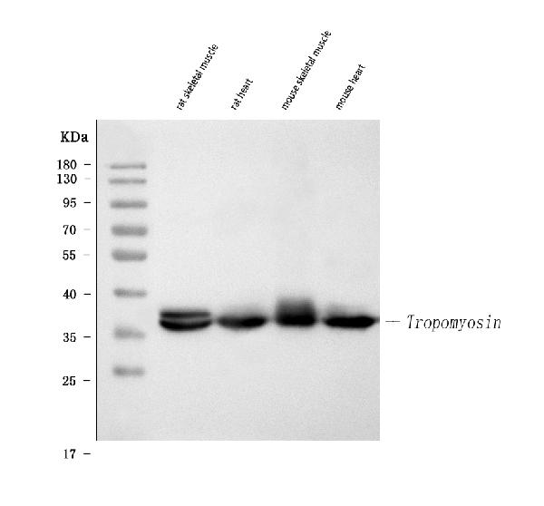
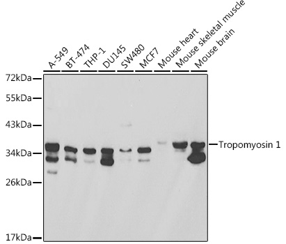
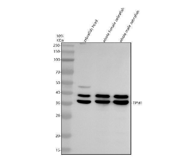
Facts about Tropomyosin alpha-1 chain.
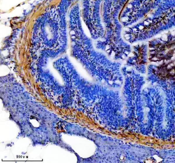
Smooth muscle contraction is regulated by interaction with caldesmon. In non-muscle cells is implicated in stabilizing cytoskeleton actin filaments.
Human | |
---|---|
Gene Name: | TPM1 |
Uniprot: | P09493 |
Entrez: | 7168 |
Belongs to: |
---|
tropomyosin family |
alpha tropomyosin; alpha-tropomyosin; C15orf13; cardiomyopathy, hypertrophic 3; chromosome 15 open reading frame 13; CMD1Y; CMH3; HTM-alpha; sarcomeric tropomyosin kappa; TMSA; tropomyosin 1 (alpha) isoform 1; tropomyosin 1 (alpha) isoform 2; tropomyosin 1 (alpha) isoform 3; tropomyosin 1 (alpha) isoform 4; tropomyosin 1 (alpha) isoform 5; tropomyosin 1 (alpha) isoform 6; tropomyosin 1 (alpha) isoform 7; tropomyosin 1 (alpha); tropomyosin alpha-1 chain; tropomyosin-1
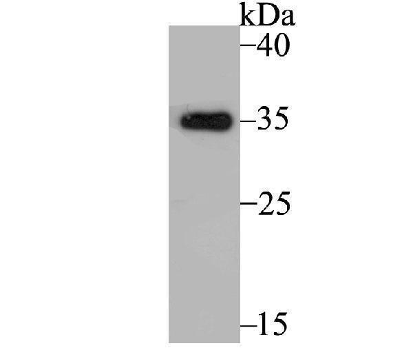
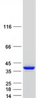
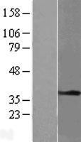
Mass (kDA):
32.709 kDA
Human | |
---|---|
Location: | 15q22.2 |
Sequence: | 15; NC_000015.10 (63042698..63071915) |
Detected in primary breast cancer tissues but undetectable in normal breast tissues in Sudanese patients. Isoform 1 is expressed in adult and fetal skeletal muscle and cardiac tissues, with higher expression levels in the cardiac tissues. Isoform 10 is expressed in adult and fetal cardiac tissues, but not in skeletal muscle.
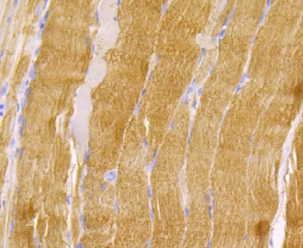
Cytoplasm, cytoskeleton. Associates with F-actin stress fibers.
Steven Boster was the inventor of his first product in 1993. He earned the nickname "he who converts science into the lavatory." Boster Bio has been developing hundreds of primary antibody products and is now the largest Chinese catalog antibody company. Today, his company uses proprietary trade secrets to develop high-sensitivity ELISA kits. You will find out some of Boster Bio’s most effective uses for the TPM1Marker.
The peptide library that was prepared for this study was composed of 24 fractionations of tryptic HeLa digest, each with a unique amino acid label. These samples were analyzed by LC-MS/MS to identify and quantify the peptides. The LC/MS/MS method was used to analyze the peptides. Results could be obtained within three to five days. The default settings were used to analyze the peptide samples. They modified some parameters to increase sensitivity. This included changing the range of covered peptides (from seven to fifty), the precursor charge to 2, and the ionization method to robust LC.
To achieve comparable results, the LC/MS/MS measurements were repeated every 5 days. The interassay analytical imprecision of each peptide was calculated by comparing results from duplicate calibration curves. The results from the peptide analysis were compared to those of immunoassays for KIM-1, IGFBP7, and NGAL.
The peptides were prepared by fractionation in an ultra-high sensitivity LC-MS workflow. The ultra-high sensitivity LC/MS/MS workflow enables single-cell proteome analysis. Single-cell proteome analysis can be done in a single cell. It is complete, stable and stochastic. By injecting cells from the early stages of their cell cycle, the peptides will be detected in the proteasome-rich environment.
LC-MS/MS and IA are complementary methods used to analyze urinary proteins. LC-MS/MS could facilitate the translation and application of new biomarkers in the clinical laboratory. LC/MS/MS confirms the peptides, proteins, whereas IA requires measurements of entire protein complexes. The method allows for multiplex quantitation, which is metrologically trackable.
The LC/MS/MS technique uses a combination immunocapture as well as peptide-specific detection techniques. Direct IA, which measures the mixture of multiple proteoforms a single protein, is the first method. The peptides obtained by IA are not as specific as those detected by LC-MS/MS. The latter uses a mass sensitve detection method. The Boster Bio peptide sample was analyzed by LC/MS/MS.
The LC/MS/MS method was used to analyze these samples. All samples were kept at room temperature for 10 years before being analysed. The LC/MS/MS analyses were precise and accurate. The sample amounts were low likely due to very little contribution from previous runs. As the LCMS sensitivity improves, the proteome coverage should increase.
The experiment revealed that the median protein levels in G1 cells ranged between 1,018 and 1,932 in G1/S, and 1,571 and 2,045 respectively in G2/M. The total protein content of a cell was calculated by adding all signals based upon peptides. The T-SCP data set reflected cellular processes, cell phases, and signal transduction.
The SQR peptide was found to contain Cys-90, a S-glutathionylated cysteine residue. It is found in the mature protein, and is highly conserved among other proteins and species. It also contains Cys90 which undergoes S-glutathionylation. This residue is involved in antioxidant defense.
Multiple samples contained CC41-reactive LITRs. Multiple phosphorylated protein presence suggests that additional signaling molecules were recruited for the synthesis CC41. Lower m.w. Lower m.w. were found on the Coomassie Blue stained gel, but not on Western blot. Peptide-based LC/MS/MS analysis identified peptides in these bands and the corresponding Galectin Protein was isolated.
The peptides were separated by hydrolysis and dried at -20 degrees C. They were then stored at a temperature that prevents the oxidation free sulfhydryl group. This method has been validated across a range of research fields, including biomedical and pharmaceutical, as well diagnostic.
For the analysis of CC41-CCV antigens, freshly isolated PBL from G14D-CCV-immunized fish were purified using goat anti-mouse IgG microbeads. PBL was then fixed using the Quantigene Flow RNA Fixation and Permeabilization Kit (Affymetrix-eBioscience). Using the Quantigene Flow RNA Preparation and Analysis Kit, the samples were sorted and stained by FACScan.
The reaction mixture was mixed with a Laemmli sample buffer and incubated at 70 degC for 10 min. After the reaction, the samples could be loaded onto a 4--20% Tris-glycine Polyacrylamide gradient gel. The samples were run for two hours at 100 V. The membranes were then blocked by 0.1% Tween20 or TTBS+ 5% dry Milk. The samples were then incubated overnight with an anti--3-nitrotyrosine polyclonal antibody (1:2000, Upstate).
The Escherichia Coli enzymes were isolated to identify the PTM enzyme. The PTM enzymes dehydrate threonine (serine), and alanine. These enzymes produce modified RumA precursors that undergo Michael-type addition cycles reactions to give lanthionine or methyllanthionine. The results of the microscale and laboratory-scale bioreactor cultivations show that the process is robust, and can be applied to industrial-scale conditions.
After extraction, the reaction mix was subjected for LC-MS/MS analysis. The samples were loaded into the trap columns at 2.5 uL/min. The column was kept at 4degC. The samples were analyzed for total peptide content using an LC-MS/MS method. The Boster bio peptides were analyzed with an LC-MS/MS.
After SDS-PAGE analysis, SQR proteins were immunoblotted with an anti-GSH monoclonal antibodies purchased from ViroGen. Western blots revealed decreased intrinsic protein S–glutathionylation in the subunit 70-kDa of SQR in post-ischemic myocardium.
PMID: 3548719 by Mische S.M., et al. Relation of streptococcal M protein with human and rabbit tropomyosin: the complete amino acid sequence of human cardiac alpha tropomyosin, a highly conserved contractile protein.
PMID: 3336357 by Lin C.-S., et al. Cloning and characterization of a cDNA encoding transformation- sensitive tropomyosin isoform 3 from tumorigenic human fibroblasts.