This website uses cookies to ensure you get the best experience on our website.


- Table of Contents
Plan chromogenic UBB IHC on paraffin sections with 0.5–1 µg/mL catalog antibody (datasheet PB9122). Expect mainly nuclear tissue staining, and account for antibody detection of protein from more than one gene (HPA tissue IHC).

Expected localisation, validated protocols, controls and antibodies — the at-a-glance facts below, then the full design guide.
| Expected localisation | Mainly nuclear in tissues (HPA tissue IHC) | |
| Staining pattern | Mainly nuclear staining across tissues (HPA tissue IHC) | |
| Antigen retrieval | Citrate pH 6 HIER, heat-mediated (datasheet PB9122) | |
| Positive control | Adrenal gland+4 more · see all | |
| Negative control | None in HPA (detected in all 45 tissues); use no-primary + isotype controls |
| Fixation | Keep fixation consistent across sections (standard IHC practice; not target-specific) | |
| Caveat | Signal may include ubiquitin from other genes (HPA tissue IHC) | |
| Regulation | Expression regulation not annotated (UniProt) | |
| Isoform / epitope | No isoforms; three ubiquitin units are processed (UniProt) |
The catalog antibody has a citrate pH 6 IHC-P protocol (datasheet: PB9122). The published protocols below include native UBB staining and mutant UBB+1 staining (PMC10930646; PMC3400757; PMC4362318; PMC5427096).
| Sample | Paraffin-embedded rat intestine tissue; fixative not specified (datasheet PB9122) |
| Fixation | Image fixative and duration unreported (datasheet PB9122); verify before use. |
| Sectioning | 4–5 µm sections on charged slides (standard) |
| Deparaffinisation | Xylene, graded ethanol series to water (standard) |
| Antigen retrieval | Heat retrieval: Citrate pH 6, 20 min (datasheet PB9122) |
| Peroxidase block | 3% H2O2, 10 min, room temperature (standard) |
| Blocking | 10% goat serum (datasheet PB9122) |
| Primary antibody | Rabbit anti-UBB, 0.5-1μg/ml (datasheet PB9122) |
| Primary incubation | Overnight at 4 °C (datasheet PB9122) |
| Detection | Streptavidin-biotin complex (SABC), DAB chromogen (datasheet PB9122) |
| Counterstain | Hematoxylin, blue, dehydrate and mount (standard) |
| Expected result | UBB-positive staining in glandular cells of adrenal gland (HPA tissue IHC: High). HPA tissue profile: Mainly nuclear expression in all tissues. No signal in the no-primary control. |
UBB-related IHC staining is mainly nuclear across tissues (HPA tissue IHC: Supported; medium consistency with RNA). Cytoplasmic staining is also biologically plausible (UniProt P0CG47: cytoplasm and nucleus). Expect strong staining in documented glandular, respiratory epithelial, glial, and cerebellar granular-layer cells (HPA tissue IHC: High). Interpret staining as ubiquitin-related rather than uniquely UBB-derived: the HPA profile cautions that its antibodies target proteins from more than one gene (HPA tissue IHC).
| Predominantly nuclear staining in breast glandular cells or bronchial respiratory epithelial cells. | This matches the reported tissue pattern; both cell populations are High (HPA tissue IHC). Cytoplasmic signal can coexist with nuclear signal (UniProt P0CG47 localization). |
| A dominant, continuous cell-surface rim with little nuclear staining. | Treat this as an atypical pattern requiring controls: HPA reports mainly nuclear tissue staining, while UniProt lists nucleus, cytoplasm, and mitochondrion outer membrane (HPA tissue IHC; UniProt P0CG47). |
| Strong staining in a cell population outside the documented examples. | Do not call it false solely because that population is unlisted: HPA reports low tissue specificity and cautions about multi-gene recognition (HPA tissue IHC). Check cellular detail and antibody controls (general IHC practice). |
| Diffuse color over tissue spaces, edges, and cells alike. | The distribution does not reproduce HPA's mainly nuclear cellular pattern (HPA tissue IHC). Check background and detection controls before assigning it to UBB-related staining (general IHC practice). |
| No cellular signal in bronchial respiratory epithelium or adrenal glandular cells. | Both are reported High (HPA tissue IHC). A blank result there calls for review of the IHC workflow and positive control before interpreting other sections as negative (general IHC practice). |
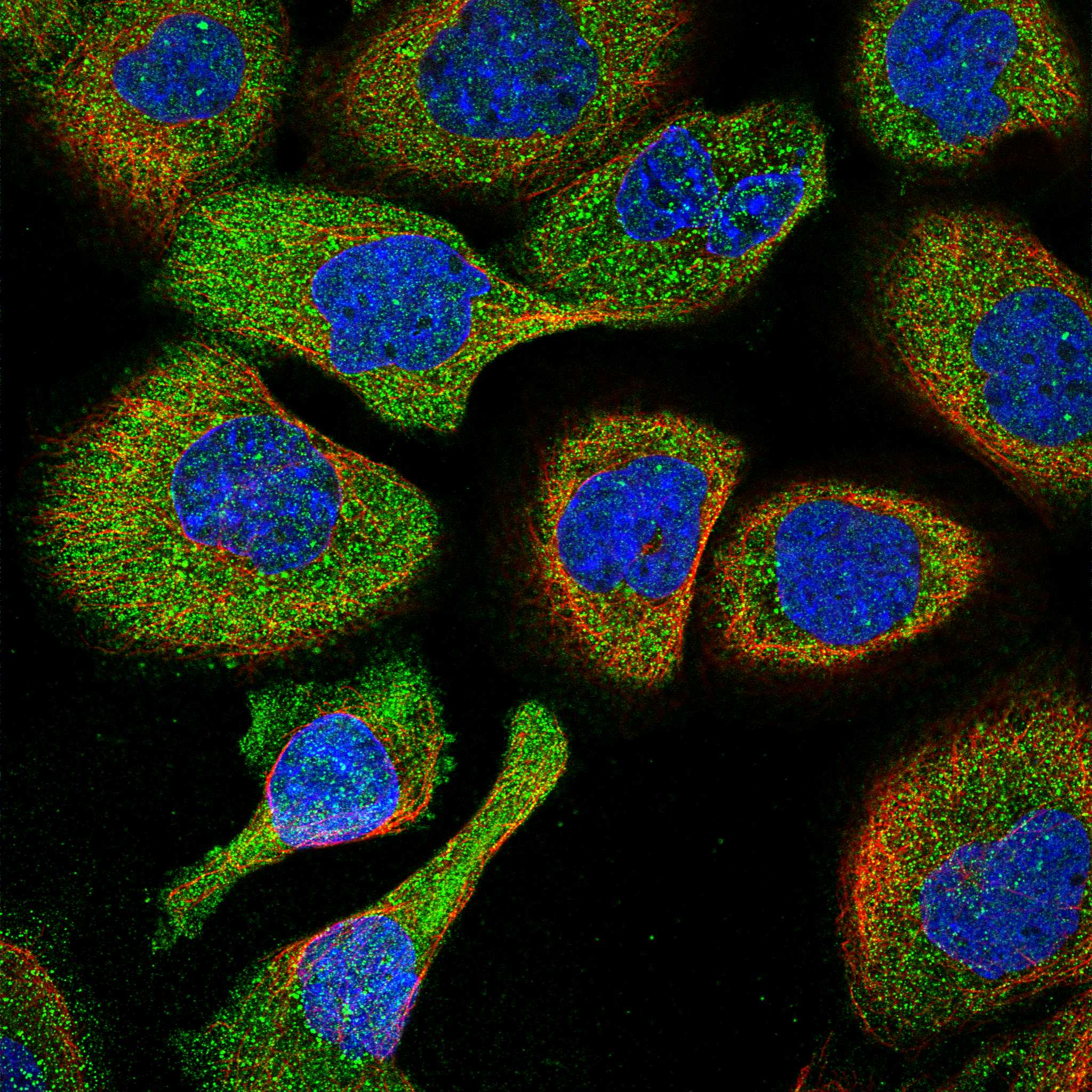
| IHC versus IF/ICC: should the compartments match? | HPA tissue IHC is mainly nuclear, whereas HPA ICC-IF reports mainly cytosol (uncertain) plus acrosome and equatorial segment (approved). Compare each result with its own assay evidence; the ICC-IF page supplies the IF workflow (HPA tissue IHC; HPA subcellular). |
| Attribution to UBB | The tissue profile cautions that antibodies target proteins from more than one gene; HPA041344, HPA049132, and CAB013048 have Supported IHC status (HPA tissue IHC; HPA antibodies). A positive stain alone cannot assign signal uniquely to UBB. |
| Precursor processing | The 229-residue precursor contains three ubiquitin units at residues 1–76, 77–152, and 153–228 (UniProt P0CG47 processing). The recognized species depends on the antibody epitope; its location is not supplied. |
| Cell-type intensity comparison | HPA reports High staining in listed glandular, respiratory epithelial, glial, and granular-layer cells, but Low in heart cardiomyocytes (HPA tissue IHC). The Low result is a comparison point, not a negative control. |
| Antigen retrieval | Retrieval conditions can be checked against the catalog antibody's IHC-P instructions as a general paraffin-IHC step (general IHC practice). Neither HPA nor UniProt establishes UBB-specific fixation sensitivity or an optimal retrieval condition. |
| Situation | Likely cause | Next action |
|---|---|---|
| Known High tissue is blank. | The result conflicts with reported High bronchial respiratory epithelial staining (HPA tissue IHC); the failed step is undetermined. | Review the positive control, catalog antibody's IHC-P instructions, retrieval, and detection reagents (general IHC practice). |
| Cytoplasm dominates and nuclei are consistently blank in tissue sections. | This departs from mainly nuclear tissue IHC, although cytoplasmic localization is recorded (HPA tissue IHC; UniProt P0CG47). | Check nuclear morphology and compare a documented High cell population on the same run (HPA tissue IHC; general IHC practice). |
| Nearly every cell and tissue space is uniformly colored. | Noncellular color is inconsistent with the mainly nuclear tissue pattern (HPA tissue IHC); background may contribute (general IHC practice). | Inspect a no-primary control and review blocking, washes, and chromogen development (general IHC practice). |
| Chromogenic signal persists without primary antibody. | The control indicates detection background; endogenous enzyme activity is one possible cause in chromogenic IHC (general IHC practice). | Review the detection control and use the appropriate endogenous-activity block for the chosen detection system (general IHC practice). |
| An unexpected cell population stains strongly. | HPA warns of recognition of proteins from multiple genes; low tissue specificity also limits cell-type exclusion (HPA tissue IHC). | Compare morphology and a documented High population, then assess specificity with an independent antibody where available (HPA tissue IHC; general IHC practice). |
| Heart cardiomyocytes stain as strongly as the chosen High control. | HPA reports Low cardiomyocyte staining, but slide processing and antibody specificity may also influence the comparison (HPA tissue IHC; general IHC practice). | Compare matched sections at the same development time and review background and antibody controls before scoring intensity (general IHC practice). |


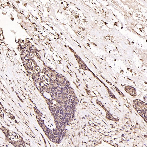
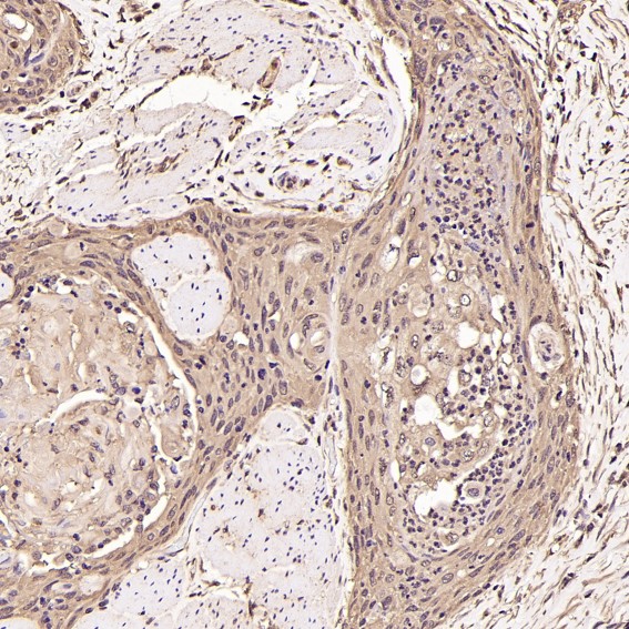


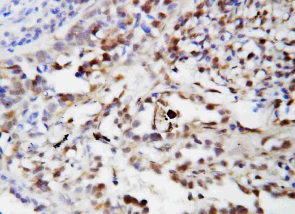


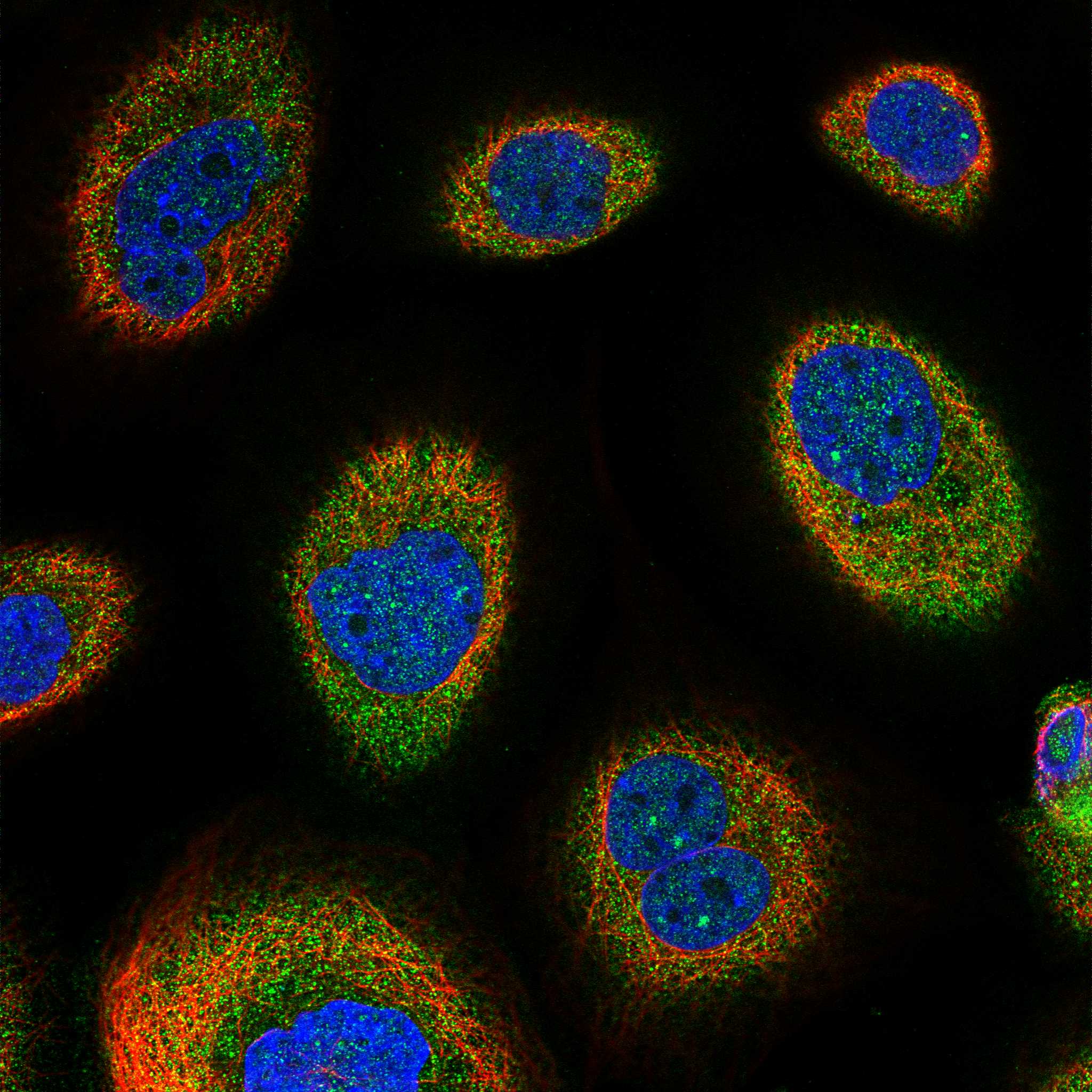
Comprehensive Human Protein Atlas IHC scoring per tissue (reliability: Supported — Medium consistency between antibody staining and RNA expression data. Caution, targets protein from more than one gene.). Rows are taken directly from the HPA tissue chart — click any row's HPA link to view the source.
| Tissue | Cell type | Level | Evidence | Source |
|---|---|---|---|---|
| Adrenal gland | Glandular cells | High | Protein (IHC) | HPA → |
| Breast | Glandular cells | High | Protein (IHC) | HPA → |
| Bronchus | Respiratory epithelial cells | High | Protein (IHC) | HPA → |
| Caudate | Glial cells | High | Protein (IHC) | HPA → |
| Cerebellum | Cells in granular layer | High | Protein (IHC) | HPA → |
| Tissue | Cell type | Level | Evidence | Source |
|---|---|---|---|---|
| None in HPA: UBB is detected in all 45 scored tissues. Use a no-primary (secondary-only) and an isotype control instead. | ||||
Use the catalog antibody’s tissue staining conditions as a starting point, then interpret UBB staining with its broad localisation and shared ubiquitin epitopes in mind.
Catalog anti-UBB antibodies have IHC images from human, mouse, and rat paraffin sections and IF images from A431, Jurkat, and Raji cells (catalog image captions).
PB9122 has IHC images of rat and mouse intestine and human mammary cancer in paraffin sections; PA1420 has an IHC image of human lung cancer tissue (respective IHC captions). M02848 has a human breast cancer IHC image and a Jurkat IF image; M02848-3 has human breast cancer, esophageal carcinoma, stomach, and colon IHC images and a Raji IF image (respective image captions).
Which to pick: For tissue IHC, PB9122 offers the clearest processing example: its rat intestine paraffin-section caption specifies citrate retrieval at pH 6 for 20 minutes, goat-serum blocking, and DAB detection; the fixative is unreported (PB9122 IHC caption). For IF/ICC, PB9122 has an A431 cell IF example, while the rabbit monoclonals M02848 and M02848-3 list IF/ICC and show Jurkat and Raji cell IF images, respectively (PB9122 IF caption; M02848 and M02848-3 catalog applications and IF captions). For cross-species IHC, PB9122 has paraffin-section examples from human, mouse, and rat; all four SKUs list reactivity with those three species, but their other IHC captions do not establish a fixative (PB9122 IHC captions; catalog reactivity; PA1420, M02848, and M02848-3 IHC captions).