This website uses cookies to ensure you get the best experience on our website.


- Table of Contents
Plan paraffin-section UHRF1 IHC around nuclear staining in thymus (HPA tissue IHC). Start with the IHC-validated antibody PB9905 at 0.5–1 μg/mL (datasheet PB9905), then score nuclear signal by cell type and intensity (standard IHC practice).

Expected localisation, validated protocols, controls and antibodies — the at-a-glance facts below, then the full design guide.
| Expected localisation | Nuclear staining in thymus (HPA tissue IHC) | |
| Staining pattern | Strong nuclear staining in thymic cortical cells (HPA tissue IHC) | |
| Antigen retrieval | EDTA pH 8.0 HIER, heat-mediated (datasheet PB9905) | |
| Positive control | Ovary+4 more · see all | |
| Negative control | Adipose tissue+4 more · see all |
| Fixation | Keep tissue fixation consistent (standard IHC practice; not target-specific) | |
| Caveat | Cell-cycle phase may shift the nuclear pattern (UniProt) | |
| Regulation | Overexpressed in breast cancer (UniProt) | |
| Isoform / epitope | 2 isoforms; epitope differences unspecified (UniProt) |
The catalog antibody’s IHC-P protocol (datasheet: PB9905) is accompanied by three published UHRF1 IHC protocols for paraffin sections (PMC2906730; PMC2713709; PMC8797458).
| Sample | Paraffin-embedded human intestinal cancer tissue; fixative not specified (datasheet PB9905) |
| Fixation | Image fixative and duration unreported (datasheet PB9905); verify before use. |
| Sectioning | 4–5 µm sections on charged slides (standard) |
| Deparaffinisation | Xylene, graded ethanol series to water (standard) |
| Antigen retrieval | Heat retrieval: EDTA pH 8.0 (datasheet PB9905); 20 min, 95–100 °C (standard) |
| Peroxidase block | 3% H2O2, 10 min, room temperature (standard) |
| Blocking | 10% goat serum (datasheet PB9905) |
| Primary antibody | Rabbit anti-UHRF1, 0.5-1μg/ml (datasheet PB9905) |
| Primary incubation | Overnight at 4 °C (datasheet PB9905) |
| Detection | Streptavidin-biotin complex (SABC), DAB chromogen (datasheet PB9905) |
| Counterstain | Hematoxylin, blue, dehydrate and mount (standard) |
| Expected result | UHRF1-positive staining in follicle cells of ovary (HPA tissue IHC: High). HPA tissue profile: Strong nuclear expression in cells in thymus. No signal in the no-primary control. |
UHRF1 should appear predominantly in nuclei on paraffin-section IHC: thymic cortical cells and ovarian follicle cells show high staining, while several other listed cell populations show medium or low staining (HPA tissue IHC). HPA rates the tissue pattern Enhanced for consistency with RNA expression (HPA tissue IHC). Nuclear and chromosome localization, with no transmembrane segment, supports a nuclear interpretation (UniProt Q96T88 localization and topology).
| Distinct nuclear staining in thymic cortical cells or ovarian follicle cells. | This fits the high-staining populations in the tissue record (HPA tissue IHC: High). Assess the stained cells and compartment together; strong color elsewhere in the section does not establish the same expected pattern (HPA tissue IHC; UniProt Q96T88 localization). |
| Nuclear staining is present but uneven across cells or regions. | Variation can be compatible with the record: lymph-node non-germinal-center cells, placental cytotrophoblasts, testicular spermatogonia and tonsillar germinal-center cells are listed at medium levels (HPA tissue IHC). UHRF1 associates with replicating DNA during S phase and is more homogeneous in non-S-phase nuclei (UniProt Q96T88 localization). |
| Predominantly cytoplasmic, membranous or extracellular color replaces nuclear staining. | Treat this as discordant with the expected compartment (UniProt Q96T88 localization; HPA subcellular: nucleoplasm supported). Review morphology, counterstain and controls before interpreting it as UHRF1; the record reports no transmembrane segment or signal peptide (UniProt Q96T88 topology and processing). |
| Strong staining appears in a cell type listed as not detected, such as adipocytes or bronchial respiratory epithelial cells. | This conflicts with those specific reference observations (HPA tissue IHC: Not detected). Cross-reactivity or endogenous detection activity is possible under general IHC practice; compare a no-primary control and the expected nuclear signal before assigning specificity. The HPA observation alone cannot identify the cause. |
| No discernible nuclear signal appears in thymic cortical cells. | This conflicts with the high thymic cortical-cell reference pattern (HPA tissue IHC: High). Check tissue identity, morphology and assay controls, then review the antibody's IHC-P instructions and detection steps (general IHC practice). A negative slide alone cannot distinguish absent antigen from an assay failure. |
| Cell and tissue context | The reference is cell-specific: cortical cells in thymus and follicle cells in ovary are high, while bone-marrow hematopoietic cells are low (HPA tissue IHC). Do not score an entire tissue by a single expected intensity. |
| Nuclear distribution and cell cycle | UHRF1 associates with replicating DNA from early to late S phase, including pericentric heterochromatin; non-S-phase nuclear distribution is described as homogeneous by similarity (UniProt Q96T88 localization). Nuclear texture alone is therefore a weak exclusion criterion. |
| Antibody validation | Two listed rabbit polyclonal antibodies, HPA049408 and HPA055446, carry Enhanced IHC status; only HPA049408 has Supported ICC status (HPA antibodies). These labels support the reported applications, but do not validate an unspecified catalog antibody. |
| Target organization | UniProt lists a single 1–793 chain, no signal peptide, no propeptide, no transmembrane segment and two isoforms (UniProt Q96T88 processing, topology and isoforms). Epitope coverage across isoforms cannot be inferred without antibody-specific epitope information. |
| Situation | Likely cause | Next action |
|---|---|---|
| Thymus shows no nuclear signal in cortical cells. | The result differs from the high reference pattern (HPA tissue IHC); tissue sampling or assay performance may need review (general IHC practice). | Confirm cortical-cell morphology, inspect positive and no-primary controls, and follow the IHC-validated antibody's documented IHC-P conditions (general IHC practice). Do not assign a UHRF1-specific fixation effect; none is supplied. |
| The section has widespread haze that obscures nuclei. | Background from blocking, washing or chromogenic detection can obscure localization (general IHC practice); the expected compartment is nuclear (UniProt Q96T88 localization). | Compare the no-primary control, review blocking and washes, and assess whether the counterstain still resolves nuclei (general IHC practice). Score only separable nuclear staining in the relevant cells (HPA tissue IHC). |
| Color is mainly cytoplasmic or outlines cell membranes. | That compartment conflicts with supported nucleoplasmic localization and nuclear tissue staining (HPA subcellular; HPA tissue IHC). The slide cannot identify the cause by appearance alone. | Check the no-primary control and morphology, then review antibody application validation and detection settings (general IHC practice; HPA antibodies for the listed antibodies). |
| Strong color occurs in adipocytes or bronchial respiratory epithelial cells. | Both are listed as not detected (HPA tissue IHC). Cross-reactivity or endogenous detection activity is possible (general IHC practice), but neither is proven by this observation. | Compare matched no-primary and positive-tissue controls; judge whether signal is nuclear and cell-specific (general IHC practice; UniProt Q96T88 localization; HPA tissue IHC). |
| Bone marrow looks weak compared with thymus. | Low staining in bone-marrow hematopoietic cells and high staining in thymic cortical cells are both recorded (HPA tissue IHC). Their different intensities alone do not indicate an assay fault. | Use thymic cortical cells as the stronger reference and score marrow hematopoietic cells against their own low reference level (HPA tissue IHC). Confirm nuclear localization before interpreting either signal (UniProt Q96T88 localization). |
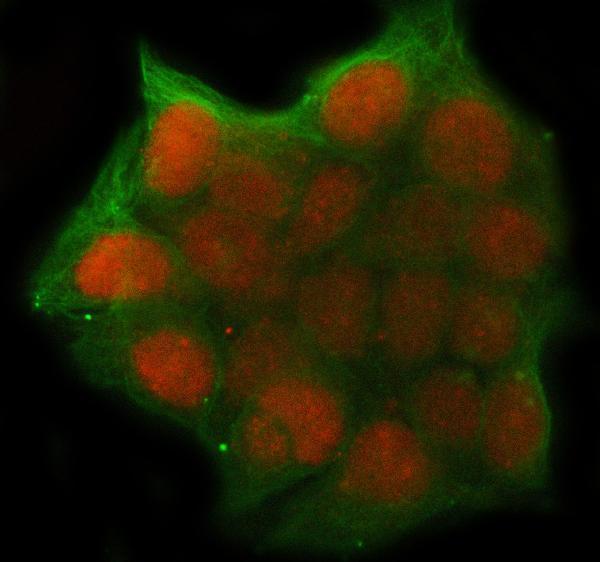
| Q: Should an IF/ICC image show the same compartment? | Supported nucleoplasmic localization is reported for ICC-IF, with images in HEK293, MCF-7 and U2OS (HPA subcellular). This establishes a localization reference, not an IF/ICC protocol. | Expect nucleoplasmic signal when interpreting IF/ICC images (HPA subcellular). For this IHC-P guide, use the tissue IHC pattern for slide interpretation; IF/ICC methods belong to the separate guide. |


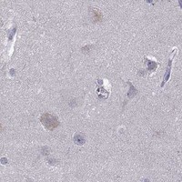

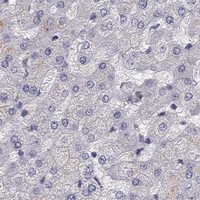


Comprehensive Human Protein Atlas IHC scoring per tissue (reliability: Enhanced — High consistency between antibody staining and RNA expression data.). Rows are taken directly from the HPA tissue chart — click any row's HPA link to view the source.
| Tissue | Cell type | Level | Evidence | Source |
|---|---|---|---|---|
| Ovary | Follicle cells | High | Protein (IHC) | HPA → |
| Thymus | Cortical cells | High | Protein (IHC) | HPA → |
| Lymph node | Non-germinal center cells | Medium | Protein (IHC) | HPA → |
| Placenta | Cytotrophoblasts | Medium | Protein (IHC) | HPA → |
| Testis | Spermatogonia cells | Medium | Protein (IHC) | HPA → |
| Tissue | Cell type | Level | Evidence | Source |
|---|---|---|---|---|
| Adipose tissue | Adipocytes | Not detected | Protein (IHC) | HPA → |
| Adrenal gland | Glandular cells | Not detected | Protein (IHC) | HPA → |
| Appendix | Endocrine cells | Not detected | Protein (IHC) | HPA → |
| Bronchus | Respiratory epithelial cells | Not detected | Protein (IHC) | HPA → |
| Caudate | Glial cells | Not detected | Protein (IHC) | HPA → |
Troubleshoot UHRF1 staining by checking nuclear localisation, tissue controls and the processing conditions documented for the catalog antibody.
Anti-UHRF1 antibodies have illustrated IHC in human paraffin-embedded intestinal cancer tissue (PB9905 IHC caption) and IF/ICC in MCF-7 cells (A01156-1 IF caption); A01156-1 also lists mouse reactivity (catalog).
PB9905 has an IHC image from a human paraffin-embedded intestinal cancer section and lists IHC and IF/ICC applications (PB9905 IHC caption; catalog). A01156-1 has an IF image from MCF-7 cells, lists IF/ICC applications, and lists human and mouse reactivity (A01156-1 IF caption; catalog).
Which to pick: Choose PB9905 for human paraffin-section IHC: its caption documents EDTA retrieval at pH 8.0 and primary antibody at 1 μg/ml (PB9905 IHC caption). For IF/ICC, A01156-1 has an MCF-7 image at 5 μg/ml and is the option with listed mouse reactivity, although that image documents human cells (A01156-1 IF caption; catalog). Neither entry reports a clone (catalog), and the PB9905 paraffin-section caption does not report the fixative (PB9905 IHC caption).