USP7 / Ubiquitin C-terminal hydrolase 7 · IHC design guide

Design Immunohistochemistry for USP7

Plan chromogenic USP7 IHC in paraffin sections around mainly nuclear tissue staining (HPA tissue IHC). Compare a medium-positive glandular compartment with smooth muscle cells, where staining was not detected (HPA tissue IHC), and start the catalog antibody at 1 μg/mL overnight at 4°C (datasheet M01239-1).

Evidence assembled Oct 2026 · For research use; verify linked source records and product datasheet before use
Immunohistochemistry protocol sheet for USP7 (IHC for USP7): expected localisation Mainly nuclear in tissue (HPA tissue IHC); cytoplasm is also annotated (UniProt), antibody M01239-1, validated IHC image, and IHC protocol steps
Printable USP7 IHC protocol sheet — expected localisation Mainly nuclear in tissue (HPA tissue IHC); cytoplasm is also annotated (UniProt), antibody M01239-1, controls and protocol steps. Open the full USP7 IHC guide →

USP7 Immunohistochemistry Experimental Design Guide

Expected localisation, validated protocols, controls and antibodies — the at-a-glance facts below, then the full design guide.

Must know before staining
Expected localisation Mainly nuclear in tissue (HPA tissue IHC); cytoplasm is also annotated (UniProt)
Staining pattern Mainly nuclear staining across several tissue cell types (HPA tissue IHC)
Antigen retrieval EDTA pH 8.0 HIER, heat-mediated (datasheet M01239-1)
Positive control ⓘ Adrenal gland+4 more · see all
Negative control ⓘ Smooth muscle+2 more · see all
Important caveats
Reasons your staining may differ from the expected pattern.
Fixation Matched tissue-IHC evidence does not establish the fixation claim. Validate the specimen-specific method before use. (selected-SKU IHC image M01239-1)
Caveat RNA breadth may overpredict detectable tissue staining (HPA tissue IHC)
Regulation Overexpressed in prostate cancer (UniProt)
Isoform / epitope 3 isoforms; check epitope coverage for the isoform of interest (UniProt)
Section 1

Recommended USP7 IHC & IF Protocols

The catalog antibody protocol (datasheet M01239-1) is followed by published USP7 IHC methods for mouse heart, ovarian tumors, and bladder tumor arrays (PMC9640964; PMC4803273; PMC6381716).

Recommended immunohistochemistry (IHC-P) protocol parameters
SampleParaffin-embedded human lung cancer tissue; fixative not specified (datasheet M01239-1)
FixationImage fixative and duration unreported (datasheet M01239-1); verify before use.
Sectioning4–5 µm sections on charged slides (standard)
DeparaffinisationXylene, graded ethanol series to water (standard)
Antigen retrievalHeat retrieval: EDTA pH 8.0 (datasheet M01239-1); 20 min, 95–100 °C (standard)
Peroxidase block3% H2O2, 10 min, room temperature (standard)
Blocking10% goat serum (datasheet M01239-1)
Primary antibodyMouse monoclonal (clone 5E2) anti-USP7, 0.5-1μg/ml (datasheet M01239-1)
Primary incubationOvernight at 4 °C (datasheet M01239-1)
DetectionStreptavidin-biotin complex (SABC), DAB chromogen (datasheet M01239-1)
CounterstainHematoxylin, blue, dehydrate and mount (standard)
Expected resultUSP7-positive staining in glandular cells of adrenal gland (HPA tissue IHC: Medium). HPA tissue profile: Mainly nuclear expression in several different tissue types. No signal in the no-primary control.
💡Decision noteStart with heat-mediated EDTA retrieval at pH 8.0 (datasheet M01239-1); the included article excerpts do not specify retrieval conditions.
Section 2

What Is the Expected USP7 Staining Pattern?

USP7 staining should be mainly nuclear in several tissue types, including glandular cells, respiratory epithelium, neurons and glia with documented medium staining (HPA tissue IHC). Cytoplasmic signal can occur, and USP7 has no transmembrane segment (UniProt Q93009). HPA rates its tissue IHC evidence Approved, with medium consistency between antibody staining and RNA expression; use the cell type and compartment together when judging a slide (HPA tissue IHC).

What am I looking at on my slide?
Clear nuclear chromogen in colon or duodenal glandular cells, with interpretable nuclear counterstain.This fits the mainly nuclear tissue pattern and the reported medium staining in those glandular cells (HPA tissue IHC). Judge whether the signal follows identifiable nuclei across intact glands; a dark patch without cellular boundaries is less persuasive (standard IHC interpretation).
Predominantly extranuclear staining with little nuclear signal in otherwise intact, expected-positive cells.Review this as a compartment mismatch: tissue IHC is mainly nuclear, although cytoplasm is also listed for USP7 (HPA tissue IHC; UniProt Q93009). Cytoplasmic signal alone cannot establish an artefact; compare cell morphology and controls before calling the pattern specific (standard IHC interpretation).
Strong staining in smooth muscle cells, soft-tissue fibroblasts or spleen red-pulp cells.These cells are reported as not detected in the supplied HPA tissue profiles (HPA tissue IHC). Unexpected staining may reflect cross-reactivity or endogenous chromogenic activity; check a no-primary control and whether the deposit follows recognizable cells (standard IHC practice).
Diffuse chromogen over cells and surrounding tissue, obscuring nuclear boundaries.A diffuse field cannot securely establish USP7 localization, even where positive cells are expected (HPA tissue IHC; standard IHC interpretation). Check the no-primary control for detection background, then review blocking, washes, antibody concentration and development time as general IHC variables (standard IHC practice).
No nuclear signal in intact adrenal glandular cells or bronchial respiratory epithelial cells.Both cell populations have reported medium staining and offer a check on a negative result (HPA tissue IHC). If the expected-positive cells are present, inspect counterstain, controls, retrieval conditions, detection and antibody working dilution before interpreting the absence as biology (standard IHC practice).
💡Expected USP7 appearanceCall a result consistent when identifiable nuclei in a documented positive cell population show clear, approximately medium USP7 staining; diffuse tissue-wide deposit or dominant staining of HPA not-detected cells is suspect (HPA tissue IHC; standard IHC interpretation).
How each factor affects the staining
Compartment and nuclear bodiesTissue IHC is mainly nuclear (HPA tissue IHC). Nucleoplasm is the supported main ICC-IF location; nuclear bodies and cytosol are supported additional locations (HPA subcellular). UniProt also lists PML bodies and reports USP7 in a minority of ND10 bodies, so puncta need cellular context (UniProt Q93009).
Cell type within the sectionHPA reports medium staining in several specific cell populations, including colon glandular cells and cerebral-cortex glial cells, but low staining in lung macrophages and kidney glomerular cells (HPA tissue IHC). Score the named cell population rather than assigning one intensity to an entire mixed tissue (standard IHC interpretation).
Evidence and antibody choiceHPA calls the tissue IHC pattern Approved with medium antibody-to-RNA consistency; HPA015641 and CAB008108 each have Approved IHC status (HPA tissue IHC; HPA antibodies). These labels support use of the reported pattern as a reference, while leaving unexpected staining to be assessed with controls (standard IHC interpretation).
Isoforms and target structureUniProt lists three USP7 isoforms, a 1–1102 chain, no signal sequence and no transmembrane segment (UniProt Q93009). The supplied record does not locate an antibody epitope or establish isoform-specific staining, so these sequence facts cannot explain a particular positive or negative slide (UniProt Q93009; HPA antibodies).
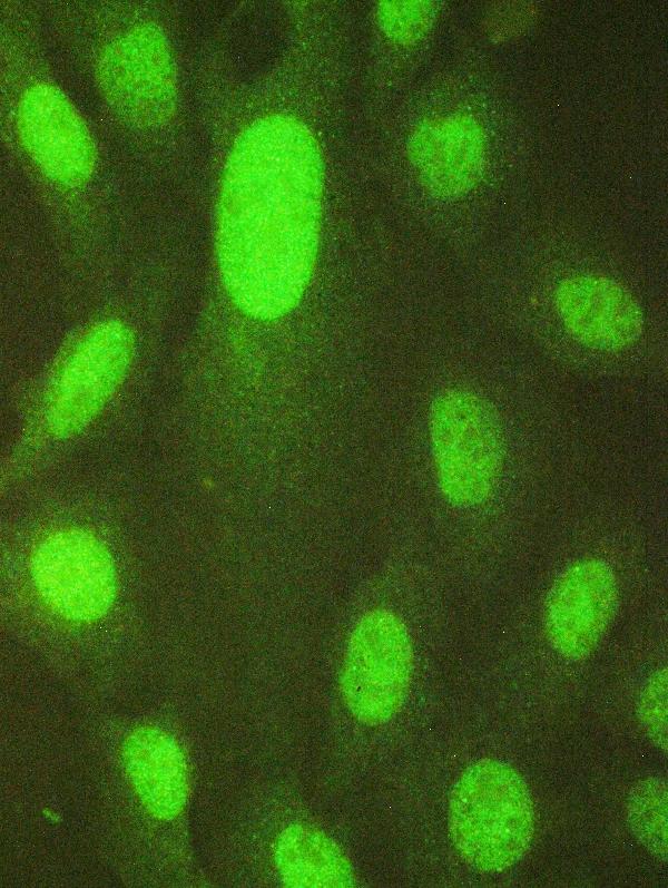
IF/ICC Q: What localization should I expect?A: Primarily nucleoplasmic fluorescence, with possible nuclear-body and cytosolic signal (HPA subcellular). HPA lists U2OS and KOLF2.1J among lines with ICC-IF images and marks CAB079904 Supported for ICC (HPA subcellular; HPA antibodies). This is localization context for IF/ICC, not an IHC-P protocol (HPA subcellular).
Why is my staining missing, weak or wrong?
SituationLikely causeNext action
A documented positive tissue has no visible USP7 signal.The result conflicts with reported medium staining in the selected positive cell population, but the HPA profile alone cannot identify the technical cause (HPA tissue IHC).Confirm the expected cells are present, then review the IHC-validated antibody's documented conditions, retrieval, detection controls and development as general IHC checks (standard IHC practice).
Nuclear signal is weak in lung macrophages or kidney glomerular cells.HPA reports low staining in those particular populations; a faint result there is less informative about assay failure than a negative result in a reported medium-staining population (HPA tissue IHC).Assess an HPA medium-staining cell population in the same run before changing conditions; keep comparisons tied to the named cells (HPA tissue IHC; standard IHC practice).
Brown deposit extends across stroma and luminal spaces.The deposit does not resolve the mainly nuclear USP7 pattern (HPA tissue IHC). Excess background or endogenous chromogenic activity is possible in an IHC run (standard IHC practice).Inspect a no-primary control, review blocking and washes, and adjust antibody concentration or chromogen development only under the assay's documented conditions (standard IHC practice).
Cytoplasmic staining dominates, with little nuclear staining.That distribution differs from the mainly nuclear tissue pattern, though cytoplasm is a documented USP7 location (HPA tissue IHC; UniProt Q93009).Compare nuclear counterstain and cell boundaries, inspect controls, and describe both compartments; do not score cytoplasmic signal alone as confirmed specificity (standard IHC interpretation).
Smooth muscle or fibroblasts stain as strongly as expected-positive cells.HPA reports USP7 as not detected in those cell populations, making the result discordant with its tissue profile (HPA tissue IHC). Cross-reactivity or detection background is possible (standard IHC interpretation).Compare the no-primary control and an HPA medium-staining population; if the unexpected cellular pattern persists, seek independent antibody confirmation (HPA tissue IHC; standard IHC practice).
A change in fixation or retrieval appears to change staining.Target-specific fixation effects are not established by the supplied assay evidence. Verify with a matched IHC source before attributing a result to fixation.Record the conditions, compare matched sections and controls, and treat any effect as assay-specific until verified; retrieval comparison is a general IHC optimization step (standard IHC practice).

Sample controls for USP7 IHC & IF

🧪Run bronchus first: respiratory epithelial cells should stain at a Medium level (HPA: bronchus, respiratory epithelial cells). Run smooth muscle as the negative tissue (HPA: smooth muscle cells, Not detected); on the bronchus slide, smooth muscle cells provide an internal negative reference and should show counterstain without specific brown nuclear signal (HPA: smooth muscle cells, Not detected; UniProt Q93009: nuclear localization).
Positive control tissue: Adrenal gland (Glandular cells, HPA Medium)
Negative control tissue: Smooth muscle (HPA Not detected)
ICC-IF cell lines (HPA subcellular resource): HPA ICC-IF images show USP7 in U2OS, KOLF2.1J, with annotated localisation: Nucleoplasm (supported) (HPA subcellular).
Technical controls: Include a no-primary, secondary-only control and a concentration-matched mouse IgG isotype control for the mouse catalog antibody (selected-SKU caption: mouse primary; standard IHC control practice); use a validated USP7 knockout specimen as a biological negative if available (standard IHC control practice). Check endogenous peroxidase and biotin background when using the caption’s biotin–streptavidin/DAB detection in bronchus (selected-SKU caption: biotinylated secondary and DAB; standard IHC control practice).
⚠️Feasibility: A target-specific fixation window and fixation effect are unreported, and the selected-SKU paraffin-section caption does not state the fixative (selected-SKU caption: fixative not stated). The reported IHC procedure uses heat retrieval in EDTA pH 8.0, but the supplied evidence does not establish whether retrieval is required or whether frozen sections are easier (selected-SKU caption: EDTA retrieval). IF/ICC has separate HPA images showing mainly nucleoplasmic signal, with nuclear bodies and cytosol also reported; the supplied evidence does not establish that IF is easier than paraffin IHC (HPA: subcellular localization and ICC-IF images).

HPA tissue IHC evidence for USP7

Comprehensive Human Protein Atlas IHC scoring per tissue (reliability: Approved — Medium consistency between antibody staining and RNA expression data.). Rows are taken directly from the HPA tissue chart — click any row's HPA link to view the source.

Positive expression · recommended positive controls

TissueCell typeLevelEvidenceSource
Adrenal gland Glandular cells Medium Protein (IHC) HPA →
Appendix Glandular cells Medium Protein (IHC) HPA →
Bronchus Respiratory epithelial cells Medium Protein (IHC) HPA →
Caudate Neuronal cells Medium Protein (IHC) HPA →
Cerebellum Cells in molecular layer Medium Protein (IHC) HPA →

Undetected expression · recommended negative controls

TissueCell typeLevelEvidenceSource
Smooth muscle Smooth muscle cells Not detected Protein (IHC) HPA →
Soft tissue Fibroblasts Not detected Protein (IHC) HPA →
Spleen Cells in red pulp Not detected Protein (IHC) HPA →
Section 3

Advanced USP7 IHC Tips

Troubleshoot USP7 staining in paraffin sections by checking retrieval, nuclear localisation, controls and scoring before interpreting changes in expression.

How should I retrieve USP7 in paraffin sections when nuclear staining is weak?
Start with heat-mediated retrieval in EDTA at pH 8.0 for paraffin sections (datasheet M01239-1). The selected tissue image used this retrieval before overnight incubation at 4°C with 1 μg/ml catalog antibody; its fixative was not reported (catalog caption M01239-1). If nuclear signal remains weak, compare a modest range of heating times on adjacent sections while keeping antibody concentration and detection constant, and inspect tissue integrity after each condition (standard IHC practice). Judge improvement by stronger staining in intact nuclei without a matching rise in diffuse background, using a positive tissue control in every run (HPA tissue IHC; standard IHC practice).
Could fixation explain variable USP7 staining between paraffin blocks?
Target-specific USP7 sensitivity to fixation is unknown because the selected paraffin-section caption does not state a fixative (catalog caption M01239-1). Record each block’s fixation history where available, then compare sections with the same EDTA pH 8.0 retrieval, 1 μg/ml primary antibody and detection conditions used in the selected image (catalog caption M01239-1; standard IHC practice). Include a concurrently processed positive control and inspect nuclear detail, section adhesion and nonspecific staining before assigning a block-to-block difference to biology (standard IHC practice). HPA’s mainly nuclear staining pattern can guide where to look, but it does not establish how fixation affects this antibody (HPA tissue IHC).
Should USP7 staining be nuclear, cytoplasmic or both?
Expect predominantly nuclear staining in tissue sections, while allowing a smaller cytoplasmic component (HPA tissue IHC; UniProt Q93009 subcellular location). USP7 is also reported in PML nuclear bodies and on chromosomes, and HPA supports nucleoplasm, nuclear bodies and cytosol in its subcellular data (UniProt Q93009 subcellular location; HPA subcellular). Score nuclear and cytoplasmic chromogen separately rather than combining them into one intensity, and compare the pattern with intact neighbouring cells on the same slide (standard IHC practice). A uniformly smeared or exclusively extracellular deposit should prompt review of section quality and detection controls before it is called USP7 localisation (standard IHC practice).
Can this stain distinguish USP7 isoforms or a modified epitope?
The record lists 3 USP7 isoforms, but the selected tissue caption does not identify this antibody’s epitope or demonstrate isoform selectivity (UniProt Q93009 isoforms; catalog caption M01239-1). USP7 contains a MATH domain at residues 68–195 and a USP domain at 214–521, with reported phosphorylation and acetylation sites elsewhere (UniProt Q93009 domains and modified residues). Those features may matter if an epitope is mapped, but they cannot establish which species produces chromogenic staining in a section (UniProt Q93009; standard IHC interpretation). For an isoform or modification claim, obtain epitope information and validate with an independent, suitably specific assay or perturbation before interpreting intensity differences (standard IHC practice).
How can I check the IHC localisation pattern by multiplex IF?
Use a separate IF validation workflow to compare USP7 with a marker for the expected cell type and a nuclear counterstain; HPA reports mainly nuclear tissue staining (HPA tissue IHC; standard IF practice). Choose fluorophores after checking the specimen’s autofluorescence, placing the weaker signal in a channel with low background and including single-stain controls (standard IF practice). USP7 has no transmembrane segment and is reported in nucleoplasm, nuclear bodies and cytosol, so choose permeabilisation to expose intracellular epitopes after fixation (UniProt Q93009 topology; HPA subcellular; standard IF practice). Compare compartment patterns across matched fields, but do not transfer the paraffin caption’s retrieval or antibody concentration to IF without validation (catalog caption M01239-1; standard IF practice).
What should I change when USP7 DAB staining is diffuse or widespread?
First compare the section with a no-primary control, looking separately for diffuse chromogen, tissue-edge staining and nuclear signal (standard IHC practice). The selected image used 10% goat serum blocking, 1 μg/ml primary antibody, a biotinylated secondary, a streptavidin–biotin complex and DAB (catalog caption M01239-1). If the control is stained, check peroxidase blocking and consider endogenous biotin as a possible contributor with this detection system; neither pattern alone establishes USP7 expression (catalog caption M01239-1; standard IHC practice). If only the primary-stained section is diffuse, titrate antibody and detection development while retaining EDTA pH 8.0 retrieval and an intact positive control (datasheet M01239-1; standard IHC practice).
How should I quantify USP7 staining across paraffin sections? ⚠ ANSWER MARKED FOR VERIFICATION
Define the cell population and compartment before scoring, because USP7 is mainly nuclear in tissue IHC but can also be cytoplasmic (HPA tissue IHC; UniProt Q93009 subcellular location). For nuclear staining, record the percentage of positive nuclei and an H-score from consistent intensity categories; report cytoplasmic staining separately if relevant (standard IHC practice). Normalise counts to the number of eligible, intact cells, or express positive-cell density per mm² of viable annotated tissue when cell counts are impractical (standard IHC practice). Apply one threshold and imaging or reading procedure across samples, excluding folds, edges and necrotic regions before comparing groups (standard IHC practice).
How do I distinguish genuine USP7 staining from artefact?
A credible pattern includes staining in intact nuclei within an appropriate cell population, consistent with HPA’s mainly nuclear tissue profile and supported nucleoplasmic location (HPA tissue IHC; HPA subcellular). Cytoplasmic signal is possible, but an extracellular deposit or staining confined to cut edges or necrosis warrants review before interpretation (UniProt Q93009 subcellular location; standard IHC practice). Compare morphology and a no-primary control, particularly when DAB appears in tissue compartments susceptible to endogenous enzyme activity (standard IHC practice). Treat the selected human lung cancer image as evidence of staining under its stated retrieval and detection conditions, not proof of specificity in every tissue or fixation condition (catalog caption M01239-1).
Boster reagents

Best USP7 / Ubiquitin C-terminal hydrolase 7 IHC Antibodies

Catalog anti-USP7 antibodies show IHC staining in human lung and mammary cancer paraffin sections (catalog IHC captions) and IF/ICC staining in U20S cells (catalog IF captions).

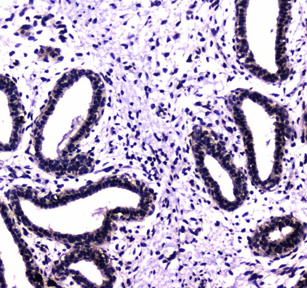
Real IHC data IHC analysis of HAUSP/USP7 using anti-HAUSP/USP7 antibody (M01239-1). HAUSP/USP7 was detected in paraffin-embedded section of human lung cancer tissue. Heat mediated antigen retrieval was performed in EDTA buffer (pH8.0, epitope retrieval solution). The tissue section was blocked with 10% goat serum. The tissue section was then incubated with 1μg/ml mouse anti-HAUSP/USP7 Antibody (M01239-1) overnight at 4°C. Biotinylated goat anti-mouse IgG was used as secondary antibody and incubated for 30 minutes at 37°C. The tissue section was developed using Strepavidin-Biotin-Complex (SABC) (Catalog # SA1021) with DAB as the chromogen.
Anti-HAUSP/USP7 Antibody ® (monoclonal, 5E2)
Cat # M01239-1
Real IHC data IHC analysis of HAUSP/USP7 using anti-HAUSP/USP7 antibody (A01239-2). HAUSP/USP7 was detected in paraffin-embedded section of human mammary cancer tissue. Heat mediated antigen retrieval was performed in citrate buffer (pH6, epitope retrieval solution) for 20 mins. The tissue section was blocked with 10% goat serum. The tissue section was then incubated with 1μg/ml rabbit anti-HAUSP/USP7 Antibody (A01239-2) overnight at 4°C. Biotinylated goat anti-rabbit IgG was used as secondary antibody and incubated for 30 minutes at 37°C. The tissue section was developed using Strepavidin-Biotin-Complex (SABC)(Catalog # SA1022) with DAB as the chromogen.
Anti-HAUSP/USP7 Antibody ®
Cat # A01239-2

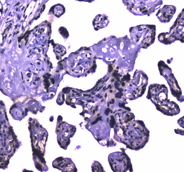
M01239-1 has IHC images from human lung cancer, human placenta and rat intestine paraffin sections, plus IF/ICC data from U20S cells (M01239-1 image captions). A01239-2 has IHC images from human mammary cancer, human placenta and mouse lymph node paraffin sections, plus IF/ICC data from U20S cells (A01239-2 image captions).

Which to pick: For tissue IHC, choose the mouse monoclonal M01239-1 for the documented EDTA retrieval at pH 8.0 (M01239-1 catalog entry and IHC caption), or A01239-2 for the documented citrate retrieval at pH 6 (A01239-2 IHC caption); neither caption reports the fixative (catalog IHC captions). For IF/ICC, both SKUs list those applications and show U20S cell staining; select the secondary antibody to match the mouse host of M01239-1 or rabbit host of A01239-2 (catalog entries and IF captions). For cross-species IHC, both list human, mouse and rat reactivity, with an additional mouse lymph node image for A01239-2 and rat intestine image for M01239-1 (catalog entries and IHC captions).

Each figure is that product's own IHC / IF validation image from its datasheet.

References

  1. UniProt Consortium. UniProt entry Q93009 (UBP7_HUMAN, Ubiquitin C-terminal hydrolase 7).
  2. Human Protein Atlas. USP7 tissue IHC expression (reliability: Approved).
  3. Human Protein Atlas. USP7 subcellular location (ICC-IF): Mainly localized to the nucleoplasm. In addition localized to the nuclear bodies and cytosol..
  4. Human Protein Atlas. USP7 antibody validation summary (3 antibodies).
  5. Administration of USP7 inhibitor P22077 inhibited cardiac hypertrophy and remodeling in Ang II-induced hypertensive mice. Frontiers in pharmacology 2022 — PMC9640964.
  6. Ubiquitin-specific protease 7 expression is a prognostic factor in epithelial ovarian cancer and correlates with lymph node metastasis. OncoTargets and therapy 2016 — PMC4803273.
  7. CCDC6 and USP7 expression levels suggest novel treatment options in high-grade urothelial bladder cancer. Journal of experimental & clinical cancer research : CR 2019 — PMC6381716.
  8. USP7 is a novel Deubiquitinase sustaining PLK1 protein stability and regulating chromosome alignment in mitosis. Journal of experimental & clinical cancer research : CR 2019 — PMC6858727.
  9. PubMed PMID:9034339 — UniProt-cited evidence.
  10. PubMed PMID:9130697 — UniProt-cited evidence.
  11. PubMed PMID:14702039 — UniProt-cited evidence.