VASP / Vasodilator-stimulated phosphoprotein · IHC design guide

Design Immunohistochemistry for VASP

Plan chromogenic VASP IHC on paraffin sections using cytoplasmic staining in most cells as the reference pattern (HPA tissue IHC). The guide covers fixation and scoring, with bone marrow hematopoietic cells showing high staining and heart cardiomyocytes showing no detected staining (HPA tissue IHC).

Evidence assembled Oct 2026 · For research use; verify linked source records and product datasheet before use
Immunohistochemistry protocol sheet for VASP (IHC for VASP): expected localisation Cytoplasm in most cells (HPA tissue IHC), antibody M00303, validated IHC image, and IHC protocol steps
Printable VASP IHC protocol sheet — expected localisation Cytoplasm in most cells (HPA tissue IHC), antibody M00303, controls and protocol steps. Open the full VASP IHC guide →

VASP Immunohistochemistry Experimental Design Guide

Expected localisation, validated protocols, controls and antibodies — the at-a-glance facts below, then the full design guide.

Must know before staining
Expected localisation Cytoplasm in most cells (HPA tissue IHC)
Staining pattern Cytoplasmic staining in most cells (HPA tissue IHC)
Antigen retrieval EDTA pH 8.0 HIER, heat-mediated (datasheet M00303)
Positive control ⓘ Appendix+4 more · see all
Negative control ⓘ Heart muscle+1 more · see all
Important caveats
Reasons your staining may differ from the expected pattern.
Fixation Keep fixation consistent across paraffin sections. (standard IHC practice; not target-specific)
Caveat Marrow peroxidase may mimic DAB signal (HPA tissue IHC; standard IHC practice)
Regulation Expression regulation not specified (UniProt)
Isoform / epitope No isoforms listed; no extracellular epitope expected (UniProt)
Section 1

Recommended VASP IHC & IF Protocols

The catalog antibody’s IHC-P protocol is accompanied by published VASP staining conditions for breast cancer and colorectal cancer sections (PMC5216765; PMC6419247).

Recommended immunohistochemistry (IHC-P) protocol parameters
SampleParaffin-embedded human liver cancer tissue; fixative not specified (datasheet M00303)
FixationImage fixative and duration unreported (datasheet M00303); verify before use.
Sectioning4–5 µm sections on charged slides (standard)
DeparaffinisationXylene, graded ethanol series to water (standard)
Antigen retrievalHeat retrieval: EDTA pH 8.0 (datasheet M00303); 20 min, 95–100 °C (standard)
Peroxidase block3% H2O2, 10 min, room temperature (standard)
Blocking10% goat serum (datasheet M00303)
Primary antibodyRabbit monoclonal (clone BHO-22) anti-VASP, 1:50 (datasheet M00303)
Primary incubationOvernight at 4 °C (datasheet M00303)
DetectionHRP-conjugated secondary, DAB chromogen (datasheet M00303)
CounterstainHematoxylin, blue, dehydrate and mount (standard)
Expected resultVASP-positive staining in glandular cells of appendix (HPA tissue IHC: High). HPA tissue profile: Cytoplasmic expression in most cells. No signal in the no-primary control.
💡Decision noteStart with the catalog antibody’s heat-mediated EDTA pH 8.0 retrieval (datasheet: M00303); adjust using the published retrieval conditions when adapting a cited protocol (PMC5216765; PMC6419247).
Section 2

What Is the Expected VASP Staining Pattern?

VASP should appear mainly in the cytoplasm of many cell types in paraffin-section IHC, with high staining reported in several glandular and hematopoietic populations (HPA tissue IHC: cytoplasmic expression in most cells; HPA: High in appendix glandular cells and bone marrow hematopoietic cells). Its actin-associated distribution can include junctions and focal adhesions (UniProt P50552 subcellular location). VASP has no transmembrane segment (UniProt P50552 topology). HPA rates tissue IHC reliability as Enhanced, with medium consistency between staining and RNA data (HPA tissue IHC).

What am I looking at on my slide?
Cytoplasmic staining in glandular cells of appendix, breast, cervix, duodenum or endometrium.This fits the reported High staining in those cell populations (HPA tissue IHC). Judge the named cells, rather than treating every structure in the section as an equally strong positive control.
Cytoplasmic staining with accentuation at cell borders or adhesion-rich edges.This can fit VASP localisation at junctions and focal adhesions (UniProt P50552 subcellular location). HPA tissue IHC describes a mainly cytoplasmic pattern; sharp edge staining alone should be interpreted alongside the cell's broader pattern (HPA tissue IHC).
Predominantly nuclear staining without a convincing cytoplasmic pattern.Treat this as a compartment mismatch requiring investigation, since the supplied localisation records place VASP in the cytoplasm, cytoskeleton and cell junctions, and at the plasma membrane in ICC-IF (UniProt P50552 subcellular location; HPA subcellular ICC-IF).
Strong staining in cardiomyocytes or skeletal myocytes while expected positive cells remain weak.These myocytes are reported as Not detected by HPA tissue IHC. Consider cross-reactivity or endogenous chromogenic activity, especially if staining is present in a no-primary control; one discordant section alone cannot identify the cause (HPA tissue IHC; general IHC practice).
Diffuse color over cells and extracellular areas, or no signal in a documented positive population.Diffuse, poorly confined color limits localisation calls (general IHC practice). A blank positive population conflicts with reported High staining in appendix glandular or bone marrow hematopoietic cells; check the run before concluding VASP is absent (HPA tissue IHC).
💡Expected VASP appearanceCall a result positive when identifiable cells show predominantly cytoplasmic staining, potentially accentuated at junctions, with High signal in an HPA-listed positive population; nuclear-only or diffuse color is suspect (HPA tissue IHC; UniProt P50552 subcellular location; general IHC practice).
How each factor affects the staining
Topology and processingVASP has no transmembrane segment or signal peptide, and UniProt lists a 2–380 protein chain (UniProt P50552 topology and processing). Interpret cytoplasmic and junction-associated staining as compatible with an intracellular actin-associated protein, without assuming a secreted or shed antigen (UniProt P50552 function and subcellular location).
Phosphorylation versus total VASPUniProt records multiple modified residues, including phosphorylation at Ser157 and Ser239 (UniProt P50552 modified residues). The supplied HPA tissue pattern does not specify phospho-VASP staining, so do not infer phosphorylation state from a total-VASP chromogenic pattern (HPA tissue IHC).
Strength of the tissue referenceHPA calls tissue IHC reliability Enhanced and reports medium consistency with RNA expression (HPA tissue IHC). Both listed antibodies have Enhanced IHC status (HPA antibodies: HPA005724 and CAB004612); this supports the reference pattern but does not establish the specificity of a different antibody or every individual section.
IF/ICC Q: Where should signal localise?A: HPA reports mainly plasma membrane, cell junction and focal adhesion localisation in ICC-IF; UniProt also describes lamellipodia, filopodial tips and stress fibers (HPA subcellular ICC-IF; UniProt P50552 subcellular location). Use this to interpret IF images, while applying the tissue IHC pattern to paraffin sections.
Fixation evidenceTarget-specific fixation effects are not established by the supplied assay evidence. Verify with a matched IHC source before attributing a result to fixation.
Why is my staining missing, weak or wrong?
SituationLikely causeNext action
No staining in appendix glandular cells or bone marrow hematopoietic cells.Both are reported High in HPA tissue IHC; a blank result may reflect a failed run or an unsuitable assay condition, but these records do not identify which.Confirm tissue identity, inspect a concurrent positive control, and check primary antibody, retrieval, detection reagent and chromogen steps (general IHC practice).
Only nuclei stain.Nuclear-only signal conflicts with the supplied cytoplasmic and adhesion-site localisation (HPA tissue IHC; UniProt P50552 subcellular location).Compare with a no-primary control and review counterstain versus chromogen appearance; reassess antibody specificity if the nuclear signal persists (general IHC practice).
Cardiomyocytes or skeletal myocytes stain strongly.HPA reports these cells as Not detected; cross-reactivity or endogenous detection activity are possibilities, not established diagnoses (HPA tissue IHC; general IHC practice).Compare an HPA-listed positive cell population and a no-primary control; review endogenous enzyme blocking for the chromogenic detection system (general IHC practice).
Color is diffuse across tissue or extends beyond cell outlines.Diffuse deposition can obscure the reported cytoplasmic pattern (HPA tissue IHC; general IHC practice).Inspect no-primary and reagent controls, then review blocking, washes, primary concentration and chromogen development time (general IHC practice).
Cell-border staining appears without clear cytoplasmic signal.Junctional enrichment is plausible, but HPA describes cytoplasmic expression in most cells (UniProt P50552 subcellular location; HPA tissue IHC).Assess identifiable positive cell types and compare matched control sections before calling an isolated border pattern specific (HPA tissue IHC; general IHC practice).
Different sections show inconsistent staining intensity.HPA reports medium consistency between antibody staining and RNA data; that rating does not diagnose a particular slide's variation (HPA tissue IHC).Compare the same cell population across sections and verify matched processing, retrieval, detection and scoring conditions (general IHC practice).

Sample controls for VASP IHC & IF

🧪Run appendix first: its glandular cells should stain (HPA: High in appendix glandular cells). Use skeletal muscle myocytes as the negative tissue (HPA: Not detected in skeletal muscle myocytes); on the appendix slide, cells outside the glandular compartment can serve as internal negatives only if they show background staining, since the supplied HPA row does not establish their VASP status (HPA: appendix glandular cells).
Positive control tissue: Appendix (Glandular cells, HPA High)
Negative control tissue: Heart muscle (HPA Not detected)
ICC-IF cell lines (HPA subcellular resource): HPA ICC-IF images show VASP in A-431, U-251MG, U2OS, U2OS, siRNA 1, U2OS, scrambled, with annotated localisation: Plasma membrane (supported), Cell Junctions (supported), Focal adhesion sites (supported) (HPA subcellular).
Technical controls: Include a no-primary, secondary-only control and a matched nonimmune rabbit IgG isotype control for the rabbit primary; confirm specificity with a VASP knockout sample if available (caption: rabbit anti-VASP M00303; standard IHC practice). Block endogenous peroxidase and check for blood-associated pigment in appendix sections before interpreting DAB signal (HPA: appendix; caption: HRP/DAB detection; standard IHC practice).
⚠️Feasibility: A target-specific fixation window or fixation effect is unreported, and the selected M00303 caption does not state a fixative (caption: fixative unreported). Paraffin-section IHC is documented with heat-mediated EDTA retrieval at pH 8.0, but the evidence does not establish that retrieval is required; neither frozen sections nor IF is shown to be easier (caption: EDTA retrieval; HPA: ICC-IF images available). Score glandular staining against local background because appendix tissue may contain blood-associated peroxidase activity that can mimic DAB signal (HPA: appendix glandular cells; standard IHC practice).

HPA tissue IHC evidence for VASP

Comprehensive Human Protein Atlas IHC scoring per tissue (reliability: Enhanced — Medium consistency between antibody staining and RNA expression data.). Rows are taken directly from the HPA tissue chart — click any row's HPA link to view the source.

Positive expression · recommended positive controls

TissueCell typeLevelEvidenceSource
Appendix Glandular cells High Protein (IHC) HPA →
Bone marrow Hematopoietic cells High Protein (IHC) HPA →
Breast Glandular cells High Protein (IHC) HPA →
Bronchus Respiratory epithelial cells High Protein (IHC) HPA →
Cervix Glandular cells High Protein (IHC) HPA →

Undetected expression · recommended negative controls

TissueCell typeLevelEvidenceSource
Heart muscle Cardiomyocytes Not detected Protein (IHC) HPA →
Skeletal muscle Myocytes Not detected Protein (IHC) HPA →
Section 3

Advanced VASP IHC Tips

Troubleshoot chromogenic VASP staining in paraffin sections by checking retrieval, compartment, controls, and cell-specific scoring (datasheet M00303; UniProt P50552; standard IHC practice).

What retrieval should I try first when VASP staining is weak?
Start with heat-mediated antigen retrieval in EDTA at pH 8.0 for the catalog antibody (datasheet M00303). The reported paraffin-section staining used this retrieval before a 1:50 primary-antibody incubation overnight at 4°C (datasheet M00303). Keep heating and cooling conditions consistent across slides, then compare staining in a documented positive cell population with a no-primary control (standard IHC practice; HPA: high in appendix glandular cells). If signal remains weak, test a second retrieval condition on serial sections while holding antibody concentration and detection constant, because the supplied evidence establishes only the EDTA condition for this antibody (standard IHC practice; datasheet M00303).
How should I troubleshoot fixation-related loss of VASP staining?
The selected product caption describes a paraffin section but does not report its fixative, so target-specific sensitivity to fixation is unknown (datasheet M00303). Record the fixative, fixation duration, tissue thickness, and processing history for each specimen before comparing staining intensity (standard IHC practice). Run similarly processed positive-control tissue beside the study sections, using a cell population with documented VASP staining, such as appendix glandular cells (standard IHC practice; HPA: high in appendix glandular cells). If processing differs between batches, compare serial sections under the same pH 8.0 EDTA retrieval and detection conditions before attributing weaker signal to biology (datasheet M00303; standard IHC practice).
Which staining patterns fit VASP in a tissue section?
Assess cytoplasmic staining first, since tissue IHC reports cytoplasmic VASP in most cells (HPA: tissue IHC profile). Peripheral or junction-associated accentuation can also fit VASP, which localizes to focal adhesions, tight junctions, lamellipodia, and filopodial tips (UniProt P50552: subcellular location). The separate subcellular imaging record supports plasma membrane, cell junction, and focal-adhesion localization, although those images do not establish how sharply these structures resolve in chromogenic sections (HPA: subcellular location; standard IHC practice). Compare the pattern with adjacent cell morphology and a no-primary control before accepting isolated nuclear color or diffuse extracellular precipitate as VASP signal (UniProt P50552: subcellular location; standard IHC practice).
Could epitope choice explain disagreement between VASP stains?
The supplied UniProt record lists 0 isoforms, so an isoform-specific explanation is unsupported by this record (UniProt P50552: isoforms). VASP has an N-terminal WH1 domain spanning residues 2–113 and reported phosphorylation sites including residues 157 and 239 (UniProt P50552: domains and modified residues). The catalog caption does not identify the antibody epitope or establish whether phosphorylation changes binding, so treat either explanation as a hypothesis requiring antibody-specific evidence (datasheet M00303; UniProt P50552: modified residues). When two stains disagree, compare their stated immunogens, retrieval conditions, and cell-level patterns on adjacent sections before interpreting the discrepancy biologically (standard IHC practice).
How can IF help investigate an ambiguous chromogenic VASP pattern?
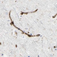
Use IF as a separate localization check, multiplexing VASP with a marker for the cell type implicated by the IHC section; colon endothelial cells are one documented high-staining population (HPA: high in colon endothelial cells; standard IF practice). Choose fluorophores after inspecting unstained tissue for autofluorescence, and place a weaker channel away from the strongest background where the available filters permit (standard IF practice). VASP lacks a transmembrane segment and can occupy the cytoplasmic side of peripheral structures, so permeabilize if the antibody epitope is intracellular; its exact epitope is unreported here (UniProt P50552: topology and subcellular location; datasheet M00303). Include single-channel and no-primary controls when judging overlap (standard IF practice).
What causes widespread brown staining with little cellular detail?
First compare a no-primary section with the complete stain to distinguish detection-system background from primary-antibody-dependent signal (standard IHC practice). For peroxidase and DAB detection, block endogenous peroxidase, check nonspecific secondary binding, and inspect for pigment or precipitate that persists outside intact cells (standard IHC practice). The reported product image used 10% goat-serum blocking, a 1:50 primary dilution, and peroxidase-based DAB development; those settings describe its demonstrated staining workflow (datasheet M00303). If background rises across multiple tissues, titrate the primary antibody and shorten chromogen development while retaining a known positive section to monitor signal loss (standard IHC practice; HPA: high in appendix glandular cells).
How should I score VASP across heterogeneous tissue sections? ⚠ ANSWER MARKED FOR VERIFICATION
Define the cell population and compartment before scoring, because VASP staining may be cytoplasmic with peripheral or junction-associated accentuation (HPA: tissue IHC profile; UniProt P50552: subcellular location). For cell-based assessment, record percent positive cells and intensity, or calculate an H-score from intensity categories 0–3, yielding a 0–300 range (standard IHC practice). For spatially restricted cells, report positive-cell density per mm² of viable, annotated tissue and specify the counted cell type (standard IHC practice). Normalize comparisons to the same cell population, viable area, staining batch, and exposure to chromogen; exclude necrosis and section edges using rules set before scoring (standard IHC practice).
When should an apparent VASP-positive cell be treated as artefact?
Favor signal within intact cells in a plausible cytoplasmic or peripheral compartment, consistent with tissue IHC and VASP localization records (HPA: tissue IHC profile; UniProt P50552: subcellular location). Check cell identity before calling an unexpected population positive: HPA reports high staining in appendix glandular cells and colon endothelial cells, while cardiomyocytes and skeletal-muscle myocytes are reported as not detected (HPA: tissue IHC cell-level observations). Staining concentrated at section edges, within necrosis, or in a no-primary control warrants investigation for processing, pigment, or endogenous-enzyme artefact (standard IHC practice). Treat discordance with the reference pattern cautiously because HPA rates tissue staining as Enhanced with medium consistency against RNA expression (HPA: reliability description).
Boster reagents

Best VASP / Vasodilator-stimulated phosphoprotein IHC Antibodies

Three anti-VASP antibodies have IHC images from paraffin-embedded human liver cancer and rat intestine, and stained human kidney (catalog: IHC image captions); one also lists human IF reactivity (catalog: M00303-3 applications, reactivity).

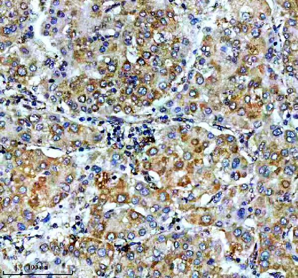
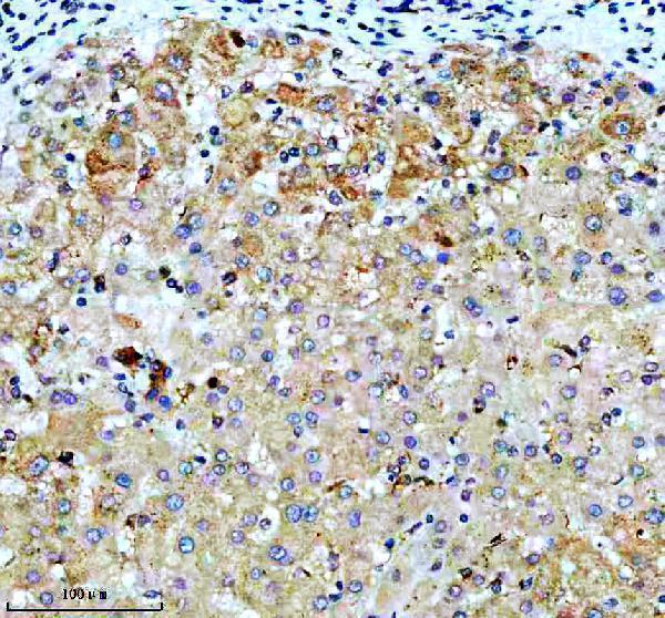
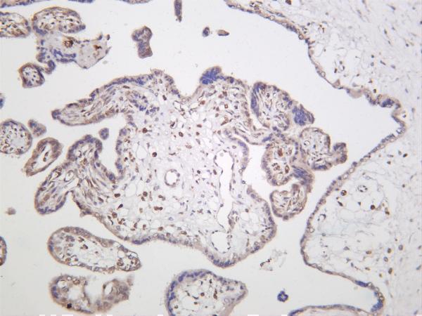
Real IHC data IHC analysis of VASP using anti-VASP antibody (M00303). VASP was detected in a paraffin-embedded section of human liver cancer tissue. Heat mediated antigen retrieval was performed in EDTA buffer (pH 8.0, epitope retrieval solution). The tissue section was blocked with 10% goat serum. The tissue section was then incubated with 1:50 rabbit anti-VASP Antibody (M00303) overnight at 4°C. Peroxidase Conjugated Goat Anti-rabbit IgG was used as secondary antibody and incubated for 30 minutes at 37°C. The tissue section was developed using HRP Conjugated Rabbit IgG Super Vision Assay Kit (Catalog # SV0002) with DAB as the chromogen.
Anti-VASP Rabbit Monoclonal Antibody
Cat # M00303
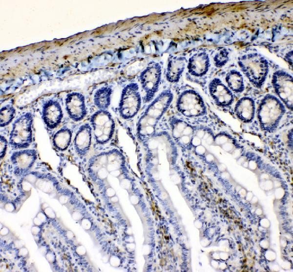
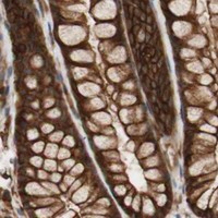
Real IHC data IHC analysis of VASP using anti-VASP antibody (A00303-1). VASP was detected in a paraffin-embedded section of rat intestine tissue. Heat mediated antigen retrieval was performed in EDTA buffer (pH 8.0, epitope retrieval solution). The tissue section was blocked with 10% goat serum. The tissue section was then incubated with 2 μg/ml rabbit anti-VASP Antibody (A00303-1) overnight at 4°C. Peroxidase Conjugated Goat Anti-rabbit IgG was used as secondary antibody and incubated for 30 minutes at 37°C. The tissue section was developed using HRP Conjugated Rabbit IgG Super Vision Assay Kit (Catalog # SV0002) with DAB as the chromogen.
Anti-VASP Antibody ®
Cat # A00303-1
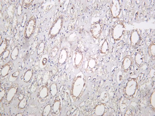
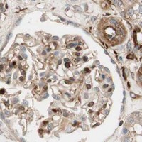
Real IHC data Human kidney was stained with anti-VASP rabbit antibody
Anti-VASP Rabbit Monoclonal Antibody
Cat # M00303-3

M00303 shows IHC staining in paraffin-embedded human liver cancer tissue (M00303 image caption). A00303-1 shows IHC staining in paraffin-embedded rat intestine, while M00303-3 shows stained human kidney and lists IHC and IF applications (A00303-1 image caption; M00303-3 image caption and applications).

Which to pick: For human tissue IHC, choose rabbit monoclonal M00303: its image documents a paraffin-embedded human section at 1:50, with the fixative unreported (catalog: M00303 clone, reactivity; M00303 image caption). For human IF/ICC planning, M00303-3 lists IF at 1:200–1:1000; its image documents human kidney staining, while IF imaging and ICC validation are unreported (catalog: M00303-3 applications, dilution, reactivity, image alts). For human and rat IHC work, A00303-1 lists both species and shows a paraffin-embedded rat intestine section at 2 μg/ml; the image caption does not report the fixative (catalog: A00303-1 reactivity; A00303-1 image caption).

Each figure is that product's own IHC / IF validation image from its datasheet.

References

  1. UniProt Consortium. UniProt entry P50552 (VASP_HUMAN, Vasodilator-stimulated phosphoprotein).
  2. Human Protein Atlas. VASP tissue IHC expression (reliability: Enhanced).
  3. Human Protein Atlas. VASP subcellular location (ICC-IF): Mainly localized to the plasma membrane, cell junctions and focal adhesion sites..
  4. Human Protein Atlas. VASP antibody validation summary (2 antibodies).
  5. A functional antiplatelet nanomotor for tri-modal ablation of acute arterial thrombus. Cell reports. Medicine 2026 — PMC13522756.
  6. Tumor suppressor berberine binds VASP to inhibit cell migration in basal-like breast cancer. Oncotarget 2016 — PMC5216765.
  7. Vasodilator-Stimulated Phosphoprotein Biomarkers Are Associated with Invasion and Metastasis in Colorectal Cancer. Biomarkers in cancer 2018 — PMC6419247.
  8. Dai Huang Fu Zi Tang could ameliorate intestinal injury in a rat model of hemorrhagic shock by regulating intestinal blood flow and intestinal expression of p-VASP and ZO-1. BMC complementary and alternative medicine 2014 — PMC3974027.
  9. PubMed PMID:7828592 — UniProt-cited evidence.
  10. PubMed PMID:10087267 — UniProt-cited evidence.
  11. PubMed PMID:14702039 — UniProt-cited evidence.