This website uses cookies to ensure you get the best experience on our website.


- Table of Contents
Plan VCP chromogenic IHC on paraffin sections around the nuclear and cytoplasmic tissue pattern (HPA tissue IHC). Purkinje cells provide a high-staining reference (HPA tissue IHC); the IHC-validated antibody PA2137 has a 0.5–1 μg/ml IHC-P range (datasheet: PA2137).

Expected localisation, validated protocols, controls and antibodies — the at-a-glance facts below, then the full design guide.
| Expected localisation | Nuclear and cytoplasmic tissue staining (HPA tissue IHC) | |
| Staining pattern | Nuclear and cytoplasmic staining across tissues (HPA tissue IHC) | |
| Antigen retrieval | Citrate pH 6 HIER, heat-mediated (datasheet PA2137) | |
| Positive control | Adipose tissue+4 more · see all | |
| Negative control | None in HPA (detected in all 45 tissues); use no-primary + isotype controls |
| Fixation | Keep formaldehyde fixation consistent across sections (standard IHC practice; not target-specific) | |
| Caveat | Pancreatic exocrine and smooth muscle cells stain weakly (HPA tissue IHC) | |
| Regulation | Expression regulation not annotated (UniProt) | |
| Isoform / epitope | No annotated isoforms; mature chain 2–806 has no membrane span (UniProt) |
Compare the catalog antibody’s IHC-P protocol with published VCP staining methods for spinal cord, cervical biopsies, and orbital B-cell lymphoma tissue (PMC4297454; PMC4951329; PMC6312057).
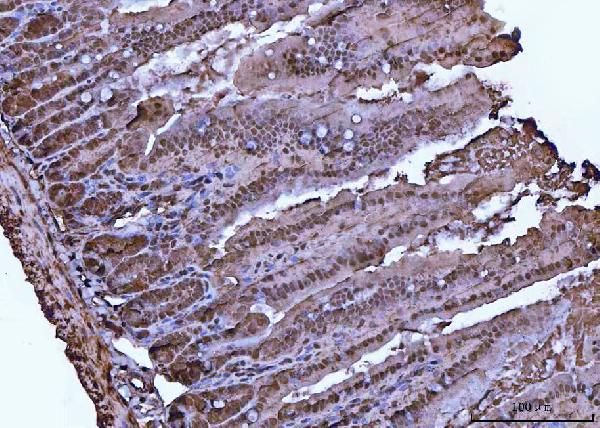
| Sample | Paraffin-embedded mouse intestine tissue; fixative not specified (datasheet PA2137) |
| Fixation | Image fixative and duration unreported (datasheet PA2137); verify before use. |
| Sectioning | 4–5 µm sections on charged slides (standard) |
| Deparaffinisation | Xylene, graded ethanol series to water (standard) |
| Antigen retrieval | Heat retrieval: Citrate pH 6, 20 min (datasheet PA2137) |
| Peroxidase block | 3% H2O2, 10 min, room temperature (standard) |
| Blocking | 10% goat serum (datasheet PA2137) |
| Primary antibody | Rabbit anti-VCP, 0.5-1μg/ml (datasheet PA2137) |
| Primary incubation | Overnight at 4 °C (datasheet PA2137) |
| Detection | Streptavidin-biotin complex (SABC), DAB chromogen (datasheet PA2137) |
| Counterstain | Hematoxylin, blue, dehydrate and mount (standard) |
| Expected result | VCP-positive staining in adipocytes of adipose tissue (HPA tissue IHC: High). HPA tissue profile: Nuclear and cytoplasmic expression in all tissues. No signal in the no-primary control. |
VCP is expected in nuclei and cytoplasm across tissues (HPA tissue IHC: Supported; medium consistency with RNA data). Cytosolic and endoplasmic reticulum localization also appears in UniProt P55072. High IHC staining is reported in adipocytes, appendix glandular cells and cerebellar Purkinje cells, among others (HPA tissue IHC). VCP has no transmembrane segment (UniProt P55072 topology).
| Nuclear and cytoplasmic staining in recognizable cells, including strong staining in adipocytes or appendix glandular cells (HPA tissue IHC). | This fits HPA’s tissue-wide pattern; those cell types are reported as High. Assess staining in its tissue context rather than requiring equal intensity in every cell (HPA tissue IHC). |
| Staining is confined to a compartment outside the expected nuclear and cytoplasmic pattern (HPA tissue IHC). | Treat an exclusively membrane-rim pattern with caution: VCP has no transmembrane segment (UniProt P55072 topology). Check morphology, detection background and a validated positive tissue before interpreting it as VCP (general IHC practice). |
| A cell population stains strongly while nearby expected cells show little or no signal (HPA tissue IHC). | Review the cell identities and compare with HPA’s cell-level observations. Unexpected staining may reflect cross-reactivity or endogenous detection activity (general IHC practice); HPA reports VCP expression across tissues, so an unlisted cell type is not automatically negative (HPA tissue IHC). |
| Signal coats tissue spaces or unrelated structures and obscures cell boundaries (general IHC practice). | Diffuse background cannot establish VCP localization. Compare a no-primary control and examine blocking, washing and detection conditions before scoring cellular staining (general IHC practice). |
| A reported High cell type has no convincing nuclear or cytoplasmic signal (HPA tissue IHC). | A failed positive control makes a negative sample difficult to interpret. Check section integrity and the antibody’s IHC-P workflow, then repeat with an HPA-reported High cell type (general IHC practice; HPA tissue IHC). |
| Tissue and cell selection | HPA reports High staining in adipocytes, appendix glandular cells, hematopoietic cells and cerebellar Purkinje cells (HPA tissue IHC). Select a recognizable reported High population as a positive reference (general IHC practice). |
| Interpreting weaker cells | Exocrine glandular cells in pancreas and smooth muscle cells are reported Low, not negative (HPA tissue IHC). Their weak signal alone is a poor reason to reject a run with a convincing High-cell control (general IHC practice). |
| Compartment and topology | Nuclear and cytoplasmic tissue staining is reported by HPA; UniProt places VCP in cytosol, endoplasmic reticulum and nucleus (HPA tissue IHC; UniProt P55072). No transmembrane segment is annotated, so a membrane-only interpretation needs scrutiny (UniProt P55072 topology). |
| Strength of tissue evidence | HPA rates tissue IHC Supported, with medium consistency between antibody staining and RNA expression (HPA tissue IHC). Use that qualification when judging subtle staining differences; it does not establish a fixation effect (HPA tissue IHC). |
| Antibody validation | HPA012728, HPA012814 and CAB005593 each have Supported IHC validation (HPA antibodies). Validation supports comparison with HPA patterns but does not guarantee that every section or detection run will reproduce them (general IHC practice). |
| IF/ICC Q&A: Where should VCP appear? | Mainly in nucleoplasm and cytosol (HPA ICC-IF: supported). Mitotic spindle and cilium-associated locations are listed as uncertain (HPA ICC-IF). Use the separate IF/ICC guide for that application. |
| Situation | Likely cause | Next action |
|---|---|---|
| No signal in an HPA-reported High cell type (HPA tissue IHC). | The run may have failed, or the chosen cells may have been misidentified (general IHC practice). | Confirm morphology and control identity; review the IHC-validated antibody’s IHC-P instructions, retrieval and detection steps, then repeat the positive control (general IHC practice). |
| Only pancreas exocrine cells or smooth muscle cells appear weak (HPA tissue IHC). | Both cell populations are reported Low, so weak staining can match the reference (HPA tissue IHC). | Judge run performance using a reported High cell population in an appropriate control section before calling the assay insensitive (HPA tissue IHC; general IHC practice). |
| Strong signal follows a membrane rim with little nuclear or cytoplasmic staining (HPA tissue IHC). | That distribution conflicts with the reported tissue pattern and VCP’s lack of a transmembrane segment (HPA tissue IHC; UniProt P55072 topology). | Inspect the no-primary control and compare staining with an HPA-reported High cell type; avoid assigning the rim to VCP until the pattern is resolved (general IHC practice). |
| Unexpected cells stain strongly while expected cells do not (HPA tissue IHC). | Possible cross-reactivity, endogenous detection activity or cell misidentification requires checking (general IHC practice). HPA provides no negative tissue in this payload (HPA tissue IHC). | Verify cell morphology and use appropriate detection controls; compare with HPA’s cell-level pattern instead of treating an unlisted tissue as negative (general IHC practice; HPA tissue IHC). |
| Diffuse deposits obscure nuclear and cytoplasmic boundaries (general IHC practice). | Background from blocking, washing or detection can conceal the expected cellular pattern (general IHC practice; HPA tissue IHC). | Review a no-primary control, reagent handling and wash steps; score VCP only where staining follows identifiable cells (general IHC practice). |
| A section is unexpectedly negative after a change in tissue processing or retrieval (general IHC practice). | Target-specific fixation effects are not established by the supplied assay evidence. Verify with a matched IHC source before attributing a result to fixation. | Check the catalog antibody’s IHC-P instructions and run a reported High cell type alongside the sample; interpret the result from observed controls, without assuming a VCP-specific fixation effect (general IHC practice; HPA tissue IHC). |




Comprehensive Human Protein Atlas IHC scoring per tissue (reliability: Supported — Medium consistency between antibody staining and RNA expression data.). Rows are taken directly from the HPA tissue chart — click any row's HPA link to view the source.
| Tissue | Cell type | Level | Evidence | Source |
|---|---|---|---|---|
| Adipose tissue | Adipocytes | High | Protein (IHC) | HPA → |
| Appendix | Glandular cells | High | Protein (IHC) | HPA → |
| Bone marrow | Hematopoietic cells | High | Protein (IHC) | HPA → |
| Breast | Adipocytes | High | Protein (IHC) | HPA → |
| Bronchus | Respiratory epithelial cells | High | Protein (IHC) | HPA → |
| Tissue | Cell type | Level | Evidence | Source |
|---|---|---|---|---|
| None in HPA: VCP is detected in all 45 scored tissues. Use a no-primary (secondary-only) and an isotype control instead. | ||||
Troubleshoot VCP staining by checking retrieval, compartment pattern, detection background and scoring against the supplied antibody protocol and tissue evidence.
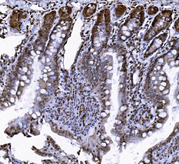
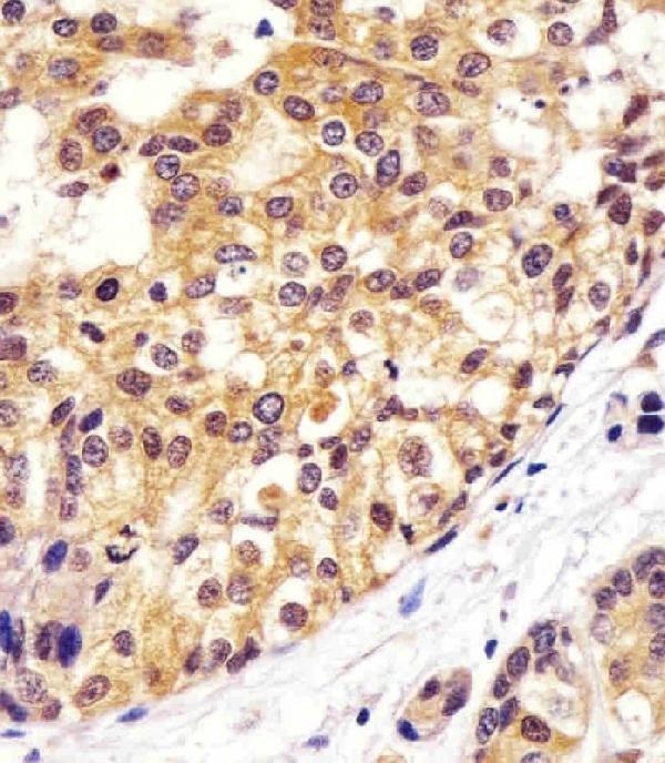
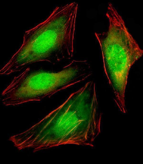
Anti-VCP antibodies have paraffin-section IHC figures from human and mouse tissues (catalog IHC captions), plus IF figures from A431 and HeLa cells (catalog IF captions).
PA2137 shows mouse intestine IHC, and PB9454 shows human glioma IHC (PA2137 and PB9454 IHC captions). A00610-2 shows mouse colon IHC, while M00610-1 shows human breast carcinoma IHC (A00610-2 and M00610-1 IHC captions).
Which to pick: For tissue IHC, choose PB9454 for human glioma sections with EDTA retrieval at pH 8.0, or PA2137 for mouse intestine sections with citrate retrieval at pH 6; neither caption reports the fixative (PB9454 and PA2137 IHC captions). For IF/ICC, M00610-1 is a mouse monoclonal antibody with an IF figure in HeLa cells; its IHC figure uses paraffin-section, paraffin-embedded breast carcinoma sections (M00610-1 catalog and figure captions). For cross-species planning, PA2137 lists human, monkey, mouse and rat reactivity, with IHC figures in human, mouse and rat tissues; its paraffin-section captions do not report a fixative (PA2137 catalog and IHC captions). The selected PA2137 tissue-IHC caption documents paraffin sections, but does not specify the fixative (selected-SKU IHC image PA2137).