This website uses cookies to ensure you get the best experience on our website.
- Table of Contents
2 Citations 16 Q&As
2 Citations 16 Q&As
1 Citations 4 Q&As
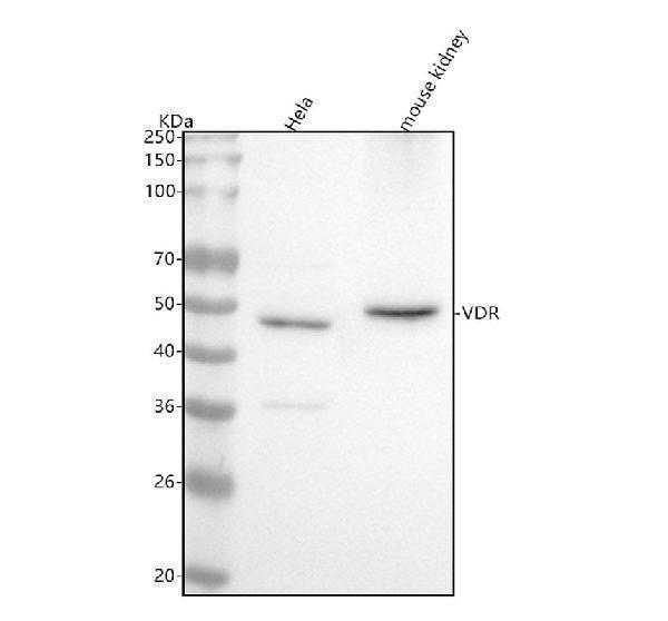
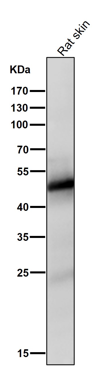
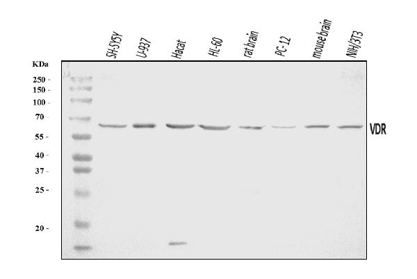
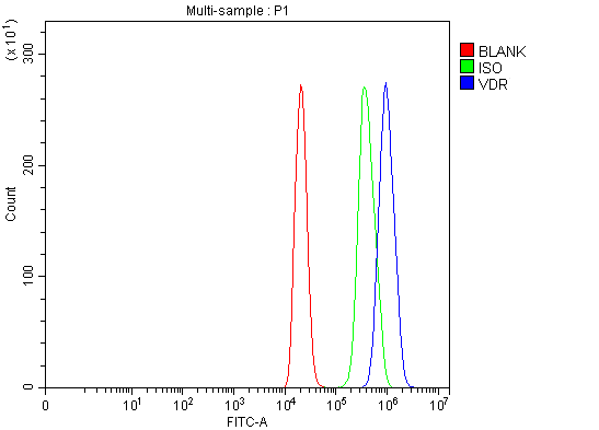
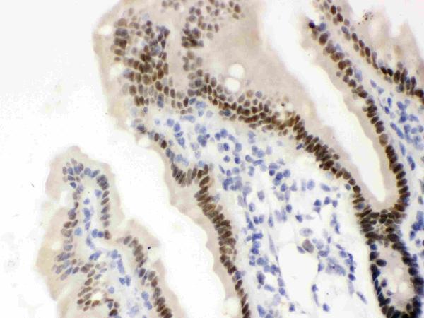
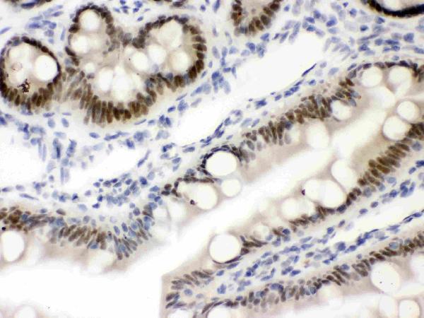
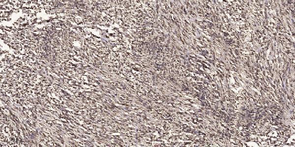
Facts about Vitamin D3 receptor.
The VDR-RXR heterodimers bind to specific response elements on DNA and activate the transcription of vitamin D3-responsive target genes (PubMed:28698609). Plays a central role in calcium homeostasis (By similarity).
Human | |
---|---|
Gene Name: | VDR |
Uniprot: | P11473 |
Entrez: | 7421 |
Belongs to: |
---|
nuclear hormone receptor family |
NR1I1; NR1I1Nuclear receptor subfamily 1 group I member 11,25-dihydroxyvitamin D3 receptor; VDR; vitamin D1,25- dihydroxyvitamin D3 receptor; vitamin D3 receptor
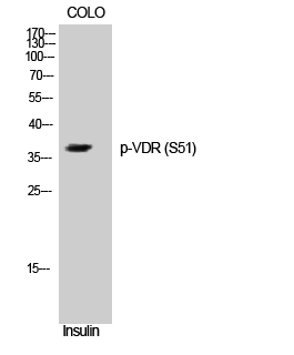
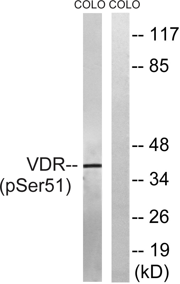
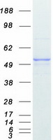
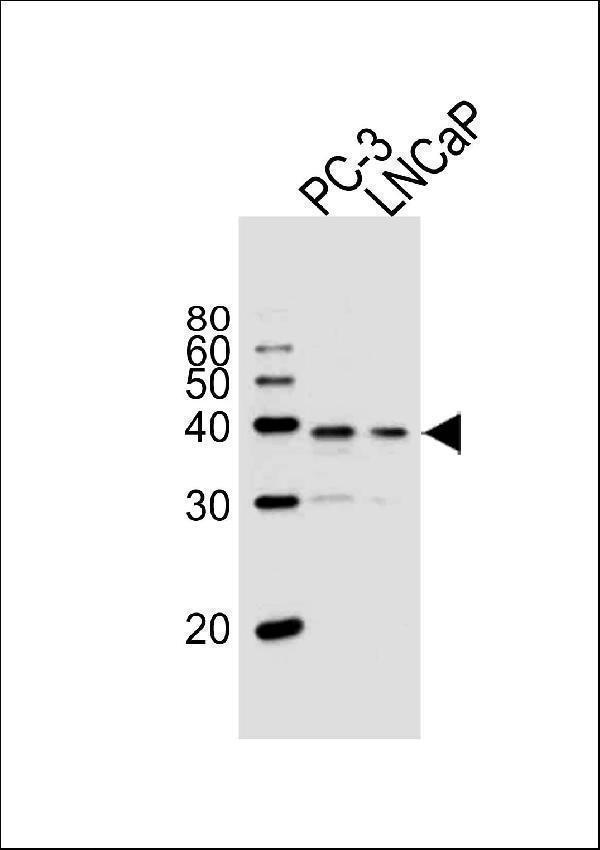
Mass (kDA):
48.289 kDA
Human | |
---|---|
Location: | 12q13.11 |
Sequence: | 12; NC_000012.12 (47841537..47905022, complement) |
Nucleus. Cytoplasm. Localizes mainly to the nucleus (PubMed:28698609, PubMed:12145331). Localization to the nucleus is enhanced by vitamin D3.
There are many applications for the VDR marker in life sciences. These include autophagy, immunofluorescence, and target transcription. This reagent makes a great instrument for your research. Here are some of the most commonly used uses of this reagent. Learn more about it! Find the best uses for the VDR marker. You'll also learn how to integrate it with other tools.
You've come to the right place if you're searching for an antibody for IHC. Boster Bio has over 16,000 antibodies to select from. They can be used in WB, FC, or ELISA. Boster offers mouse and rabbit polyclonal antibodies, as monoclonal antibodies. Boster also offers free secondary antibodies for purchase. And because they are so widely used in scientific applications, Boster is one of the most trusted sources of high-quality antibodies.
IHC is a highly popular method of analyzing tissues in many labs. This technique allows users to see the distribution of antigens inside cells and within tissues. Enzymes, which produce colors-producing reactions, can show the binding of antigens to antibodies. There are many different methods to determine the localization of antigens, and sample preparation is important. The appearance of antigens is influenced by the quality of the tissue sample. Here are some helpful tips to help you choose the right method for you.
Sample preparation is the first step in the IHC process. You will need a fresh tissue sample to complete this. This can be accomplished via biopsy, surgery, or even animal models. Autopsy samples are typically taken following the death of an animal. They may be left alone for up to two hours. The sample is therefore comparable to a postmortem examination. Since the antigens will denature after two hours, it is important to fixate your sample as quickly as possible after death.
The next step of IHC is detection. To detect proteins it is necessary to use a secondary antibody. employed. It bonds to the primary antibody. The secondary antibody may then be used to analyze samples for Western Blotting or ICC/IF. These are only one of the many IHC methods Boster Bio offers. They are able to identify numerous proteins, and Boster Bio also has a array of secondary antibodies for IHC and ICC/IF.
The VDR marker was found in the mouse kidney, and is a great diagnostic tool to detect kidney cell damage. Boster Bio, a Chinese company located in Hubei Province, manufactured this antibody. The antibody recognizes Human Antigen and has been tested for Western Blot analyses. First, determine the kind of the tissue sample to determine whether the VDR marker is effective.
The VDR gene contains instructions for the production of the vitamin D receptor protein. Vitamin D is a vital vitamin for human health and is essential to maintain the balance between calcium and phosphate in our body. Vitamin D regulates absorption of calcium from the intestinal tract. Vitamin D also plays an important part in other processes, not related to bone growth. However it is believed that the VDR marker can assist in identifying the presence of disease and creating effective treatments.
It is important to know that VDR expression in melanoma cell lines has no evident connection with metastatic stages or the invasion of vascular vessels. VDR expression does not correlate with prognosis, vascular invade or melanoma familiarity. Researchers can employ VDR markers to assess the tumor's response to vitamin D. This is documented in a paper titled "Best Uses of VDR Markers in Melanoma cells."
The optical density (OD), of the tubes was determined at approximately 450 nm with a multi-functional microplate reader. Immunohistochemistry was performed on 5 um thick paraffin sections, dewaxing and immunohistochemistry. The VDRmice displayed a "full house" phenomenon, with a high amount of immunological markers, including IgG, IgA, IgM, C3 and C1q. The mice with VDR showed a marked increase of deposition as compared to the control mice.
Intestinal VDR regulates autophagy, paneth cells, microbial homeostasis, and bacterial collection. Its reduced expression correlates with an altered Paneth cell function and dysbiosis. In the end the intestinal VDR could be used as a biomarker for dysbiosis as well as inflammatory bowel disease.
The VDR is a gene expressed in the cytoplasm and is responsible for mediating the majority of functions of 1,25-dihydroxyvitamin D. It heteromerizes with retinoid X receptors and regulates gene transcription by binding to a specific vitamin D response element. The VDR targets include CAMP/LL-37 (cathelicidin antimicrobial peptide), DEFB/b defensin, CLDN2 (claudin 2) and the autophagy regulator ATG16L1.
Research into cancer is among the most effective uses of VDR Marker in human tissues. This gene is a key factor in the development of NAFLD and the development of atherosclerosis. Furthermore it has been proven to be involved in oxLDL degradation in the liver. Additionally, this gene could also play a role in the development of NASH.
The inflammasome NLRP3 is a key regulator of cell death. It is located in the tubular cells of the LN. Its activation inhibits the NLRP3/caspase-1/IL-18 axis and suppresses the nuclear translocation of NF-kB. Paricalcitol also exerts negative effects on the NFKB nuclear transfer pathway.
The boster bio.vdr antibody recognizes Human, Mouse and Rat VDR antigen. It has been tested for Western Blot. This antibody is suitable for a variety of types of Western Blot applications, including immunofluorescence. The most effective uses of the VDR marker are described below. Below are a few of the most common boster bio applications. VDR immunohistochemistry is a method to detect vitamin D expression.
Boster bio.vdr can be used for immunocytochemistry and immunohistochemistry to color cells and tissue sections. The product is stored at -20degC and lasts for one year. The Boster guarantee covers the use of this product in the above-listed applications. The company manufactures this antibody at its Wuhan, China facility. It offers a variety of sample preparation solutions for immunocytochemistry and immunohistochemistry.
The VDR gene has been found to be a possible candidate for developing a new drug. The VDR gene has been implicated in numerous human diseases and is found in various tumors. VDR-deficient mice are more at risk for hypertension and oxidative stress as also autophagy and an increase in oxidative stress. This genetic mutation can also cause diabetes and cancer. It is important to detect genetic diseases early.
The median expression levels of VDR were not statistically significant. In the Boster Bio: Best Uses of the VDR marker
The VDR marker was discovered to inhibit Gli1, which is a b catenin-regulated protein. This gene is essential in the process of hair follicle differentiation and its silence reduces the activity of b-catenin. It also blocks the expression of the b-catenin target gene, PADI1. The activity of bcatenin is diminished when VDR is eliminated.
The 1,25(OH)2D3 and b-catenin responses elements are located next to one another. Despite their distinct features, these genes are believed to function together by reducing TCF/LEF activity. These two proteins interact to encourage differentiation and increase the number of hair follicles in hair follicles. In addition to blocking these signaling pathways, VDR and b catenin can compete with each other.
The VDR marker can be used for a variety of purposes such as the diagnosis and treatment of autoimmune diseases as well as cancer. It can also be used to monitor beta-catenin levels in immune cells. However, the precise mechanism isn't fully understood. Researchers are still working to understand the role of VDR in the context of autoimmunity, as well as cancer progression. These findings will aid in understanding the role of VDR in the regulation of beta-catenin.
A study of keratinocytes that have been transformed by Ras suggests that VDR/RXR/DRIP205 collaborate and regulate beta-catenin. This interaction provides an important basis for optimizing vitamin D treatment for patients suffering from skin cancer. These findings will help in the identification of new treatment options for moderate/mild psoriasis.
When working with the VDR marker for beta-catenin, a regulator, it is essential to understand how this gene controls gene expression. The VDR antagonizes transcription factors such as IL-2. It also blocks the expression of 1,25(OH)2D3 synthesizing enzyme the CYP24A1 enzyme, which is needed for IL-17 production.
PMID: 2835767 by Baker A.R., et al. Cloning and expression of full-length cDNA encoding human vitamin D receptor.
PMID: 1324736 by Goto H., et al. A single receptor identical with that from intestine/T47D cells mediates the action of 1,25-dihydroxyvitamin D-3 in HL-60 cells.
*More publications can be found for each product on its corresponding product page