This website uses cookies to ensure you get the best experience on our website.


- Table of Contents
Real validated WNT1 Western blot protocols, expected-band and isoform facts, troubleshooting for weak or shifted signal, and recommended anti-WNT1 WB antibodies. Everything you need to plan the experiment before you commit precious samples.
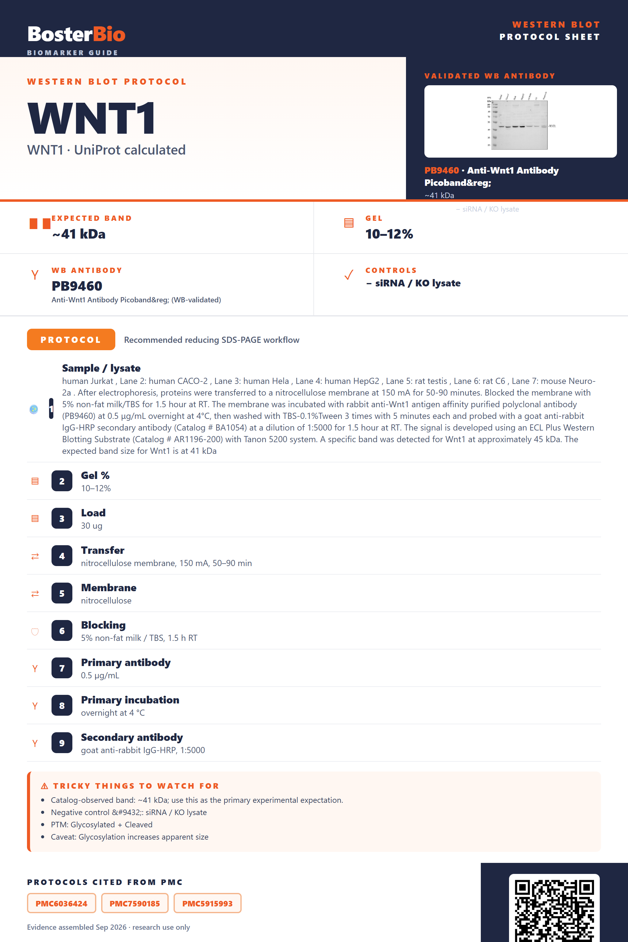
Expected bands, validated protocols, controls and antibodies — the at-a-glance facts below, then the full design guide.
| Expected band | ~41 kDa | |
| Gel | 10–12% | |
| Negative control | siRNA / KO lysate |
| PTM | Glycosylated + Cleaved | |
| Caveat | Glycosylation increases apparent size | |
| Regulation | Wnt beta catenin signaling | |
| Isoform | 1 isoform(s) |
Literature-validated Western blot parameters for WNT1 — gel percentage, transfer, blocking, antibody incubation and detection, extracted from published methods.
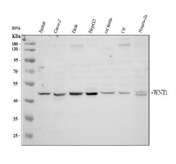
| Sample / lysate | human Jurkat , Lane 2: human CACO-2 , Lane 3: human Hela , Lane 4: human HepG2 , Lane 5: rat testis , Lane 6: rat C6 , Lane 7: mouse Neuro-2a . After electrophoresis, proteins were transferred to a nitrocellulose membrane at 150 mA for 50-90 minutes. Blocked the membrane with 5% non-fat milk/TBS for 1.5 hour at RT. The membrane was incubated with rabbit anti-Wnt1 antigen affinity purified polyclonal antibody (PB9460) at 0.5 μg/mL overnight at 4°C, then washed with TBS-0.1%Tween 3 times with 5 minutes each and probed with a goat anti-rabbit IgG-HRP secondary antibody (Catalog # BA1054) at a dilution of 1:5000 for 1.5 hour at RT. The signal is developed using an ECL Plus Western Blotting Substrate (Catalog # AR1196-200) with Tanon 5200 system. A specific band was detected for Wnt1 at approximately 45 kDa. The expected band size for Wnt1 is at 41 kDa |
| Gel % | 10–12% |
| Load | 30 ug |
| Transfer | nitrocellulose membrane, 150 mA, 50–90 min |
| Membrane | nitrocellulose |
| Blocking | 5% non-fat milk / TBS, 1.5 h RT |
| Primary antibody | 0.5 µg/mL |
| Primary incubation | overnight at 4 °C |
| Secondary antibody | goat anti-rabbit IgG-HRP, 1:5000 |
| Wash | TBS-0.1% Tween, 3 × 5 min |
| Detection | ECL |
| Exposure / imaging | Tanon 5200 |
| Observed band | 45 kDa |
WNT1's 370 aa precursor predicts 41 kDa, but signal-peptide cleavage and four N-glycosylation sites shift the mature secreted band variably above the ~38 kDa unmodified backbone.
| Band appearing somewhat larger than the ~38 kDa unmodified mature backbone | the four N-linked glycosylation sites add carbohydrate mass to the secreted, signal-peptide-cleaved chain |
| Diffuse or smeared band instead of one crisp line | heterogeneous glycan occupancy across the four N-linked sites spreads the apparent mass |
| Mature band smaller than the 41 kDa full-length translation product | the 27-residue N-terminal signal peptide is cleaved before secretion, removing about 3 kDa from the precursor |
| Weak or absent band in whole-cell lysate but present in conditioned medium or ECM fraction | WNT1 is annotated as secreted and extracellular matrix-associated, so most mature protein leaves the cell rather than accumulating intracellularly |
| Band shifts to a smaller, sharper size only under fully reducing, heat-denatured conditions | the eleven intra-chain disulfide bonds of the cysteine-rich Wnt fold keep the protein compact unless fully reduced, so incomplete reduction produces anomalous migration |
| Predicted mass (UniProt) | the 370 aa full-length precursor calculates to 41 kDa, the reference point before any processing or modification is accounted for |
| N-terminal signal peptide (residues 1-27) | cleaved during secretion, removing roughly 3 kDa so the mature extracellular chain runs smaller than the 41 kDa full-length precursor |
| N-linked glycosylation at Asn29, Asn316, Asn346, Asn359 | adds variable carbohydrate mass to the mature chain, pushing the apparent band above the unmodified backbone and broadening it |
| Secreted, extracellular matrix localization | shifts most mature protein out of the cell, so intracellular lysates show weaker signal than conditioned medium or ECM-enriched fractions |
| Eleven intra-chain disulfide bonds | stabilize a compact tertiary fold that can migrate anomalously, often faster or smaller, unless samples are fully reduced and denatured |
| Situation | Likely cause | Next action |
|---|---|---|
| No band in lysate | WNT1 is secreted and ECM-associated, so most mature protein leaves the cell instead of accumulating in whole-cell lysate | probe concentrated conditioned medium or an ECM-enriched fraction rather than relying on standard whole-cell lysate alone |
| Band higher than expected | heavy N-glycosylation at the four annotated sites adds carbohydrate mass above the unmodified backbone | treat the sample with PNGase F to deglycosylate and confirm the shift back toward the predicted backbone size |
| Broad smear instead of sharp band | heterogeneous glycan occupancy across the four N-linked sites produces a spread of closely spaced masses | run a longer, lower-percentage gel or deglycosylate the sample before electrophoresis to sharpen the band |
| Band lower than expected | incomplete reduction of the eleven intra-chain disulfide bonds leaves a compact, faster-migrating species | increase reducing agent concentration with fresh DTT or BME and fully heat-denature the sample before loading |
| Weak or no signal | as a low-abundance secreted morphogen, WNT1 may be diluted below detection in unconcentrated lysate or medium | concentrate conditioned medium, for example by TCA precipitation or spin filters, and load a larger amount of total protein |
Comprehensive Human Protein Atlas IHC scoring per tissue. Rows are taken directly from the HPA tissue chart — click any row's HPA link to view the source.
| Tissue | Cell type | Level | Evidence | Source |
|---|
| Tissue | Cell type | Level | Evidence | Source |
|---|
Deeper troubleshooting and optimisation questions for WNT1, answered from its protein features.
BosterBio's WNT1 antibodies are among the best-performing WB antibodies on the market — well cited, thoroughly validated, and orthogonally cross-validated against negative tissues and complementary methods.
For WNT1 detection, we recommend antibodies selected for top Western blot performance, extensive literature citation, and rigorous validation, including orthogonal confirmation against negative-control tissue and complementary detection methods, ensuring confident, reproducible results in your blot design.
Which to pick: Only one Boster WNT1 antibody is catalogued here, PB9460, which includes an actual Western blot validation image, making it the clear, single choice for your WNT1 blot.