This website uses cookies to ensure you get the best experience on our website.


- Table of Contents
Real validated WNT5A Western blot protocols, expected-band and isoform facts, troubleshooting for weak or shifted signal, and recommended anti-WNT5A WB antibodies. Everything you need to plan the experiment before you commit precious samples.
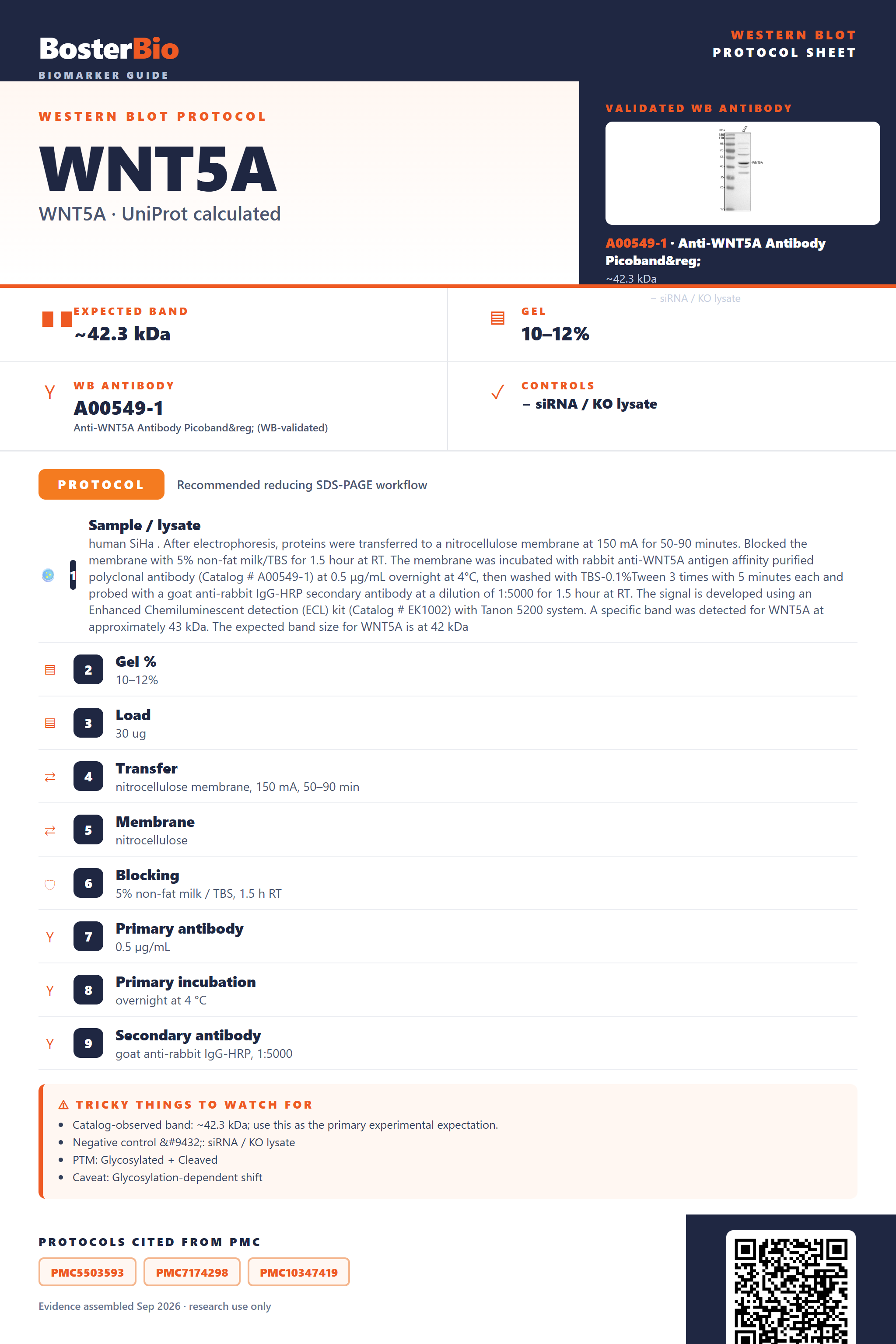
Expected bands, validated protocols, controls and antibodies — the at-a-glance facts below, then the full design guide.
| Expected band | ~42.3 kDa | |
| Observed band | ~43 kDa | |
| Gel | 10–12% | |
| Negative control | siRNA / KO lysate |
| PTM | Glycosylated + Cleaved | |
| Caveat | Glycosylation-dependent shift | |
| Regulation | Epithelial mesenchymal transition | |
| Isoform | 2 isoform(s) |
Literature-validated Western blot parameters for WNT5A — gel percentage, transfer, blocking, antibody incubation and detection, extracted from published methods.
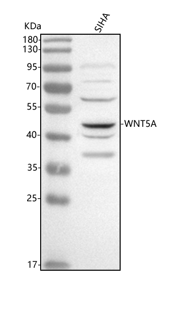
| Sample / lysate | human SiHa . After electrophoresis, proteins were transferred to a nitrocellulose membrane at 150 mA for 50-90 minutes. Blocked the membrane with 5% non-fat milk/TBS for 1.5 hour at RT. The membrane was incubated with rabbit anti-WNT5A antigen affinity purified polyclonal antibody (Catalog # A00549-1) at 0.5 μg/mL overnight at 4°C, then washed with TBS-0.1%Tween 3 times with 5 minutes each and probed with a goat anti-rabbit IgG-HRP secondary antibody at a dilution of 1:5000 for 1.5 hour at RT. The signal is developed using an Enhanced Chemiluminescent detection (ECL) kit (Catalog # EK1002) with Tanon 5200 system. A specific band was detected for WNT5A at approximately 43 kDa. The expected band size for WNT5A is at 42 kDa |
| Gel % | 10–12% |
| Load | 30 ug |
| Transfer | nitrocellulose membrane, 150 mA, 50–90 min |
| Membrane | nitrocellulose |
| Blocking | 5% non-fat milk / TBS, 1.5 h RT |
| Primary antibody | 0.5 µg/mL |
| Primary incubation | overnight at 4 °C |
| Secondary antibody | goat anti-rabbit IgG-HRP, 1:5000 |
| Wash | TBS-0.1% Tween, 3 × 5 min |
| Detection | ECL |
| Exposure / imaging | Tanon 5200 |
| Observed band | 43 kDa |
WNT5A has a 42.3 kDa predicted precursor mass but runs at ~43 kDa on blots, reflecting N-glycosylation at four sites offsetting signal peptide/propeptide cleavage.
| single band near 43 kDa | mature, N-glycosylated Wnt-5a monomer, consistent with the empirical ~43 kDa band reported on real blots |
| band sitting above an unmodified ~35 kDa mature core | N-linked glycans added at the four annotated N-glycosylation sites increase the apparent mass of the cleaved mature chain |
| broad or fuzzy band spanning roughly 40-45 kDa rather than a crisp line | heterogeneous occupancy or processing across the four N-glycosylation sites (Asn114, Asn120, Asn312, Asn326) |
| two distinct bands at different apparent masses | co-detection of splice isoforms 1 and 2, which differ in sequence and are expected to migrate differently |
| little or no band in whole-cell lysate | Wnt-5a is secreted into the extracellular space/matrix, so most of the protein is exported rather than retained intracellularly |
| a smaller band well below the ~43 kDa mature form | an unprocessed precursor-to-mature transition (signal peptide/propeptide, residues 1-61) or degradation of the secreted chain |
| predicted precursor mass (UniProt) | sets the 42.3 kDa baseline for the full-length, unprocessed 380-residue Wnt-5a chain |
| N-glycosylation at Asn114, Asn120, Asn312, Asn326 | adds carbohydrate mass that pushes the mature band up toward the observed ~43 kDa and can broaden it if glycoform occupancy varies |
| signal peptide (1-35) and propeptide (36-61) cleavage | removes about 61 N-terminal residues, which lowers the mature backbone mass before glycosylation adds mass back |
| alternative splicing (isoforms 1 and 2) | isoform 2 is expected to run at a different apparent size than isoform 1, though relative size is only qualitative |
| secreted/extracellular matrix localization | reduces signal in whole-cell lysate since most protein is exported rather than retained inside the cell |
| Situation | Likely cause | Next action |
|---|---|---|
| No band in lysate | Wnt-5a is a secreted, extracellular matrix protein, so cell lysate only captures the small intracellular pool | probe conditioned media or an ECM-enriched fraction instead of, or in addition to, whole-cell lysate |
| Band higher than expected | N-linked glycosylation at up to four sites adds mass above the 42.3 kDa predicted precursor | treat a parallel sample with PNGase F and confirm the band shifts down toward the unmodified mass |
| Broad smear instead of sharp band | heterogeneous glycan occupancy or processing across the four N-glycosylation sites | run a higher-percentage gel with a longer separation, or deglycosylate the sample before loading |
| Multiple bands | co-expression of splice isoforms 1 and 2 from alternative splicing | check which isoform the antibody epitope maps to before interpreting the extra band |
| Band lower than expected | incomplete glycosylation, or use of a non-glycosylated bacterial/recombinant standard that runs lower than native protein | compare against a mammalian-expressed positive control lane |
| Fragments below expected size | an unprocessed signal peptide/propeptide intermediate or proteolytic degradation of the secreted chain during lysate preparation | add protease inhibitors during lysis and check whether the smaller band corresponds to a known processing intermediate |

Comprehensive Human Protein Atlas IHC scoring per tissue. Rows are taken directly from the HPA tissue chart — click any row's HPA link to view the source.
| Tissue | Cell type | Level | Evidence | Source |
|---|
| Tissue | Cell type | Level | Evidence | Source |
|---|
Deeper troubleshooting and optimisation questions for WNT5A, answered from its protein features.
BosterBio's WNT5A antibodies are among the best-performing WB antibodies on the market — well cited, thoroughly validated, and orthogonally cross-validated against negative tissues and complementary methods.
The anti-WNT5A antibodies featured below are best-performing choices for Western blot, extensively cited in the literature and rigorously validated, including orthogonal confirmation against negative tissue controls and complementary detection methods, ensuring specific, reproducible detection of WNT5A protein in your samples.
Which to pick: Both A00549-1 and PB9063 carry real WB validation images, so either is a solid pick. A00549-1 is labeled for WNT5A and PB9063 for Wnt5a; check each product's stated species reactivity to match your sample before choosing.