XDH / Xanthine dehydrogenase/oxidase · IHC design guide

Design Immunohistochemistry for XDH

Plan XDH paraffin IHC around cytoplasmic staining in lactating glandular cells and additional plasma positivity (HPA tissue IHC). Use lactating breast as a positive control (HPA tissue IHC), and start the catalog antibody at 2–5 μg/ml (datasheet A01884-2).

Evidence assembled Oct 2026 · For research use; verify linked source records and product datasheet before use
Immunohistochemistry protocol sheet for XDH (IHC for XDH): expected localisation Cytoplasmic, with plasma positivity in lactating breast (HPA tissue IHC), antibody A01884-2, validated IHC image, and IHC protocol steps
Printable XDH IHC protocol sheet — expected localisation Cytoplasmic, with plasma positivity in lactating breast (HPA tissue IHC), antibody A01884-2, controls and protocol steps. Open the full XDH IHC guide →

XDH Immunohistochemistry Experimental Design Guide

Expected localisation, validated protocols, controls and antibodies — the at-a-glance facts below, then the full design guide.

Must know before staining
Expected localisation Cytoplasmic, with plasma positivity in lactating breast (HPA tissue IHC)
Staining pattern Lactating glandular cells: cytoplasmic; plasma positive (HPA tissue IHC)
Antigen retrieval EDTA pH 8.0 HIER, heat-mediated (datasheet A01884-2)
Positive control ⓘ Lactating breast
Negative control ⓘ Adipose tissue+4 more · see all
Important caveats
Reasons your staining may differ from the expected pattern.
Fixation Keep fixation consistent across sections (standard IHC practice; not target-specific); Matched tissue-IHC evidence does not establish the fixation claim. Validate the specimen-specific method before use. (selected-SKU IHC image A01884-2)
Caveat Plasma positivity may complicate glandular cell scoring (HPA tissue IHC)
Regulation No specific regulator reported (UniProt)
Isoform / epitope No isoforms or cleavage; epitope position is unspecified (UniProt)
Section 1

Recommended XDH IHC & IF Protocols

The catalog antibody’s IHC-P protocol (datasheet: A01884-2) is followed by published protocols for urothelial carcinoma (PMC13309574), chicken liver (PMC10770749), and hepatocellular carcinoma (PMC5623907).

Recommended immunohistochemistry (IHC-P) protocol parameters
SampleParaffin-embedded human colon adenocarcinoma tissue; fixative not specified (datasheet A01884-2)
FixationImage fixative and duration unreported (datasheet A01884-2); verify before use.
Sectioning4–5 µm sections on charged slides (standard)
DeparaffinisationXylene, graded ethanol series to water (standard)
Antigen retrievalHeat retrieval: EDTA pH 8.0 (datasheet A01884-2); 20 min, 95–100 °C (standard)
Peroxidase block3% H2O2, 10 min, room temperature (standard)
Blocking10% goat serum (datasheet A01884-2)
Primary antibodyRabbit anti-XDH, 2-5 μg/ml (datasheet A01884-2)
Primary incubationOvernight at 4 °C (datasheet A01884-2)
DetectionHRP-conjugated secondary, DAB chromogen (datasheet A01884-2)
CounterstainHematoxylin, blue, dehydrate and mount (standard)
Expected resultXDH-positive staining in lactating glandular cells of lactating breast (HPA tissue IHC: Medium). HPA tissue profile: Cytoplasmic expression with additional plasma positivity in lactating breast. No signal in the no-primary control.
💡Decision noteStart with heat-mediated EDTA pH 8.0 retrieval for the catalog antibody (datasheet: A01884-2); the published excerpts do not specify retrieval conditions (PMC13309574; PMC10770749; PMC5623907).
Section 2

What Is the Expected XDH Staining Pattern?

In paraffin-section IHC, expect XDH mainly in the cytoplasm of lactating breast glandular cells, with additional plasma positivity in that tissue (HPA: tissue IHC, Supported). XDH has no transmembrane segment; UniProt also lists peroxisomal and secreted locations (UniProt P47989: topology and subcellular location). HPA reports medium staining in lactating glandular cells, but its reliability note says no tissue staining or RNA data were available for comparison (HPA: tissue IHC).

What am I looking at on my slide?
Cytoplasmic staining in lactating glandular cells, with some plasma positivity.This matches the reported tissue pattern; medium staining is the clearest supplied positive reference (HPA: lactating breast, Medium; tissue IHC profile). Assess the cell-associated signal separately from plasma positivity.
Predominantly nuclear staining in a paraffin section, without the expected cytoplasmic pattern.Treat this as a possible staining artefact and validate it against the lactating breast pattern (HPA: tissue IHC). HPA reports uncertain nucleoplasmic and nucleolar localisation in ICC-IF, so nuclear signal alone cannot settle the IHC interpretation (HPA: subcellular).
Strong staining in adipocytes while adjacent expected-positive glandular cells show little signal.Consider cross-reactivity or endogenous detection activity before calling this XDH: HPA reports adipocytes as not detected in breast and adipose tissue, while lactating glandular cells stain at medium intensity (HPA: tissue IHC).
Even colour across cells, stroma and areas lacking a defined cellular pattern.Interpret this as background until controls establish a specific signal. The reported positive pattern is cytoplasmic in lactating glandular cells, with additional plasma positivity, rather than uniform staining throughout the section (HPA: tissue IHC profile).
No signal in lactating breast glandular cells.A negative result here conflicts with the supplied medium-intensity reference (HPA: lactating breast, Medium). Check the section and detection controls before concluding that XDH is absent; HPA's Supported rating carries its stated comparison-data limitation (HPA: tissue IHC reliability note).
💡Expected XDH appearanceCall a section positive when lactating breast glandular cells show discernible medium cytoplasmic staining, allowing additional plasma positivity; isolated strong adipocyte staining is suspect (HPA: tissue IHC).
How each factor affects the staining
Tissue and cell selectionLactating glandular cells provide the supplied medium-staining reference; duodenal and small-intestinal enterocytes and kidney tubule cells are reported as low, while listed adipocytes are not detected (HPA: tissue IHC). Compare the same cell type when scoring.
Compartment and protein topologyXDH has no transmembrane segment or signal peptide, and UniProt lists cytoplasmic, peroxisomal and secreted locations (UniProt P47989). For paraffin IHC, use the observed cytoplasmic pattern and additional plasma positivity as the tissue reference (HPA: tissue IHC).
Antibody evidenceHPA062641 is Supported for IHC, whereas HPA069323 has no IHC status in the supplied antibody record (HPA: antibodies). An ICC approval does not establish an IHC staining pattern or a paraffin-section working dilution.
IF/ICC Q&A: should nuclear fluorescence define the IHC result?No. HPA marks its nucleoplasmic and additional nucleolar ICC-IF locations uncertain (HPA: subcellular). Interpret IF/ICC on its own guide page; the supplied paraffin IHC tissue reference is cytoplasmic, with additional plasma positivity in lactating breast (HPA: tissue IHC).
Why is my staining missing, weak or wrong?
SituationLikely causeNext action
Lactating glandular cells are blank.The observed result conflicts with the supplied medium-positive tissue reference (HPA: lactating breast). A failed detection step is one general IHC possibility.Confirm section identity, then inspect positive-control and detection-control results. Review retrieval and detection conditions as general IHC workflow checks; no XDH-specific retrieval or fixation effect is supplied.
Only a faint signal appears in intestinal enterocytes or kidney tubules.Low staining is reported for duodenal and small-intestinal enterocytes and kidney tubule cells (HPA: tissue IHC).Score those cells against their low reference level. Use lactating glandular cells, reported as medium, when a clearer positive comparison is needed (HPA: tissue IHC).
Adipocytes stain strongly.HPA lists adipocytes in breast and adipose tissue as not detected (HPA: tissue IHC). Cross-reactivity or endogenous detection activity may explain an unexpected signal.Inspect a no-primary control and the spatial pattern. If chromogenic detection uses an enzyme, check the relevant endogenous-enzyme control as standard IHC practice.
Colour is diffuse across the section.Nonspecific background or a detection-control problem is possible; diffuse staining does not match the cell-associated tissue profile (HPA: tissue IHC).Compare with a no-primary control. Review blocking, antibody concentration and washes as general IHC checks, then rescore only clearly localised staining.
Nuclei dominate the stain.The paraffin IHC pattern is reported as cytoplasmic (HPA: tissue IHC). HPA's nuclear ICC-IF locations are uncertain and come from a different application (HPA: subcellular).Check counterstain and detection controls, then compare cytoplasmic staining in lactating glandular cells before assigning the nuclear signal to XDH (HPA: tissue IHC).
Two antibodies give different tissue patterns.The supplied validation statuses differ: HPA062641 is IHC Supported; HPA069323 has no IHC status listed (HPA: antibodies).Keep antibody-specific results separate and compare each with the reported cell and compartment pattern. Do not treat ICC approval as paraffin IHC validation (HPA: antibodies; tissue IHC).

Sample controls for XDH IHC & IF

🧪Run lactating breast first and score lactating glandular cells for XDH staining (HPA: Medium in lactating glandular cells). Use adipose tissue adipocytes as the negative tissue (HPA: Not detected in adipocytes); on the positive slide, compare staining with other cells, whose XDH status is not established by the supplied HPA rows.
Positive control tissue: Lactating breast (Lactating glandular cells, HPA Medium)
Negative control tissue: Adipose tissue (HPA Not detected)
ICC-IF cell lines (HPA subcellular resource): HPA ICC-IF images show XDH in BJ [Human fibroblast], U-251MG, U2OS, with annotated localisation: Nucleoplasm (uncertain) (HPA subcellular).
Technical controls: Include a no-primary, secondary-only control and a concentration-matched rabbit IgG isotype control (selected-SKU caption: rabbit anti-XDH primary). Use a matched XDH knockout specimen or validated antigen competition as a biological specificity control; block endogenous peroxidase for chromogenic IHC and assess tissue autofluorescence if using IF (standard IHC/IF practice).
⚠️Feasibility: No target-specific fixation window or fixation effect is reported in the supplied evidence, and the fixative is unreported in the selected-SKU tissue-IHC caption. The caption uses heat-mediated EDTA retrieval at pH 8.0 on a paraffin section, but does not establish that retrieval is required or that frozen sections or IF are easier (selected-SKU tissue-IHC caption). XDH is detected in milk, so luminal material in lactating breast could complicate assignment of staining to glandular cells (UniProt P47989: detected in milk; HPA: Medium in lactating glandular cells).

HPA tissue IHC evidence for XDH

Comprehensive Human Protein Atlas IHC scoring per tissue (reliability: Supported — No tissue staining or RNA expression data available for comparison.). Rows are taken directly from the HPA tissue chart — click any row's HPA link to view the source.

Positive expression · recommended positive controls

TissueCell typeLevelEvidenceSource
Lactating breast Lactating glandular cells Medium Protein (IHC) HPA →

Undetected expression · recommended negative controls

TissueCell typeLevelEvidenceSource
Adipose tissue Adipocytes Not detected Protein (IHC) HPA →
Adrenal gland Glandular cells Not detected Protein (IHC) HPA →
Appendix Endocrine cells Not detected Protein (IHC) HPA →
Bone marrow Hematopoietic cells Not detected Protein (IHC) HPA →
Breast Adipocytes Not detected Protein (IHC) HPA →
Section 3

Advanced XDH IHC Tips

Troubleshoot XDH staining in paraffin sections by checking retrieval, cellular distribution, controls and scoring against the supplied tissue evidence.

How should I optimize retrieval when XDH staining is weak in paraffin sections?
Begin with heat-mediated EDTA at pH 8.0 for paraffin sections (datasheet A01884-2). The selected image used this retrieval before 2 μg/mL primary antibody overnight at 4°C, so retain those conditions when diagnosing weak signal (caption A01884-2). Compare an adjacent section processed identically without primary antibody, and inspect morphology after heating because excessive retrieval can damage tissue and create misleading edge staining (general IHC practice). If staining remains weak, titrate heating duration on serial sections, keeping EDTA and pH 8.0 fixed before trying another retrieval buffer as a fallback (general IHC practice).
Could fixation explain inconsistent XDH staining between paraffin blocks?
Target-specific fixation sensitivity is unknown because the selected paraffin-section caption does not report a fixative (caption A01884-2). Record fixative, fixation interval, specimen thickness and processing delay for each block, then compare matched sections under identical retrieval and detection conditions (general IHC practice). When optimizing new material, use a consistent laboratory fixation workflow and inspect both tissue morphology and cytoplasmic signal before adjusting antibody concentration (general IHC practice; HPA tissue IHC). Check apparent signal loss against a concurrently processed positive control and a no-primary control, since changes in antigen accessibility or background can mimic target loss (general IHC practice).
Which XDH staining compartment should I expect in tissue sections?
For chromogenic tissue sections, expect mainly cytoplasmic staining, with additional plasma positivity reported in lactating breast (HPA tissue IHC). XDH is annotated in cytoplasm, peroxisomes and secreted material, with no transmembrane segment, so a crisp, continuous membrane rim needs independent confirmation (UniProt P47989 subcellular; UniProt P47989 topology). Use glandular-cell signal in lactating breast as a reference and compare nearby adipocytes, which were not detected, on a section processed in the same run (HPA tissue IHC). Do not score a nuclear pattern as established tissue localisation solely because HPA ICC/IF lists nucleoplasm and nucleoli: both locations are labeled uncertain (HPA subcellular).
Can different XDH patterns reflect isoforms or epitope masking?
The record lists 0 isoforms and one 1–1333 chain; avoid assigning differing IHC patterns to alternate XDH isoforms (UniProt P47989 isoforms; UniProt P47989 processing). The A01884-2 epitope is unspecified, so its accessibility and cross-reactivity cannot be predicted from the supplied evidence (caption A01884-2). Compare serial sections with and without primary antibody, then titrate around 2 μg/mL under the same retrieval conditions before attributing pattern changes to biology (caption A01884-2; general IHC practice). The record lists 0 glycosylation sites and 0 modified residues, providing no basis for a modification-specific explanation of staining differences (UniProt P47989 PTMs).
How can IF help resolve ambiguous XDH cell localisation?
For an IF follow-up to chromogenic IHC, multiplex XDH with a glandular epithelial marker in lactating breast to test whether signal belongs to the expected cells (HPA tissue IHC; general IF practice). Choose spectrally separated fluorophores and inspect unstained tissue in each channel before assigning dim XDH fluorescence, since tissue autofluorescence can imitate a positive signal (general IF practice). Because XDH has no transmembrane segment and is annotated in cytoplasm and peroxisomes, permeabilize as needed to expose an intracellular epitope and titrate detergent gently (UniProt P47989 topology; UniProt P47989 subcellular; general IF practice). Confirm channel separation with single-stain controls (general IF practice).
What should I check when XDH DAB staining looks diffuse?
Compare a no-primary section with the full stain, and check whether brown signal follows tissue edges or damaged areas (general IHC practice). Apply an endogenous peroxidase block before HRP detection, because endogenous activity can produce DAB signal without bound anti-XDH antibody (general chromogenic IHC practice). The selected image used 10% goat serum blocking, 2 μg/mL primary overnight at 4°C, and HRP/DAB detection; use these conditions as a reference while changing one variable at a time (caption A01884-2). Check a secondary-only section and rinse thoroughly before DAB development to assess nonspecific secondary binding (general IHC practice).
How should I quantify XDH-positive cells across tissue sections? ⚠ ANSWER MARKED FOR VERIFICATION
Score cytoplasmic DAB in defined cell populations, recording percentage positive and intensity 0–3 for each specimen (HPA tissue IHC; general IHC scoring practice). An H-score from 0–300 sums each intensity level multiplied by its percentage of scored cells; report the threshold and number of cells or fields assessed (general IHC scoring practice). Normalize positive counts to evaluable cells of the same class, or report stained-cell density per mm² of viable tissue when architecture varies (general IHC practice). Exclude necrosis, folds and section edges consistently, and compare matched processing batches because handling and detection affect intensity (general IHC practice).
How can I distinguish true XDH staining from artefact?
A plausible tissue pattern is mainly cytoplasmic in lactating glandular cells, with nearby adipocytes negative (HPA tissue IHC). XDH is also annotated in peroxisomes and secreted material; extracellular staining needs context, while a sharp membrane-only or dominant nuclear pattern needs confirmation (UniProt P47989 subcellular; UniProt P47989 topology; HPA subcellular). Inspect edges, folds, necrosis and no-primary controls before interpreting brown precipitate, and block endogenous peroxidase during HRP/DAB staining (general IHC practice). The selected colon adenocarcinoma image shows DAB after EDTA pH 8.0 retrieval; that image alone does not establish specificity across tissues (caption A01884-2; general IHC interpretation).
Boster reagents

Best XDH / Xanthine dehydrogenase/oxidase IHC Antibodies

Real paraffin-section IHC images cover human colon, liver cancer and thyroid papillary carcinoma (A01884-2 captions), and mouse and rat intestine (A01884 and M01884-2 captions). A01884 lists ICC, but has no IF image (A01884 catalog).

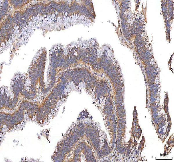
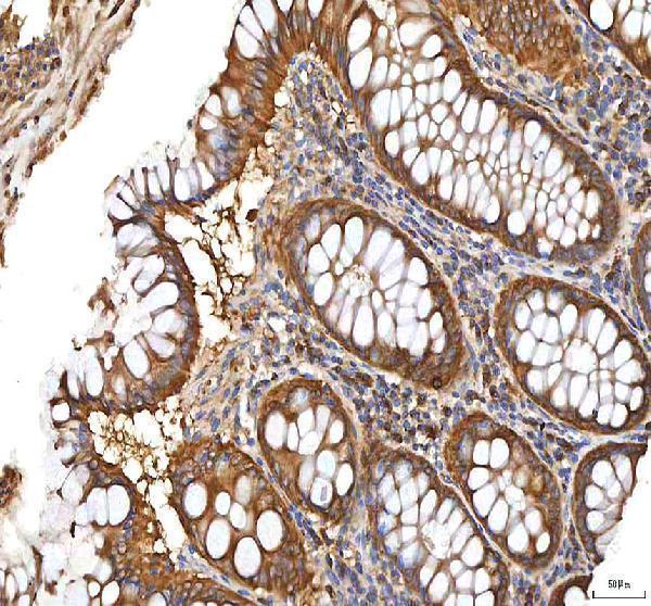
Real IHC data IHC analysis of XDH using anti-XDH antibody (A01884-2). XDH was detected in a paraffin-embedded section of human colon adenocarcinoma tissue. Heat mediated antigen retrieval was performed in EDTA buffer (pH 8.0, epitope retrieval solution). The tissue section was blocked with 10% goat serum. The tissue section was then incubated with 2 μg/ml rabbit anti-XDH Antibody (A01884-2) overnight at 4°C. Peroxidase Conjugated Goat Anti-rabbit IgG was used as secondary antibody and incubated for 30 minutes at 37°C. The tissue section was developed using HRP Conjugated Rabbit IgG Super Vision Assay Kit (Catalog # SV0002) with DAB as the chromogen.
Anti-XDH Antibody ®
Cat # A01884-2
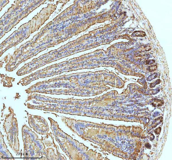
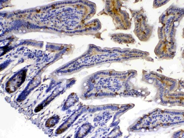
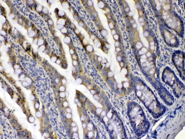
Real IHC data IHC analysis of Xanthine Oxidase using anti-Xanthine Oxidase antibody (M01884-2). Xanthine Oxidase was detected in a paraffin-embedded section of mouse small intestine tissue. Heat mediated antigen retrieval was performed in EDTA buffer (pH 8.0, epitope retrieval solution). The tissue section was blocked with 10% goat serum. The tissue section was then incubated with 1:50 rabbit anti-Xanthine Oxidase Antibody (M01884-2) overnight at 4°C. Peroxidase Conjugated Goat Anti-rabbit IgG was used as secondary antibody and incubated for 30 minutes at 37°C. The tissue section was developed using HRP Conjugated Rabbit IgG Super Vision Assay Kit (Catalog # SV0002) with DAB as the chromogen.
Anti-Xanthine Oxidase Rabbit Monoclonal Antibody
Cat # M01884-2
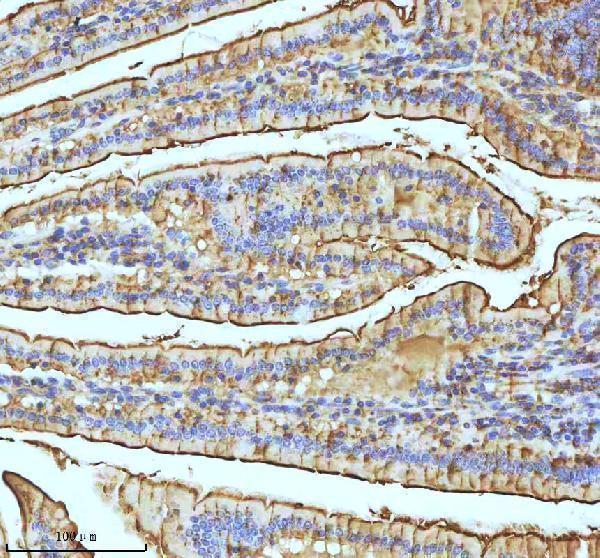
Real IHC data IHC analysis of Xanthine Oxidase using anti-Xanthine Oxidase antibody (A01884). Xanthine Oxidase was detected in paraffin-embedded section of mouse intestine tissue . Heat mediated antigen retrieval was performed in citrate buffer (pH6, epitope retrieval solution) for 20 mins. The tissue section was blocked with 10% goat serum. The tissue section was then incubated with 1μg/ml rabbit anti-Xanthine Oxidase Antibody (A01884) overnight at 4°C. Biotinylated goat anti-rabbit IgG was used as secondary antibody and incubated for 30 minutes at 37°C. The tissue section was developed using Strepavidin-Biotin-Complex (SABC)(Catalog # SA1022) with DAB as the chromogen.
Anti-Xanthine Oxidase/XDH Antibody ®
Cat # A01884

A01884-2 shows paraffin-section IHC in human colon, colon adenocarcinoma, liver cancer and thyroid papillary carcinoma (A01884-2 captions); M01884-2 shows mouse and rat small intestine (M01884-2 captions). A01884 shows mouse and rat intestine in paraffin sections (A01884 captions) and lists IHC-F and ICC applications (A01884 catalog).

Which to pick: For human tissue IHC, start with A01884-2 at 2–5 μg/ml (A01884-2 catalog); its human paraffin-section examples use EDTA retrieval at pH 8.0 (A01884-2 captions). For IF/ICC, choose A01884 because it lists ICC and IHC-F at 0.5–1 μg/ml (A01884 catalog); its paraffin-section IHC example uses citrate retrieval at pH 6 (A01884 caption). For mouse and rat IHC, A01884 has images in both species (A01884 captions), while M01884-2 offers rabbit monoclonal clone 19X78 (M01884-2 catalog) with EDTA retrieval at pH 8.0 in both (M01884-2 captions); fixation is unreported in these captions (A01884-2, A01884 and M01884-2 captions).

Each figure is that product's own IHC / IF validation image from its datasheet.

References

  1. UniProt Consortium. UniProt entry P47989 (XDH_HUMAN, Xanthine dehydrogenase/oxidase).
  2. Human Protein Atlas. XDH tissue IHC expression (reliability: Supported).
  3. Human Protein Atlas. XDH subcellular location (ICC-IF): Mainly localized to the nucleoplasm. In addition localized to the nucleoli..
  4. Human Protein Atlas. XDH antibody validation summary (2 antibodies).
  5. Xanthine dehydrogenase rewires metabolism and the survival of nutrient deprived lung adenocarcinoma cells by facilitating UPR and autophagic degradation. International journal of biological sciences 2023 — PMC9909990.
  6. Exploring XDH expression as a prognostic marker for urothelial carcinoma. Discover oncology 2026 — PMC13309574.
  7. Screening of heat stress-related biomarkers in chicken serum through label-free quantitative proteomics. Poultry science 2024 — PMC10770749.
  8. Xanthine dehydrogenase downregulation promotes TGFβ signaling and cancer stem cell-related gene expression in hepatocellular carcinoma. Oncogenesis 2017 — PMC5623907.
  9. PubMed PMID:8224915 — UniProt-cited evidence.
  10. PubMed PMID:8135849 — UniProt-cited evidence.
  11. PubMed PMID:7575623 — UniProt-cited evidence.