This website uses cookies to ensure you get the best experience on our website.


- Table of Contents
Plan chromogenic XRCC4 IHC in paraffin sections around a broad nuclear staining pattern (HPA tissue IHC). Start the IHC-validated antibody A00787-2 at 2–5 μg/ml (datasheet A00787-2), and score nuclear staining with matched controls.

Expected localisation, validated protocols, controls and antibodies — the at-a-glance facts below, then the full design guide.
| Expected localisation | Nuclear staining in tissue IHC (HPA tissue IHC) | |
| Staining pattern | Broad nuclear staining across tissue cell types (HPA tissue IHC) | |
| Antigen retrieval | EDTA pH 8.0 HIER, heat-mediated (datasheet A00787-2) | |
| Positive control | Adipose tissue+4 more · see all | |
| Negative control | Liver+2 more · see all |
| Fixation | Keep fixation consistent across sections. (standard IHC practice; not target-specific) | |
| Caveat | Cholangiocytes may lack detectable staining (HPA tissue IHC) | |
| Regulation | Widely expressed (UniProt) | |
| Isoform / epitope | 3 isoforms; epitope coverage is unspecified (UniProt) |
Compare the catalog antibody’s IHC-P protocol (datasheet A00787-2) with three published XRCC4 tissue IHC methods (PMC5675685; PMC8425215; PMC7661505).
| Sample | Paraffin-embedded human lung squamous cell carcinoma tissue; fixative not specified (datasheet A00787-2) |
| Fixation | Image fixative and duration unreported (datasheet A00787-2); verify before use. |
| Sectioning | 4–5 µm sections on charged slides (standard) |
| Deparaffinisation | Xylene, graded ethanol series to water (standard) |
| Antigen retrieval | Heat retrieval: EDTA pH 8.0 (datasheet A00787-2); 20 min, 95–100 °C (standard) |
| Peroxidase block | 3% H2O2, 10 min, room temperature (standard) |
| Blocking | 10% goat serum (datasheet A00787-2) |
| Primary antibody | Rabbit anti-XRCC4, 2-5 μg/ml (datasheet A00787-2) |
| Primary incubation | Overnight at 4 °C (datasheet A00787-2) |
| Detection | HRP-conjugated secondary, DAB chromogen (datasheet A00787-2) |
| Counterstain | Hematoxylin, blue, dehydrate and mount (standard) |
| Expected result | XRCC4-positive staining in adipocytes of adipose tissue (HPA tissue IHC: High). HPA tissue profile: Ubiquitous nuclear expression. No signal in the no-primary control. |
XRCC4 should appear mainly in nuclei across many tissues (HPA tissue IHC: ubiquitous nuclear expression; Supported reliability). UniProt also lists chromosome and cytoplasm localization, including recruitment to double-strand breaks (UniProt Q13426: subcellular location). It has no transmembrane segment (UniProt Q13426: topology). Expect staining in documented positive cells, including bone marrow hematopoietic cells and breast glandular cells (HPA tissue IHC: High).
| Nuclear staining in many cell types, with clear signal in bone marrow hematopoietic cells. | This fits the reported ubiquitous nuclear pattern and a documented High cell population (HPA tissue IHC: profile; bone marrow, High). Score the relevant cells and nuclear compartment together; a positive-looking section alone does not establish antibody specificity. |
| Predominantly cytoplasmic, membranous, or extracellular staining with little nuclear signal. | The dominant tissue pattern should be nuclear (HPA tissue IHC: ubiquitous nuclear expression). UniProt lists cytoplasm as an additional location, so limited cytoplasmic signal is not automatically false (UniProt Q13426: subcellular location). Strong membrane or extracellular signal lacks support from the supplied localization and topology record (UniProt Q13426: no transmembrane segment). |
| Strong signal in liver cholangiocytes, ovarian stroma, or smooth muscle cells. | These populations were reported as Not detected in the supplied tissue observations (HPA tissue IHC: liver; ovary; smooth muscle). Check cell identification and controls, then consider cross-reactivity or endogenous chromogenic activity. A discrepancy with HPA is a reason to investigate, not proof that an individual cell cannot express XRCC4. |
| Broad chromogen haze covers nuclei, cytoplasm, and surrounding tissue. | A haze that obscures cell boundaries cannot be scored as the reported nuclear pattern (HPA tissue IHC: profile). In chromogenic IHC, nonspecific antibody binding, insufficient blocking or washing, and endogenous detection activity can produce background (general IHC practice); compare with a no-primary control. |
| No nuclear signal in bone marrow hematopoietic cells or breast glandular cells. | Both are listed as High in the supplied tissue observations (HPA tissue IHC: bone marrow; breast). First check that the expected cells are present and the staining run worked. HPA's overall tissue reliability is Supported with medium consistency against RNA, so a single negative section requires controls and repeat assessment (HPA tissue IHC: reliability). |
| How strong is the tissue-pattern evidence? | HPA calls the tissue IHC result Supported and describes medium consistency between staining and RNA expression (HPA tissue IHC: reliability). Its positive and Not detected entries are observations for named cell populations, not universal intensity cutoffs; score each compartment and cell type against the relevant entry. |
| Which antibody evidence applies to IHC? | HPA006801 is listed as IHC Supported; HPA051538 has no IHC validation entry in the supplied antibody record (HPA antibodies: IHC status). Both have ICC Enhanced entries (HPA antibodies: ICC status). Do not transfer an ICC validation label to an IHC result. |
| Could isoforms or modifications affect interpretation? | UniProt lists three XRCC4 isoforms and several modified residues, including phosphorylation sites (UniProt Q13426: isoforms; modified residues). The supplied record gives no antibody epitope or variant-specific staining data, so an unusual tissue pattern cannot be assigned to an isoform or modification from these sources alone. |
| Is a processed or shed extracellular pool expected? | The listed full-length chain spans residues 1–336, with a C-terminal 266–336 entry; no signal peptide, propeptide, glycosylation sites, or transmembrane segment are listed (UniProt Q13426: processing; glycosylation; topology). These annotations do not support interpreting extracellular chromogen as a normal shed XRCC4 pattern. |
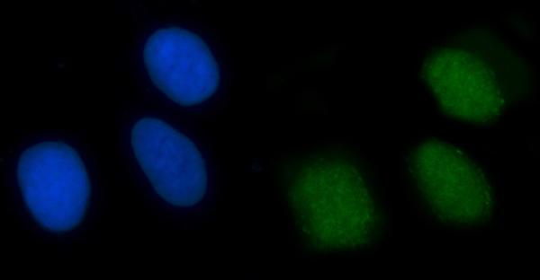
| IF/ICC Q&A: where should signal appear? | In the separate IF/ICC application, HPA reports enhanced nucleoplasmic localization and images from A-431, U-251MG, and U2OS cells (HPA subcellular: location; imaged cell lines). That supports a nucleoplasmic IF expectation; it does not supply an IHC staining protocol or change the tissue IHC reliability rating. |
| Situation | Likely cause | Next action |
|---|---|---|
| A documented High population is unstained while the section appears otherwise intact. | Target-specific fixation effects are not established by the supplied assay evidence. Verify with a matched IHC source before attributing a result to fixation. | Confirm cell identity and a functioning positive control, then review retrieval, primary incubation, detection, and counterstain using the IHC-validated antibody's established IHC-P instructions (general IHC practice). |
| Signal is mostly cytoplasmic with weak nuclei. | Some cytoplasmic localization is annotated, but ubiquitous nuclear staining is the observed tissue pattern (UniProt Q13426: subcellular location; HPA tissue IHC: profile). The observed balance may reflect background or a genuine difference that needs verification. | Compare nuclear and cytoplasmic signal in documented High cells and inspect a no-primary control; repeat with an IHC-supported reagent if the compartment pattern remains discordant (HPA antibodies: HPA006801 IHC Supported; general IHC practice). |
| Membranes or extracellular material stain prominently. | That distribution lacks support from the nuclear tissue profile and the absence of a transmembrane segment (HPA tissue IHC: profile; UniProt Q13426: topology). Nonspecific binding or detection background is possible (general IHC practice). | Check a no-primary control, blocking, and washing; score only cellular signal with a defensible compartment assignment (general IHC practice). |
| Cells listed as Not detected are strongly positive. | The result differs from HPA observations for liver cholangiocytes, ovarian stroma, or smooth muscle cells (HPA tissue IHC: Not detected entries). Misidentified cells, cross-reactivity, or endogenous activity are possible (general IHC practice). | Verify cell identity and compare nearby documented positive cells, a no-primary control, and repeat sections before treating the discordance as biological (HPA tissue IHC: profile; general IHC practice). |
| The entire section has diffuse chromogen background. | Nonlocalized color can result from nonspecific binding, incomplete washing, or endogenous detection activity (general IHC practice). It cannot establish the nuclear XRCC4 pattern reported by HPA (HPA tissue IHC: profile). | Inspect the no-primary control, improve blocking and washes as appropriate to the detection system, and adjust detection exposure under the established IHC workflow (general IHC practice). |
| IF/ICC appears nucleoplasmic, but matched tissue IHC is inconclusive. | The applications have different HPA evidence: enhanced nucleoplasmic ICC localization versus Supported tissue IHC with medium RNA consistency (HPA subcellular: main location; HPA tissue IHC: reliability). Agreement in IF does not validate the tissue section. | Evaluate the IHC section using its own positive cell populations and controls; use the separate IF/ICC guide for fluorescence workflow details (HPA tissue IHC: High populations; general IHC practice). |
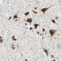

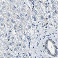


Comprehensive Human Protein Atlas IHC scoring per tissue (reliability: Supported — Medium consistency between antibody staining and RNA expression data.). Rows are taken directly from the HPA tissue chart — click any row's HPA link to view the source.
| Tissue | Cell type | Level | Evidence | Source |
|---|---|---|---|---|
| Adipose tissue | Adipocytes | High | Protein (IHC) | HPA → |
| Adrenal gland | Glandular cells | High | Protein (IHC) | HPA → |
| Appendix | Glandular cells | High | Protein (IHC) | HPA → |
| Bone marrow | Hematopoietic cells | High | Protein (IHC) | HPA → |
| Breast | Glandular cells | High | Protein (IHC) | HPA → |
Troubleshoot XRCC4 staining in paraffin section IHC by checking retrieval, nuclear localisation, controls and scoring before interpreting biological differences.
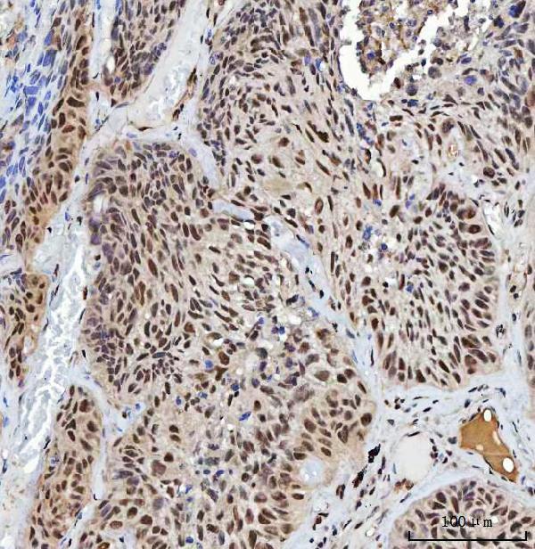
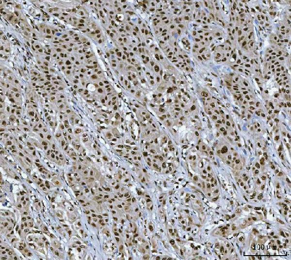
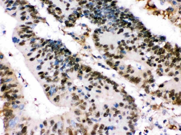
The human-reactive XRCC4 catalog antibodies have IHC images from paraffin-embedded cancer sections (catalog IHC captions); A00787-2 also has IF images from U20S cells and a paraffin-embedded tissue section (A00787-2 IF captions).
A00787-2 shows IHC in human lung squamous cell, breast duct, colorectal and liver cancer sections, plus IF in U20S cells and a human colorectal adenocarcinoma section (A00787-2 image captions). PB9908 shows IHC in a human intestinal cancer section (PB9908 IHC caption).
Which to pick: For paraffin-section IHC, both have chromogenic examples: A00787-2 uses 2 μg/ml with EDTA pH 8.0 retrieval and DAB in human lung squamous cell carcinoma (A00787-2 IHC caption); PB9908 uses 1 μg/ml with the same retrieval buffer and chromogen in human intestinal cancer (PB9908 IHC caption). For IF/ICC, choose A00787-2: both applications are listed and IF images are provided (A00787-2 applications and IF captions); PB9908 lists neither application (PB9908 applications). Neither has documented cross-species reactivity here because both list Human only (catalog reactivity); clonality is unreported (catalog entries), as is the fixative used for either pictured IHC section (IHC captions).