Click image to see more details
Product Info Summary
| SKU: | DZ41260 |
|---|---|
| Size: | 200 μl/vial |
| Reactive Species: | Zebrafish |
| Host: | Rabbit |
| Application: | IHC |
Customers Who Bought This Also Bought

Product info
Product Name
Anti-Zebrafish col12a1a Antibody
SKU/Catalog Number
DZ41260
Size
200 μl/vial
Form
Liquid
Description
Boster Bio Anti-col12a1a Antibody catalog # DZ41260. This antibody reacts with Zebrafish.
Storage & Handling
At -20°C for one year, at 4°C for one month. Avoid repeated freezing and thawing.
Cite This Product
Anti-Zebrafish col12a1a Antibody (Boster Biological Technology, Pleasanton CA, USA, Catalog # DZ41260)
Host
Rabbit
Contents
Each vial contains 20mM PBS, 50% glycerol, 0.02% NaN3.
Clonality
Polyclonal
Isotype
Rabbit IgG
Reactive Species
DZ41260 is reactive to col12a1a in Zebrafish
Application & Images
Applications
DZ41260 is guaranteed for IHC Boster Guarantee
Recommend Dilution
| Application | Dilution | Species |
|---|---|---|
| Immunohistochemistry(Paraffin-embedded Section) | 2-5μg/ml |
Tested application
Use TE buffer pH 9.0 for antigen retrieval; (*) citrate buffer pH 6.0 is an alternative.
Validation Images & Assay Conditions
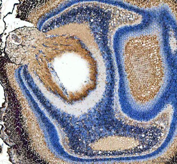
Click image to see more details
IHC analysis of Col12a1a using anti-Col12a1a antibody (DZ41260).
Col12a1a was detected in a paraffin-embedded section of zebrafish eye tissue. Heat mediated antigen retrieval was performed in EDTA buffer (pH 8.0, epitope retrieval solution). The tissue section was blocked with 10% goat serum. The tissue section was then incubated with 2 μg/ml rabbit Col12a1a Antibody (DZ41260) overnight at 4°C. Peroxidase Conjugated Goat Anti-rabbit IgG was used as secondary antibody and incubated for 30 minutes at 37°C. The tissue section was developed using HRP Conjugated Rabbit IgG Super Vision Assay Kit (Catalog # SV0002) with DAB as the chromogen.

Click image to see more details
Mmp14b is a regulator of CM protrusion. A Collagen hybridizing peptide (CHP) and phalloidin staining of a wild-type ventricle at 10 dpci. Yellow box denotes the area in the zoomed image. B In situ hybridization of mmp14b expression in a wild-type ventricle at 7 dpci. Black dashed line denotes the approximate injury border. C Colocalization of mmp14b (HCR, magenta) with CMs (MHC immunostaining), endocardial cells (vwf HCR), macrophages (GFP immunostaining in Tg(mpeg1:EGFP) ventricles, and fibroblasts ( col12a1a (HCR, green) and postnb (HCR, cyan) in the cortical BZ region at 10 dpci. Yellow box in the schematic of the heart marks the cortical CM region depicted in the zoomed images. Created in BioRender. Beisaw, A. (2025) . D Schematic depicting the exon structure of the mmp14b locus and CRISPR/Cas9-induced full-length deletion between exons 2 and 9 of mmp14b . Created in BioRender. Beisaw, A. (2025) . E Mmp14b wild-type and putative mutant protein domain structure (left). RT-PCR of the mmp14b open reading frame from wild-type and mmp14b Δ/Δ mutant embryos (right). SP signal peptide, Pro propeptide, Cat catalytic domain, H hinge region, TM transmembrane domain, C C-terminal tail. F RT-qPCR of mmp14b and mmp14a expression in single ventricles from mmp14b Δ/Δ ( n = 5 ventricles) and wild-type siblings ( n = 5 ventricles) at 10 dpci. Data are presented as mean ± SD. P -values were calculated using an unpaired two-sided t -test. Source data are presented in the Source Data file. G Phalloidin staining of thick cryosections from mmp14b Δ/Δ and wild-type sibling ventricles at 10 dpci (left). Quantification of CM protrusion length (right, mmp14b Δ/Δ n = 1124 CM protrusions from 8 ventricles, wild-type sibling n = 1480 CM protrusions from 9 ventricles). Data are presented as violin plots of all points with solid gray lines indicating the median and dotted gray lines indicating 25th and 75th percentile. P -values were calculated using a two-sided Mann–Whitney test. Source data are presented in the Source Data file. H Picrosirius red staining of collagen in mmp14b Δ/Δ ( n = 8 ventricles) and wild-type sibling ( n = 10 ventricles) at 60 dpci (left). Quantification of scar area (% of ventricle area) on the right. Data are presented as mean ± SD. P -value was calculated using an unpaired two-sided t -test. Source data are presented in the Source Data file. I Quantification of CM proliferation within 100 μm of the wound border from PCNA/Mef2 immunostaining in mmp14b Δ/Δ ( n = 3 ventricles) and wild-type sibling ( n = 4 ventricles) at 7 dpci. Data are presented as mean ± SD. P -value was calculated using an unpaired two-sided t -test. Source data are presented in the Source Data file. Scale bars: 100 μm in ( A , B , G , and H ), 20 μm in zoomed image in ( A and C ).
Index in PubMed under a CC BY license. PMID: 40268967

Click image to see more details
Mmp14b is essential for macrophage presence and ECM remodeling at the border zone. A Collagen hybridizing peptide (CHP) and mScarlet immunostaining in Tg(mpeg1:EGFP); Tg(myl7:lck-mScarlet) ventricles from mmp14b Δ/Δ and wild-type siblings at 10 dpci. White dashed lines indicate the wound border and yellow boxes contain zoomed images from the wound border zone. B Quantification of CHP intensity at the border zone (arb. units, arbitrary units) in Tg(mpeg1:EGFP); Tg(myl7:lck-mScarlet) ventricles from mmp14b Δ/Δ ( n = 5 ventricles) and wild-type siblings ( n = 4 ventricles) at 10 dpci. Data are presented as mean ± SD. P -value was calculated using an unpaired two-sided t -test. Source data are presented in the Source Data file. C GFP and mScarlet immunostaining in Tg(mpeg1:EGFP); Tg(myl7:lck-mScarlet) ventricles from mmp14b Δ/Δ and wild-type siblings at 10 dpci. White dashed lines indicate the approximate wound border. D Quantification of mpeg1 :EGFP+ cells 50 μm proximal and distal to the wound border in ventricles from mmp14b Δ/Δ ( n = 6 ventricles) and wild-type siblings (7 dpci n = 7 ventricles, 10 dpci n = 6 ventricles) at 7 and 10 dpci. Data are presented as mean ± SD. P -values were calculated using unpaired two-sided t -tests and corrected for multiple comparisons using the Holm-Sidak method. Source data are presented in the Source Data file. E RT-qPCR analysis of fibroblast marker genes in mmp14b Δ/Δ mutant ( n = 5 ventricles) and wild-type sibling ( n = 5 ventricles) at 10 dpci. Data are presented as mean ± SD. P -values were calculated using an unpaired two-sided t -test or a two-sided Mann–Whitney test ( col1a1a ). Source data are presented in the Source Data file. F col12a1a in situ hybridization chain reaction (HCR) in mmp14b Δ/Δ mutant and wild-type sibling ventricles at 10 dpci. Yellow boxes denote zoomed images at the cortical BZ, blue boxes denote zoomed images at the apex of the wound. Epi, epicardium. Scale bars: 100 μm, 20 μm in the zoomed images in ( F ).
Index in PubMed under a CC BY license. PMID: 40268967

Click image to see more details
IHC analysis of Col12a1a using anti-Col12a1a antibody (DZ41260).
Col12a1a was detected in a paraffin-embedded section of zebrafish gill tissue. Heat mediated antigen retrieval was performed in EDTA buffer (pH 8.0, epitope retrieval solution). The tissue section was blocked with 10% goat serum. The tissue section was then incubated with 2 μg/ml rabbit Col12a1a Antibody (DZ41260) overnight at 4°C. Peroxidase Conjugated Goat Anti-rabbit IgG was used as secondary antibody and incubated for 30 minutes at 37°C. The tissue section was developed using HRP Conjugated Rabbit IgG Super Vision Assay Kit (Catalog # SV0002) with DAB as the chromogen.
Specific Publications For Anti-Zebrafish col12a1a Antibody (DZ41260)
Loading publications
Recommended Resources
Here are featured tools and databases that you might find useful.
- Boster's Pathways Library
- Protein Databases
- Bioscience Research Protocol Resources
- Data Processing & Analysis Software
- Photo Editing Software
- Scientific Literature Resources
- Research Paper Management Tools
- Molecular Biology Software
- Primer Design Tools
- Bioinformatics Tools
- Phylogenetic Tree Analysis
Customer Reviews
Have you used Anti-Zebrafish col12a1a Antibody?
Share your experimental results or join a short interview to earn up to $1,000 in product credits or other rewards.
0 Reviews For Anti-Zebrafish col12a1a Antibody
Customer Q&As
Have a question?
Find answers in Q&As, reviews.
Can't find your answer?
Submit your question
