Click image to see more details
Product Info Summary
| SKU: | AZA0A8M6YTF7 |
|---|---|
| Size: | 100 μg/vial |
| Reactive Species: | Zebrafish |
| Host: | Rabbit |
| Application: | IF, IHC |
Customers Who Bought This Also Bought

Product info
Product Name
Anti-Zebrafish IGF1RA Antibody
SKU/Catalog Number
AZA0A8M6YTF7
Size
100 μg/vial
Form
Lyophilized
Description
Boster Bio Anti-Zebrafish IGF1RA Antibody catalog # AZA0A8M6YTF7. Tested in IHC, IF applications. This antibody reacts with Zebrafish.
Storage & Handling
At -20°C for one year from date of receipt. After reconstitution, at 4°C for one month. It can also be aliquotted and stored frozen at -20°C for six months. Avoid repeated freezing and thawing.
Cite This Product
Anti-Zebrafish IGF1RA Antibody (Boster Biological Technology, Pleasanton CA, USA, Catalog # AZA0A8M6YTF7)
Host
Rabbit
Contents
Each vial contains 4 mg Trehalose, 0.9 mg NaCl, 0.2 mg Na2HPO4.
Clonality
Polyclonal
Immunogen
E.coli-derived Zebrafish IGF1RA recombinant protein (Position: H36-R886).
Reactive Species
AZA0A8M6YTF7 is reactive to IGF1RA in Zebrafish
Background of IGF1RA
Enables cysteine-type endopeptidase inhibitor activity involved in apoptotic process and insulin-like growth factor binding activity. Acts upstream of or within several processes, including embryonic organ development; fin regeneration; and insulin-like growth factor receptor signaling pathway. Predicted to be located in membrane. Predicted to be part of insulin receptor complex. Predicted to be active in axon and plasma membrane. Is expressed in several structures, including digestive system; female organism; male organism; musculature system; and reproductive system. Human ortholog(s) of this gene implicated in several diseases, including IgA glomerulonephritis; dementia (multiple); kidney cancer (multiple); liver disease (multiple); and neurodegenerative disease (multiple). Orthologous to human IGF1R (insulin like growth factor 1 receptor).
Antibody Validation
Boster validates all antibodies on WB, IHC, ICC, Immunofluorescence, and ELISA with known positive control and negative samples to ensure specificity and high affinity, including thorough antibody incubations.
Application & Images
Applications
AZA0A8M6YTF7 is guaranteed for IF, IHC Boster Guarantee
Assay Dilutions Recommendation
The recommendations below provide a starting point for assay optimization. The actual working concentration varies and should be decided by the user.
Immunohistochemistry(Paraffin-embedded Section), 2-5 μg/ml, Zebrafish
Immunofluorescence, 2 μg/ml, Zebrafish
Validation Images & Assay Conditions
Click image to see more details
IHC analysis of IGF1RA using anti-IGF1RA antibody (AZA0A8M6YTF7).
IGF1RA was detected in a paraffin-embedded section of zebrafish colon tissue. Heat mediated antigen retrieval was performed in EDTA buffer (pH 8.0, epitope retrieval solution). The tissue section was blocked with 10% goat serum. The tissue section was then incubated with 2 μg/ml rabbit anti-IGF1RA Antibody (AZA0A8M6YTF7) overnight at 4°C. Peroxidase Conjugated Goat Anti-rabbit IgG was used as secondary antibody and incubated for 30 minutes at 37°C. The tissue section was developed using HRP Conjugated Rabbit IgG Super Vision Assay Kit (Catalog # SV0002) with DAB as the chromogen.

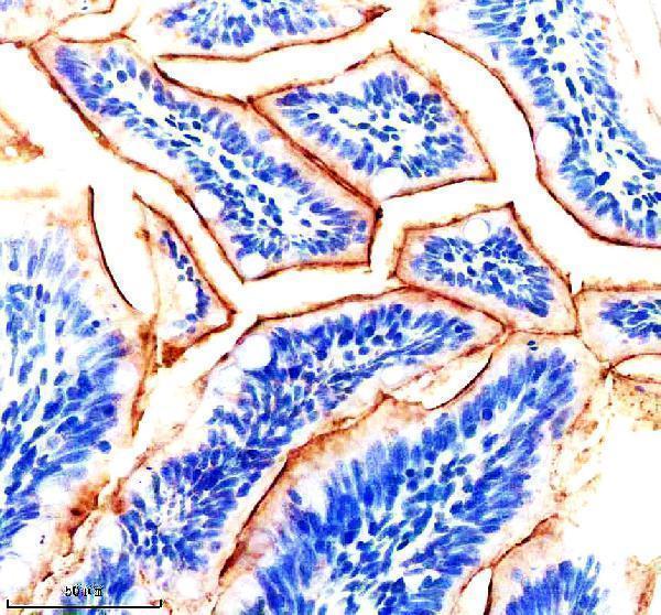
Click image to see more details
IHC analysis of IGF1RA using anti-IGF1RA antibody (AZA0A8M6YTF7).
IGF1RA was detected in a paraffin-embedded section of zebrafish muscle tissue. Heat mediated antigen retrieval was performed in EDTA buffer (pH 8.0, epitope retrieval solution). The tissue section was blocked with 10% goat serum. The tissue section was then incubated with 2 μg/ml rabbit anti-IGF1RA Antibody (AZA0A8M6YTF7) overnight at 4°C. Peroxidase Conjugated Goat Anti-rabbit IgG was used as secondary antibody and incubated for 30 minutes at 37°C. The tissue section was developed using HRP Conjugated Rabbit IgG Super Vision Assay Kit (Catalog # SV0002) with DAB as the chromogen.

Click image to see more details
IF analysis of IGF1RA using anti-IGF1RA antibody (AZA0A8M6YTF7).
IGF1RA was detected in a paraffin-embedded section of zebrafish embryo tissue. Heat mediated antigen retrieval was performed in EDTA buffer (pH 8.0, epitope retrieval solution). The tissue section was blocked with 10% goat serum. The tissue section was then incubated with 5 μg/mL rabbit anti-IGF1RA Antibody (AZA0A8M6YTF7) overnight at 4°C. DyLight®488 Conjugated Goat Anti-Rabbit IgG (BA1127) was used as secondary antibody at 1:500 dilution and incubated for 30 minutes at 37°C. The section was counterstained with DAPI. Visualize using a fluorescence microscope and filter sets appropriate for the label used.
Specific Publications For Anti-Zebrafish IGF1RA Antibody (AZA0A8M6YTF7)
Loading publications
Recommended Resources
Here are featured tools and databases that you might find useful.
- Boster's Pathways Library
- Protein Databases
- Bioscience Research Protocol Resources
- Data Processing & Analysis Software
- Photo Editing Software
- Scientific Literature Resources
- Research Paper Management Tools
- Molecular Biology Software
- Primer Design Tools
- Bioinformatics Tools
- Phylogenetic Tree Analysis
Customer Reviews
Have you used Anti-Zebrafish IGF1RA Antibody?
Share your experimental results or join a short interview to earn up to $1,000 in product credits or other rewards.
0 Reviews For Anti-Zebrafish IGF1RA Antibody
Customer Q&As
Have a question?
Find answers in Q&As, reviews.
Can't find your answer?
Submit your question
