Click image to see more details
-

-
-
-
-
+2
Product Info Summary
| SKU: | DZ41119 |
|---|---|
| Size: | 200 μl/vial |
| Reactive Species: | Zebrafish |
| Host: | Rabbit |
| Application: | IF, IHC |
Customers Who Bought This Also Bought

Product info
Product Name
Anti-Zebrafish Tfap2a Antibody
SKU/Catalog Number
DZ41119
Size
200 μl/vial
Form
Liquid
Description
Boster Bio Anti-Tfap2a Antibody catalog # DZ41119. This antibody reacts with Zebrafish.
Storage & Handling
At -20°C for one year, at 4°C for one month. Avoid repeated freezing and thawing.
Cite This Product
Anti-Zebrafish Tfap2a Antibody (Boster Biological Technology, Pleasanton CA, USA, Catalog # DZ41119)
Host
Rabbit
Contents
Each vial contains 20mM PBS, 50% glycerol, 0.02% NaN3.
Clonality
Polyclonal
Isotype
Rabbit IgG
Reactive Species
DZ41119 is reactive to tfap2a in Zebrafish
Application & Images
Applications
DZ41119 is guaranteed for IF, IHC Boster Guarantee
Assay Dilutions Recommendation
The recommendations below provide a starting point for assay optimization. The actual working concentration varies and should be decided by the user.
Immunohistochemistry(Paraffin-embedded Section), 2-5μg/ml
Immunofluorescence, 4 μg/ml
Positive Control
IHC: zebrafish esophagus tissue, zebrafish brain tissue, zebrafish kidney tissue
ICC/IF: zebrafish embryos tissue
Validation Images & Assay Conditions
Click image to see more details
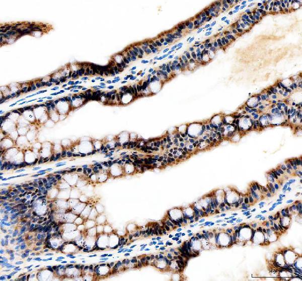
IHC analysis of Tfap2a using anti-Tfap2a antibody (DZ41119).
Tfap2a was detected in a paraffin-embedded section of zebrafish esophagus tissue. Heat mediated antigen retrieval was performed in EDTA buffer (pH 8.0, epitope retrieval solution). The tissue section was blocked with 10% goat serum. The tissue section was then incubated with 2 μg/ml rabbit anti-Tfap2a Antibody (DZ41119) overnight at 4°C. Peroxidase Conjugated Goat Anti-rabbit IgG was used as secondary antibody and incubated for 30 minutes at 37°C. The tissue section was developed using HRP Conjugated Rabbit IgG Super Vision Assay Kit (Catalog # SV0002) with DAB as the chromogen.

Click image to see more details
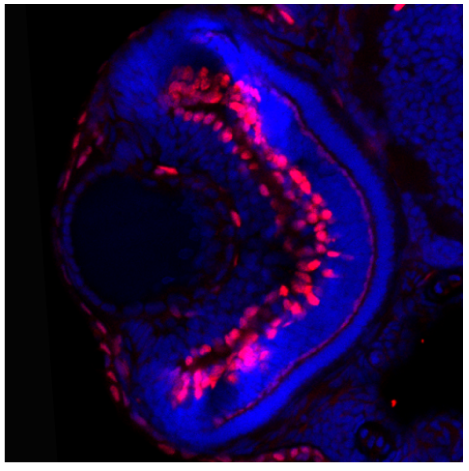
IF analysis of Tfap2a using anti-Tfap2a antibody (DZ41119).
Tfap2a was detected in a OCT-embedded section of Zebrafish retinal cryo-section. The tissue section was then incubated with 1:100 rabbit anti-Tfap2a Antibody (DZ41119) overnight at 4°C. Visualize using a confocol microscope.

Click image to see more details
IHC analysis of Tfap2a using anti-Tfap2a antibody (DZ41119).
Tfap2a was detected in a paraffin-embedded section of zebrafish brain tissue. Heat mediated antigen retrieval was performed in EDTA buffer (pH 8.0, epitope retrieval solution). The tissue section was blocked with 10% goat serum. The tissue section was then incubated with 2 μg/ml rabbit anti-Tfap2a Antibody (DZ41119) overnight at 4°C. Peroxidase Conjugated Goat Anti-rabbit IgG was used as secondary antibody and incubated for 30 minutes at 37°C. The tissue section was developed using HRP Conjugated Rabbit IgG Super Vision Assay Kit (Catalog # SV0002) with DAB as the chromogen.

Click image to see more details
IHC analysis of Tfap2a using anti-Tfap2a antibody (DZ41119).
Tfap2a was detected in a paraffin-embedded section of zebrafish kidney tissue. Heat mediated antigen retrieval was performed in EDTA buffer (pH 8.0, epitope retrieval solution). The tissue section was blocked with 10% goat serum. The tissue section was then incubated with 2 μg/ml rabbit anti-Tfap2a Antibody (DZ41119) overnight at 4°C. Peroxidase Conjugated Goat Anti-rabbit IgG was used as secondary antibody and incubated for 30 minutes at 37°C. The tissue section was developed using HRP Conjugated Rabbit IgG Super Vision Assay Kit (Catalog # SV0002) with DAB as the chromogen.

Click image to see more details
IF analysis of Tfap2a using anti-Tfap2a antibody (DZ41119).
Tfap2a was detected in a paraffin-embedded section of zebrafish embryos tissue. Heat mediated antigen retrieval was performed in EDTA buffer (pH 8.0, epitope retrieval solution). The tissue section was blocked with 10% goat serum. The tissue section was then incubated with 4 μg/mL rabbit anti-Tfap2a Antibody (DZ41119) overnight at 4°C. DyLight®594 Conjugated Goat Anti-Rabbit IgG (BA1142) was used as secondary antibody at 1:500 dilution and incubated for 30 minutes at 37°C. Visualize using a fluorescence microscope and filter sets appropriate for the label used.
Specific Publications For Anti-Zebrafish Tfap2a Antibody (DZ41119)
Loading publications
Recommended Resources
Here are featured tools and databases that you might find useful.
- Boster's Pathways Library
- Protein Databases
- Bioscience Research Protocol Resources
- Data Processing & Analysis Software
- Photo Editing Software
- Scientific Literature Resources
- Research Paper Management Tools
- Molecular Biology Software
- Primer Design Tools
- Bioinformatics Tools
- Phylogenetic Tree Analysis
Customer Reviews
Have you used Anti-Zebrafish Tfap2a Antibody?
Share your experimental results or join a short interview to earn up to $1,000 in product credits or other rewards.
1 Reviews For Anti-Zebrafish Tfap2a Antibody
Was able to detect tfap2a expression in the retinal ganglion cells and anterior segment at 3dpf.
Excellent
| SKU | DZ41119 |
|---|---|
| Application | Immunofluorescence |
| Sample | Zebrafish retinal cryo-section |
| Sample Processing Description | Embryos fixed in 4% PFA for 4h. Embryos washed in PBST and 30% then 50% sucrose. Embedded in OCT and cryo-sectioned at 20nm |
| Primary Antibody | Zebrafish Tfap2a Antibody |
| Primary Incubation | 1:100, overnight at 4 ℃ |
| Detection | Used a Nikon C2+ confocal microscope |
| Results Summary | Was able to detect tfap2a expression in the retinal ganglion cells and anterior segment at 3dpf. |
Jakub Famulski
Verified customer
Submitted 2025-11-03
Customer Q&As
Have a question?
Find answers in Q&As, reviews.
Can't find your answer?
Submit your question
