ECHS1 antibody (A04508-1) performs well in ELISA, Flow Cytometry, IF, IHC, ICC, WB and shows reactivity with Human, Mouse, Rat.
  • Validated for inter-lot consistency, specificity, and reproducibility
  • Committed to quality since 1993
Filter:
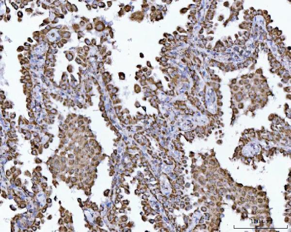
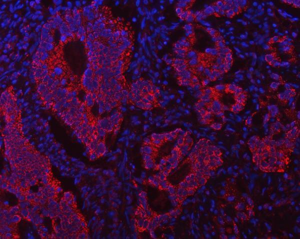
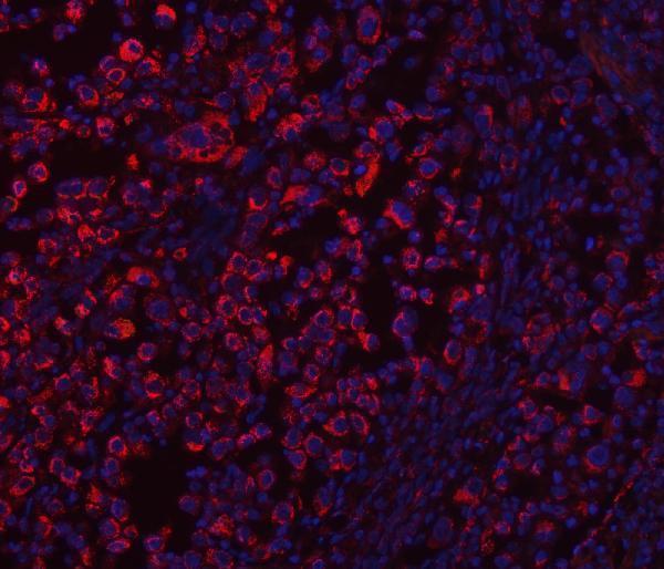
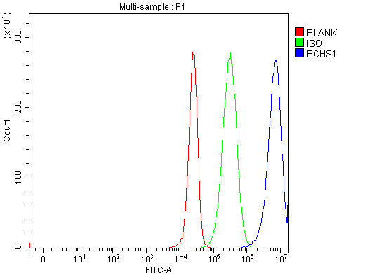
a04508 1 echs1 primary antibodies wb testing 1a04508 1 echs1 primary antibodies wb testing 2a04508 1 echs1 primary antibodies ihc testing 3a04508 1 echs1 primary antibodies ihc testing 4a04508 1 echs1 primary antibodies ihc testing 5a04508 1 echs1 primary antibodies ihc testing 6a04508 1 echs1 primary antibodies ihc testing 7a04508 1 echs1 primary antibodies ihc testing 8a04508 1 echs1 primary antibodies ihc testing 9a04508 1 echs1 primary antibodies if testing 10a04508 1 echs1 primary antibodies if testing 11a04508 1 echs1 primary antibodies if testing 12a04508 1 echs1 primary antibodies fcm testing 13

Anti-ECHS1 Antibody Picoband®

ECHS1 antibody

Reactive SpeciesHostApplication
Human, Mouse, RatRabbitELISA, Flow Cytometry, IF, IHC, ICC, WB

SKU: A04508-1

Size: 100 μg

Bundle to save: I do not want free secondary ab
Price:
$370/100 μg

In stock. Ships Next Business Day.

Quantity: 1

1
Get A Quote
Guarantee / Product Promise

Tested Applications

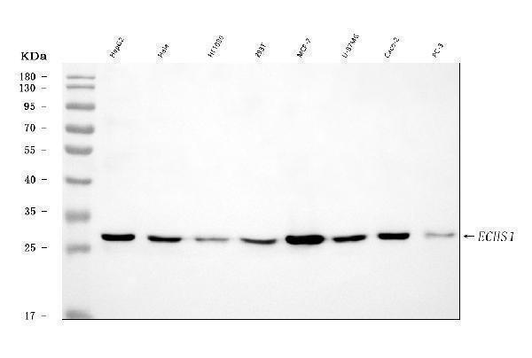
ECHS1 antibody was positively detected by WB in:

human HepG2 whole cell, human Hela whole cell, human HT1080 whole cell, human 293T whole cell, human MCF-7 whole cell, human U-87MG whole cell, human CACO-2 whole cell, human PC-3 whole cell, rat liver tissue, rat kidney tissue, rat RH35 whole cell, mouse liver tissue, mouse kidney tissue, mouse HEPA1-6 whole cell

Suggested blocking solution with 5% non-fat milk or BSA; (*)Recommended protein loading: 20-40 ug per lane

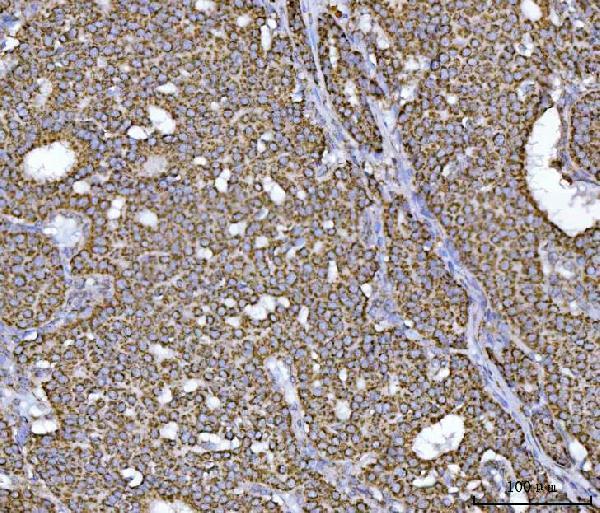
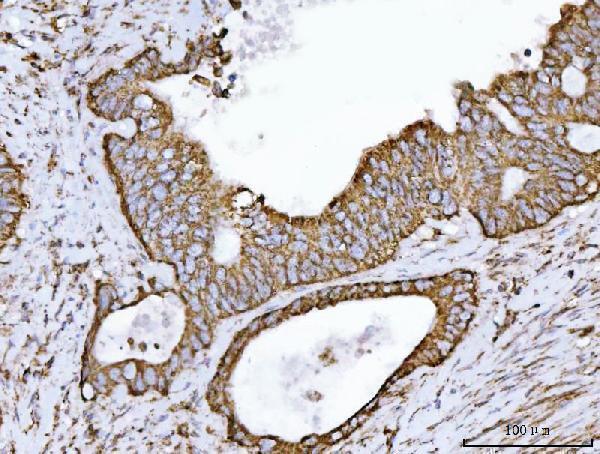
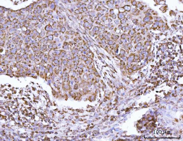
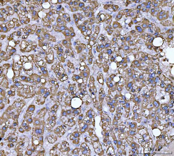
ECHS1 antibody was positively detected by IHC in:

human breast cancer tissue, human colorectal adenocarcinoma tissue, human esophageal squamous carcinoma tissue, human liver cancer tissue, human lung cancer tissue, human ovarian cancer tissue, rat liver tissue

Use TE buffer pH 9.0 for antigen retrieval; (*) citrate buffer pH 6.0 is an alternative.

ECHS1 antibody was positively detected by ICC/IF in:

CACO-2 cell

ECHS1 antibody was positively detected by IF in:

human intestinal cancer tissue, human lung cancer tissue

ECHS1 antibody was positively detected by FC in:

293T cell

Recommend Dilution

Application
Dilution
Species
Western blot
0.25-0.5 μg/ml
Human, Mouse, Rat
Immunohistochemistry(Paraffin-embedded Section)
2-5 μg/ml
Human, Rat
Immunocytochemistry/Immunofluorescence
5 μg/ml
Human
Immunofluorescence
5 μg/ml
Human
Flow Cytometry (Fixed)
1-3 μg/1x10^6 cells
Human
ELISA
0.1-0.5 μg/ml
-

* It is recommended to optimize the amount of this reagent in each experimental system to achieve the best results.

Note: For flow cytometry (FCM), validated for fixed cells only. Not validated for FACS or surface flow cytometry using live cells.

Reactive Data

Filter:
Boster Guarantee:What is this?
WB
IHC
IF
ELISA
Human
Tested
Tested
Tested
Recommend
Rat
Tested
Tested
Recommend
Recommend
Mouse
Tested
Recommend
Monkey
Predicted
Predicted
Predicted
Predicted

Product Information

Product Name
Anti-ECHS1 Antibody Picoband®
View all ECHS1 Antibodies
Form
Lyophilized
Uniprot
Gene ID
Storage & Handling
At -20°C for one year from date of receipt. After reconstitution, at 4°C for one month. It can also be aliquotted and stored frozen at -20°C for six months. Avoid repeated freezing and thawing.
Host
Rabbit
Contents
Each vial contains 4 mg Trehalose, 0.9 mg NaCl, 0.2 mg Na2HPO4.
Clonality
Polyclonal
Isotype
Rabbit IgG
Immunogen
E.coli-derived human ECHS1 recombinant protein (Position: R12-Q290).
Cross-Reactivity
No cross-reactivity with other proteins.
Reconstitution
Adding 0.2 ml of distilled water will yield a concentration of 500 μg/ml.
Observed Molecular Weight
29 kDa
Calculated Molecular Weight

31.4 kDa

Background of ECHS1
Enoyl Coenzyme A hydratase, short chain, 1, mitochondrial, also known as ECHS1, is a human gene. The protein encoded by this gene functions in the second step of the mitochondrial fatty acid beta-oxidation pathway. It catalyzes the hydration of 2-trans-enoyl-coenzyme A (CoA) intermediates to L-3-hydroxyacyl-CoAs. The gene product is a member of the hydratase/isomerase superfamily. It localizes to the mitochondrial matrix. Transcript variants utilizing alternative transcription initiation sites have been described in the literature.
Cite This Product
Anti-ECHS1 Antibody Picoband® (Boster Biological Technology, Pleasanton CA, USA, Catalog # A04508-1)

Protocols

Western Blot protocol for ECHS1 antibody
Immunohistochemistry (IHC) protocol for ECHS1 antibody
Immunocytochemistry/Immunofluorescence (ICC/IF) protocol for ECHS1 antibody
Immunofluorescence (IF) protocol for ECHS1 antibody
Flow Cytometry (FC) protocol for ECHS1 antibody

Review

Have you used Anti-ECHS1 Antibody Picoband®?

Submit a review and receive $370 in product credits, a $185 Amazon gift card, or our carefully curated gift bundles.

Product Promise

We promise Anti-ECHS1 Antibody Picoband® performs excellently in relevant applications for ELISA, Flow Cytometry, IF, IHC, ICC, WB. If it doesn't perform as promised, we will refund the product. Learn more

In stock
Order Product
A04508-1
Buy one primary antibody get one 0.5ml HRP or Biotin secondary antibody for free.
*Sample sizes are prepared on demand and will take extra lead time. (cannot be conjugated)
$370.00

Troubleshooting

troubleshooting-box-image

Download troubleshooting handbooks for IHC, Western blot and ELISA for FREE.

Download Free PDFs Now

Boster Guarantee

Guaranteed product quality

Guaranteed product quality

We promise all of our products perform as described in datasheets.

This product is for Research Use Only. Not for use in diagnostic procedures.

Update date: Oct 4, 2026