Click image to see more details
-

-
-
-
-
+8
Product Info Summary
| SKU: | A01633-1 |
|---|---|
| Size: | 100 µg/vial |
| Reactive Species: | Mouse, Rat |
| Host: | Rabbit |
| Application: | IF, IHC |
Customers Who Bought This Also Bought

Product info
Product Name
Anti-HCRT Antibody
SKU/Catalog Number
A01633-1
Size
100 µg/vial
Form
Lyophilized
Description
Boster Bio Anti-HCRT Antibody catalog # A01633-1. Tested in IF, IHC application. This antibody reacts with Mouse, Rat.
Storage & Handling
At -20°C for one year from date of receipt. After reconstitution, at 4°C for one month. It can also be aliquotted and stored frozen at -20°C for six months. Avoid repeated freezing and thawing.
Cite This Product
Anti-HCRT Antibody (Boster Biological Technology, Pleasanton CA, USA, Catalog # A01633-1)
Host
Rabbit
Contents
Each vial contains 4 mg Trehalose, 0.9 mg NaCl, 0.2 mg Na2HPO4.
Clonality
Polyclonal
Isotype
IgG
Immunogen
A synthetic peptide corresponding to a sequence in the middle region of human HCRT, identical to the related mouse and rat sequences.
Cross-reactivity
No cross reactivity with other proteins.
Reactive Species
A01633-1 is reactive to HCRT in Mouse, Rat
Calculated molecular weight
13.4 kDa
Background of HCRT
This gene encodes a hypothalamic neuropeptide precursor protein that gives rise to two mature neuropeptides, orexin A and orexin B, by proteolytic processing. Orexin A and orexin B, which bind to orphan G-protein coupled receptors HCRTR1 and HCRTR2, function in the regulation of sleep and arousal. This neuropeptide arrangement may also play a role in feeding behavior, metabolism, and homeostasis.
Antibody Validation
Boster validates all antibodies on WB, IHC, ICC, Immunofluorescence, and ELISA with known positive control and negative samples to ensure specificity and high affinity, including thorough antibody incubations.
Application & Images
Applications
A01633-1 is guaranteed for IF, IHC Boster Guarantee
Recommend Dilution
| Application | Dilution | Species |
|---|---|---|
| Immunohistochemistry(Paraffin-embedded Section) | 2-5 µg/ml | Mouse, Rat |
| Immunofluorescence | 5 μg/ml | Mouse, Rat |
Tested application
Use TE buffer pH 9.0 for antigen retrieval; (*) citrate buffer pH 6.0 is an alternative.
Validation Images & Assay Conditions
Click image to see more details
IHC analysis of HCRT using anti-HCRT antibody (A01633-1).
HCRT was detected in a paraffin-embedded section of mouse brain tissue. Heat mediated antigen retrieval was performed in EDTA buffer (pH 8.0, epitope retrieval solution). The tissue section was blocked with 10% goat serum. The tissue section was then incubated with 2 μg/ml rabbit anti-HCRT Antibody (A01633-1) overnight at 4°C. Peroxidase Conjugated Goat Anti-rabbit IgG was used as secondary antibody and incubated for 30 minutes at 37°C. The tissue section was developed using HRP Conjugated Rabbit IgG Super Vision Assay Kit (Catalog # SV0002) with DAB as the chromogen.

Click image to see more details
IHC analysis of HCRT using anti-HCRT antibody (A01633-1).
HCRT was detected in a paraffin-embedded section of rat brain tissue. Heat mediated antigen retrieval was performed in EDTA buffer (pH 8.0, epitope retrieval solution). The tissue section was blocked with 10% goat serum. The tissue section was then incubated with 2 μg/ml rabbit anti-HCRT Antibody (A01633-1) overnight at 4°C. Peroxidase Conjugated Goat Anti-rabbit IgG was used as secondary antibody and incubated for 30 minutes at 37°C. The tissue section was developed using HRP Conjugated Rabbit IgG Super Vision Assay Kit (Catalog # SV0002) with DAB as the chromogen.

Click image to see more details
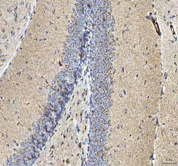
IHC analysis of HCRT using anti-HCRT antibody (A01633-1).
HCRT was detected in a paraffin-embedded section of rat cerebellum tissue. Heat mediated antigen retrieval was performed in EDTA buffer (pH 8.0, epitope retrieval solution). The tissue section was blocked with 10% goat serum. The tissue section was then incubated with 2 μg/ml rabbit anti-HCRT Antibody (A01633-1) overnight at 4°C. Peroxidase Conjugated Goat Anti-rabbit IgG was used as secondary antibody and incubated for 30 minutes at 37°C. The tissue section was developed using HRP Conjugated Rabbit IgG Super Vision Assay Kit (Catalog # SV0002) with DAB as the chromogen.

Click image to see more details
IF analysis of HCRT using anti-HCRT antibody (A01633-1).
HCRT was detected in a paraffin-embedded section of mouse brain tissue. Heat mediated antigen retrieval was performed in EDTA buffer (pH 8.0, epitope retrieval solution). The tissue section was blocked with 10% goat serum. The tissue section was then incubated with 5 μg/mL rabbit anti-HCRT Antibody (A01633-1) overnight at 4°C. Cy3 Conjugated Goat Anti-Rabbit IgG (BA1032) was used as secondary antibody at 1:500 dilution and incubated for 30 minutes at 37°C. The section was counterstained with DAPI. Visualize using a fluorescence microscope and filter sets appropriate for the label used.

Click image to see more details
IF analysis of HCRT using anti-HCRT antibody (A01633-1).
HCRT was detected in a paraffin-embedded section of rat brain tissue. Heat mediated antigen retrieval was performed in EDTA buffer (pH 8.0, epitope retrieval solution). The tissue section was blocked with 10% goat serum. The tissue section was then incubated with 5 μg/mL rabbit anti-HCRT Antibody (A01633-1) overnight at 4°C. Cy3 Conjugated Goat Anti-Rabbit IgG (BA1032) was used as secondary antibody at 1:500 dilution and incubated for 30 minutes at 37°C. The section was counterstained with DAPI. Visualize using a fluorescence microscope and filter sets appropriate for the label used.
Specific Publications For Anti-HCRT Antibody (A01633-1)
Loading publications
Recommended Resources
Here are featured tools and databases that you might find useful.
- Boster's Pathways Library
- Protein Databases
- Bioscience Research Protocol Resources
- Data Processing & Analysis Software
- Photo Editing Software
- Scientific Literature Resources
- Research Paper Management Tools
- Molecular Biology Software
- Primer Design Tools
- Bioinformatics Tools
- Phylogenetic Tree Analysis
Customer Reviews
Have you used Anti-HCRT Antibody?
Share your experimental results or join a short interview to earn up to $1,000 in product credits or other rewards.
0 Reviews For Anti-HCRT Antibody
Customer Q&As
Have a question?
Find answers in Q&As, reviews.
Can't find your answer?
Submit your question
