Click image to see more details
Product Info Summary
| SKU: | P00151 |
|---|---|
| Size: | 100 μl |
| Reactive Species: | Human, Mouse, Rat |
| Host: | Rabbit |
| Application: | IF, ICC, WB |
Customers Who Bought This Also Bought

Product info
Product Name
Anti-Phospho-FAK (Y397) PTK2 Rabbit Monoclonal Antibody
SKU/Catalog Number
P00151
BM4426 is an alternative SKU for this antibody, used in previous lots.
Size
100 μl
Form
Liquid
Description
Boster Bio Anti-Phospho-FAK (Y397) PTK2 Rabbit Monoclonal Antibody catalog # P00151. Tested in WB, ICC/IF applications. This antibody reacts with Human, Mouse, Rat.
Storage & Handling
Store at -20°C for one year. For short term storage and frequent use, store at 4°C for up to one month. Avoid repeated freeze-thaw cycles.
Cite This Product
Anti-Phospho-FAK (Y397) PTK2 Rabbit Monoclonal Antibody (Boster Biological Technology, Pleasanton CA, USA, Catalog # P00151)
Host
Rabbit
Contents
Rabbit IgG in stabilizing components, phosphate buffered saline, pH 7.4, 150mM NaCl, 0.02% sodium azide and 50% glycerol.
*This antibody is supplied in a stabilized formulation.
Compatibility with conjugation reactions depends on the chemistry of the conjugation method used.
For conjugation methods that are not compatible with the stabilizing components present in this formulation, a carrier-free antibody format is required.
Clonality
Monoclonal
Clone Number
EFO-16
Isotype
Rabbit IgG
Immunogen
A synthesized peptide derived from human Phospho-FAK (Y397)
Reactive Species
P00151 is reactive to PTK2 in Human, Mouse, Rat
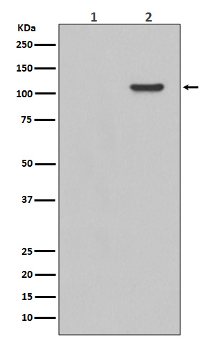
Observed Molecular Weight
119 kDa
Calculated molecular weight
119.2 kDa
Antibody Validation
Boster validates all antibodies on WB, IHC, ICC, Immunofluorescence, and ELISA with known positive control and negative samples to ensure specificity and high affinity, including thorough antibody incubations.
Application & Images
Applications
P00151 is guaranteed for IF, ICC, WB Boster Guarantee
Assay Dilutions Recommendation
The recommendations below provide a starting point for assay optimization. The actual working concentration varies and should be decided by the user.
WB 1:500-2000
ICC/IF 1:50-200
Positive Control
WB: EGF treated 293 whole cell
Validation Images & Assay Conditions
Click image to see more details
Western blot analysis of Phospho-FAK (Y397) expression in EGF treated 293 whole cell lysates,The lane on the left is treated with the antigen-specific peptide.
Specific Publications For Anti-Phospho-FAK (Y397) PTK2 Rabbit Monoclonal Antibody (P00151)
Loading publications
Recommended Resources
Here are featured tools and databases that you might find useful.
- Boster's Pathways Library
- Protein Databases
- Bioscience Research Protocol Resources
- Data Processing & Analysis Software
- Photo Editing Software
- Scientific Literature Resources
- Research Paper Management Tools
- Molecular Biology Software
- Primer Design Tools
- Bioinformatics Tools
- Phylogenetic Tree Analysis
Customer Reviews
Have you used Anti-Phospho-FAK (Y397) PTK2 Rabbit Monoclonal Antibody?
Share your experimental results or join a short interview to earn up to $1,000 in product credits or other rewards.
0 Reviews For Anti-Phospho-FAK (Y397) PTK2 Rabbit Monoclonal Antibody
Customer Q&As
Have a question?
Find answers in Q&As, reviews.
Can't find your answer?
Submit your question
4 Customer Q&As for Anti-Phospho-FAK (Y397) PTK2 Rabbit Monoclonal Antibody
Question
We have tried in the past anti-Phospho-FAK (Y397) Rabbit Monoclonal antibody for IF on cervix carcinoma in the past. I am using mouse, and I plan to use the antibody for ICC next. We need examining cervix carcinoma as well as brain in our next experiment. Could give a recommendation on which antibody would work the best for ICC?
E. Bhatt
Verified customer
Asked: 2020-02-24
Answer
I have checked the website and datasheets of our anti-Phospho-FAK (Y397) Rabbit Monoclonal antibody and I see that P00151 has been validated on mouse in both IF and ICC. Thus P00151 should work for your application. Our Boster satisfaction guarantee will cover this product for ICC in mouse even if the specific tissue type has not been validated. We do have a comprehensive range of products for ICC detection and you can check out our website bosterbio.com to find out more information about them.
Boster Scientific Support
Answered: 2020-02-24
Question
My colleagues were happy with the WB result of your anti-Phospho-FAK (Y397) Rabbit Monoclonal antibody. However we have been able to see positive staining in trachea cell junction using this antibody. Is that expected? Could you tell me where is PTK2 supposed to be expressed?
Verified Customer
Verified customer
Asked: 2017-09-06
Answer
From literature, trachea does express PTK2. Generally PTK2 expresses in cell junction, focal adhesion. cell. Regarding which tissues have PTK2 expression, here are a few articles citing expression in various tissues:
Brain, Pubmed ID: 8422239, 23503467
Cervix carcinoma, Pubmed ID: 16964243, 17081983, 18669648, 18691976, 20068231
Cervix carcinoma, and Erythroleukemia, Pubmed ID: 23186163
Melanocyte, Pubmed ID: 8247543, 9422762
Placenta, Pubmed ID: 15489334
T-cell, Pubmed ID: 7692878
Trachea, Pubmed ID: 14702039
Boster Scientific Support
Answered: 2017-09-06
Question
We are currently using anti-Phospho-FAK (Y397) Rabbit Monoclonal antibody P00151 for rat tissue, and we are well pleased with the IF results. The species of reactivity given in the datasheet says human, mouse, rat. Is it possible that the antibody can work on zebrafish tissues as well?
P. Rodriguez
Verified customer
Asked: 2015-10-01
Answer
The anti-Phospho-FAK (Y397) Rabbit Monoclonal antibody (P00151) has not been tested for cross reactivity specifically with zebrafish tissues, though there is a good chance of cross reactivity. We have an innovator award program that if you test this antibody and show it works in zebrafish you can get your next antibody for free. Please contact me if I can help you with anything.
Boster Scientific Support
Answered: 2015-10-01
Question
We have observed staining in mouse corpus callosum. Do you have any suggestions? Is anti-Phospho-FAK (Y397) Rabbit Monoclonal antibody supposed to stain corpus callosum positively?
O. Wu
Verified customer
Asked: 2014-08-11
Answer
Based on literature corpus callosum does express PTK2. Based on Uniprot.org, PTK2 is expressed in corpus callosum, t-cell, brain, trachea, placenta, osteosarcoma, melanocyte, cervix carcinoma, cervix carcinoma erythroleukemia, among other tissues. Regarding which tissues have PTK2 expression, here are a few articles citing expression in various tissues:
Brain, Pubmed ID: 8422239, 23503467
Cervix carcinoma, Pubmed ID: 16964243, 17081983, 18669648, 18691976, 20068231
Cervix carcinoma, and Erythroleukemia, Pubmed ID: 23186163
Melanocyte, Pubmed ID: 8247543, 9422762
Placenta, Pubmed ID: 15489334
T-cell, Pubmed ID: 7692878
Trachea, Pubmed ID: 14702039
Boster Scientific Support
Answered: 2014-08-11
