Click image to see more details
Product Info Summary
| SKU: | A00950-1 |
|---|---|
| Size: | 100 μg/vial |
| Reactive Species: | Human |
| Host: | Rabbit |
| Application: | ELISA, Flow Cytometry, IHC |
Customers Who Bought This Also Bought

Product info
Product Name
Anti-TIRAP Antibody
SKU/Catalog Number
A00950-1
Size
100 μg/vial
Form
Lyophilized
Description
Boster Bio Anti-TIRAP Antibody catalog # A00950-1. Tested in IHC, Flow Cytometry, ELISA applications. This antibody reacts with Human.
Storage & Handling
At -20°C for one year from date of receipt. After reconstitution, at 4°C for one month. It can also be aliquotted and stored frozen at -20°C for six months. Avoid repeated freezing and thawing.
Cite This Product
Anti-TIRAP Antibody (Boster Biological Technology, Pleasanton CA, USA, Catalog # A00950-1)
Host
Rabbit
Contents
Each vial contains 4 mg Trehalose, 0.9 mg NaCl, 0.2 mg Na2HPO4.
Clonality
Polyclonal
Immunogen
E.coli-derived human TIRAP recombinant protein (Position: P10-S221). Human TIRAP shares 74% amino acid (aa) sequence identity with mouse TIRAP.
Reactive Species
A00950-1 is reactive to TIRAP in Human
Calculated molecular weight
23.9 kDa
Background of TIRAP
The innate immune system recognizes microbial pathogens through Toll-like receptors (TLRs), which identify pathogen-associated molecular patterns. Different TLRs recognize different pathogen-associated molecular patterns and all TLRs have a Toll-interleukin 1 receptor (TIR) domain, which is responsible for signal transduction. The protein encoded by this gene is a TIR adaptor protein involved in the TLR4 signaling pathway of the immune system. It activates NF-kappa-B, MAPK1, MAPK3 and JNK, which then results in cytokine secretion and the inflammatory response. Alternative splicing of this gene results in several transcript variants; however, not all variants have been fully described.
Antibody Validation
Boster validates all antibodies on WB, IHC, ICC, Immunofluorescence, and ELISA with known positive control and negative samples to ensure specificity and high affinity, including thorough antibody incubations.
Application & Images
Applications
A00950-1 is guaranteed for ELISA, Flow Cytometry, IHC Boster Guarantee
Recommend Dilution
| Application | Dilution | Species |
|---|---|---|
| Immunohistochemistry(Paraffin-embedded Section) | 2-5 μg/ml | Human |
| Flow Cytometry (Fixed) | 1-3 μg/1x106 cells | Human |
| ELISA | 0.1-0.5 μg/ml |
Validation Images & Assay Conditions
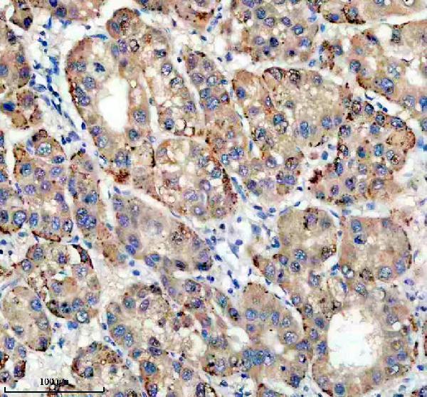
Click image to see more details
IHC analysis of TIRAP using anti-TIRAP antibody (A00950-1).
TIRAP was detected in a paraffin-embedded section of human liver cancer tissue. Heat mediated antigen retrieval was performed in EDTA buffer (pH 8.0, epitope retrieval solution). The tissue section was blocked with 10% goat serum. The tissue section was then incubated with 2 μg/ml rabbit anti-TIRAP Antibody (A00950-1) overnight at 4°C. Peroxidase Conjugated Goat Anti-rabbit IgG was used as secondary antibody and incubated for 30 minutes at 37°C. The tissue section was developed using HRP Conjugated Rabbit IgG Super Vision Assay Kit (Catalog # SV0002) with DAB as the chromogen.

Click image to see more details
IHC analysis of TIRAP using anti-TIRAP antibody (A00950-1).
TIRAP was detected in a paraffin-embedded section of human liver tissue. Heat mediated antigen retrieval was performed in EDTA buffer (pH 8.0, epitope retrieval solution). The tissue section was blocked with 10% goat serum. The tissue section was then incubated with 2 μg/ml rabbit anti-TIRAP Antibody (A00950-1) overnight at 4°C. Peroxidase Conjugated Goat Anti-rabbit IgG was used as secondary antibody and incubated for 30 minutes at 37°C. The tissue section was developed using HRP Conjugated Rabbit IgG Super Vision Assay Kit (Catalog # SV0002) with DAB as the chromogen.

Click image to see more details
Flow Cytometry analysis of HepG2 cells using anti-TIRAP antibody (A00950-1).
Overlay histogram showing HepG2 cells stained with A00950-1 (Blue line). The cells were fixed with 4% paraformaldehyde and blocked with 10% normal goat serum. And then incubated with rabbit anti-TIRAP Antibody (A00950-1, 1 μg/1x106 cells) for 30 min at 20°C. DyLight®488 conjugated goat anti-rabbit IgG (BA1127, 5-10 μg/1x106 cells) was used as secondary antibody for 30 minutes at 20°C. Isotype control antibody (Green line) was rabbit IgG (1 μg/1x106) used under the same conditions. Unlabelled sample without incubation with primary antibody and secondary antibody (Red line) was used as a blank control.
Specific Publications For Anti-TIRAP Antibody (A00950-1)
Loading publications
Recommended Resources
Here are featured tools and databases that you might find useful.
- Boster's Pathways Library
- Protein Databases
- Bioscience Research Protocol Resources
- Data Processing & Analysis Software
- Photo Editing Software
- Scientific Literature Resources
- Research Paper Management Tools
- Molecular Biology Software
- Primer Design Tools
- Bioinformatics Tools
- Phylogenetic Tree Analysis
Customer Reviews
Have you used Anti-TIRAP Antibody?
Share your experimental results or join a short interview to earn up to $1,000 in product credits or other rewards.
0 Reviews For Anti-TIRAP Antibody
Customer Q&As
Have a question?
Find answers in Q&As, reviews.
Can't find your answer?
Submit your question
