Click image to see more details
Product Info Summary
| SKU: | AZQ9I8N6 |
|---|---|
| Size: | 100 μg/vial |
| Reactive Species: | Zebrafish |
| Host: | Rabbit |
| Application: | IHC |
Customers Who Bought This Also Bought

Product info
Product Name
Anti-Zebrafish M-CSFR/CSF1R Antibody
SKU/Catalog Number
AZQ9I8N6
Size
100 μg/vial
Form
Lyophilized
Description
Boster Bio Anti-Zebrafish M-CSFR/CSF1R Antibody catalog #AZQ9I8N6. Tested in IHC applications. This antibody reacts with Zebrafish.
Storage & Handling
At -20°C for one year from date of receipt. After reconstitution, at 4°C for one month. It can also be aliquotted and stored frozen at -20°C for six months. Avoid repeated freezing and thawing.
Cite This Product
Anti-Zebrafish M-CSFR/CSF1R Antibody (Boster Biological Technology, Pleasanton CA, USA, Catalog # AZQ9I8N6)
Host
Rabbit
Contents
Each vial contains 4 mg Trehalose, 0.9 mg NaCl, 0.2 mg Na2HPO4.
Clonality
Polyclonal
Immunogen
E.coli-derived zebrafish M-CSFR/CSF1R recombinant protein (Position: D409-C977)
Reactive Species
AZQ9I8N6 is reactive to CSF1R in Zebrafish
Background of CSF1R
CSF1R(Colony-Stimulating Factor 1 Receptor) also known as MCSFR, FMS, c-FMS, CD115, ONCOGENE FMS or CD115 ANTIGEN, encodes a tyrosine kinase growth factor receptor for colony-stimulating factor-1, the macrophage-and monocyte-specific growth factor. The gene is located on long arm of chromosome 5(5q32) on the Crick(minus) strand. The as-yet-unidentified°CSF1 Rpromoter/enhancer sequences may be confined to the nucleotides separating the 2 genes or could potentially lie within the PDGFR gene itself. The encoded protein is a single pass type I membrane protein and acts as the receptor for°Colony stimulating factor 1, a cytokine which controls the production, differentiation, and function of macrophages. The encoded protein is atyrosine kinase transmembrane receptor and member of the CSF1/PDGF receptor family of tyrosine-protein kinases. Kondo et al.(2000) showed that the endogenous myelomonocytic cytokine receptors for GM-CSF and macrophage colony-stimulating factor(CSF1R) are expressed at low to moderate levels on the more primitive hematopoietic stem cells, are absent on common lymphoid progenitors, and are upregulated after myeloid lineage induction by IL2.
Antibody Validation
Boster validates all antibodies on WB, IHC, ICC, Immunofluorescence, and ELISA with known positive control and negative samples to ensure specificity and high affinity, including thorough antibody incubations.
Application & Images
Applications
AZQ9I8N6 is guaranteed for IHC Boster Guarantee
Recommend Dilution
| Application | Dilution | Species |
|---|---|---|
| Immunohistochemistry(Paraffin-embedded Section) | 2-5 μg/ml | Zebrafish |
Tested application
Use TE buffer pH 9.0 for antigen retrieval; (*) citrate buffer pH 6.0 is an alternative.
Validation Images & Assay Conditions
Click image to see more details
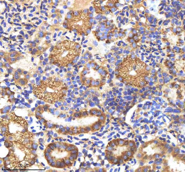
IHC analysis of M-CSFR/CSF1R using anti-M-CSFR/CSF1R antibody (AZQ9I8N6).
M-CSFR/CSF1R was detected in a paraffin-embedded section of zebrafish kidney tissue. Heat mediated antigen retrieval was performed in EDTA buffer (pH 8.0, epitope retrieval solution). The tissue section was blocked with 10% goat serum. The tissue section was then incubated with 2 μg/ml rabbit anti-M-CSFR/CSF1R Antibody (AZQ9I8N6) overnight at 4°C. Peroxidase Conjugated Goat Anti-rabbit IgG was used as secondary antibody and incubated for 30 minutes at 37°C. The tissue section was developed using HRP Conjugated Rabbit IgG Super Vision Assay Kit (Catalog # SV0002) with DAB as the chromogen.

Click image to see more details
IHC analysis of M-CSFR/CSF1R using anti-M-CSFR/CSF1R antibody (AZQ9I8N6).
M-CSFR/CSF1R was detected in a paraffin-embedded section of zebrafish pancreas tissue. Heat mediated antigen retrieval was performed in EDTA buffer (pH 8.0, epitope retrieval solution). The tissue section was blocked with 10% goat serum. The tissue section was then incubated with 2 μg/ml rabbit anti-M-CSFR/CSF1R Antibody (AZQ9I8N6) overnight at 4°C. Peroxidase Conjugated Goat Anti-rabbit IgG was used as secondary antibody and incubated for 30 minutes at 37°C. The tissue section was developed using HRP Conjugated Rabbit IgG Super Vision Assay Kit (Catalog # SV0002) with DAB as the chromogen.

Click image to see more details
IHC analysis of M-CSFR/CSF1R using anti-M-CSFR/CSF1R antibody (AZQ9I8N6).
M-CSFR/CSF1R was detected in a paraffin-embedded section of zebrafish spinal cord tissue. Heat mediated antigen retrieval was performed in EDTA buffer (pH 8.0, epitope retrieval solution). The tissue section was blocked with 10% goat serum. The tissue section was then incubated with 2 μg/ml rabbit anti-M-CSFR/CSF1R Antibody (AZQ9I8N6) overnight at 4°C. Peroxidase Conjugated Goat Anti-rabbit IgG was used as secondary antibody and incubated for 30 minutes at 37°C. The tissue section was developed using HRP Conjugated Rabbit IgG Super Vision Assay Kit (Catalog # SV0002) with DAB as the chromogen.
Specific Publications For Anti-Zebrafish M-CSFR/CSF1R Antibody (AZQ9I8N6)
Loading publications
Recommended Resources
Here are featured tools and databases that you might find useful.
- Boster's Pathways Library
- Protein Databases
- Bioscience Research Protocol Resources
- Data Processing & Analysis Software
- Photo Editing Software
- Scientific Literature Resources
- Research Paper Management Tools
- Molecular Biology Software
- Primer Design Tools
- Bioinformatics Tools
- Phylogenetic Tree Analysis
Customer Reviews
Have you used Anti-Zebrafish M-CSFR/CSF1R Antibody?
Share your experimental results or join a short interview to earn up to $1,000 in product credits or other rewards.
0 Reviews For Anti-Zebrafish M-CSFR/CSF1R Antibody
Customer Q&As
Have a question?
Find answers in Q&As, reviews.
Can't find your answer?
Submit your question
