Click image to see more details
Product Info Summary
| SKU: | AZA0A8M9PE33 |
|---|---|
| Size: | 100 μg/vial |
| Reactive Species: | Zebrafish |
| Host: | Rabbit |
| Application: | IHC |
Customers Who Bought This Also Bought

Product info
Product Name
Anti-Zebrafish PABPC4 Antibody
SKU/Catalog Number
AZA0A8M9PE33
Size
100 μg/vial
Form
Lyophilized
Description
Boster Bio Anti-Zebrafish PABPC4 Antibody catalog # AZA0A8M9PE33. Tested in IHC applications. This antibody reacts with Zebrafish.
Storage & Handling
At -20°C for one year from date of receipt. After reconstitution, at 4°C for one month. It can also be aliquotted and stored frozen at -20°C for six months. Avoid repeated freezing and thawing.
Cite This Product
Anti-Zebrafish PABPC4 Antibody (Boster Biological Technology, Pleasanton CA, USA, Catalog # AZA0A8M9PE33)
Host
Rabbit
Contents
Each vial contains 4 mg Trehalose, 0.9 mg NaCl, 0.2 mg Na2HPO4.
Clonality
Polyclonal
Immunogen
A synthetic peptide corresponding to a sequence in the middle region of zebrafish PABPC4.
Reactive Species
AZA0A8M9PE33 is reactive to PABPC4 in Zebrafish
Background of PABPC4
Polyadenylate-binding protein 4 (PABPC4) is a protein that in humans is encoded by the PABPC4 gene. Poly(A)-binding proteins (PABPs) bind to the poly(A) tail present at the 3-prime ends of most eukaryotic mRNAs. PABPC4 or IPABP (inducible PABP) was isolated as an activation-induced T-cell mRNA encoding a protein. Activation of T cells increased PABPC4 mRNA levels in T cells approximately 5-fold. PABPC4 contains 4 RNA-binding domains and proline-rich C terminus. PABPC4 is localized primarily to the cytoplasm. It is suggested that PABPC4 might be necessary for regulation of stability of labile mRNA species in activated T cells. PABPC4 was also identified as an antigen, APP1 (activated-platelet protein-1), expressed on thrombin-activated rabbit platelets. PABPC4 may also be involved in the regulation of protein translation in platelets and megakaryocytes or may participate in the binding or stabilization of polyadenylates in platelet dense granules. Alternatively spliced transcript variants encoding different isoforms have been found for this gene. This protein has also been found to interact with coronavirus nucleocapsid proteins and is thought to inhibit coronavirus replication.
Antibody Validation
Boster validates all antibodies on WB, IHC, ICC, Immunofluorescence, and ELISA with known positive control and negative samples to ensure specificity and high affinity, including thorough antibody incubations.
Application & Images
Applications
AZA0A8M9PE33 is guaranteed for IHC Boster Guarantee
Recommend Dilution
| Application | Dilution | Species |
|---|---|---|
| Immunohistochemistry(Paraffin-embedded Section) | 2-5 μg/ml | Zebrafish |
Validation Images & Assay Conditions
Click image to see more details
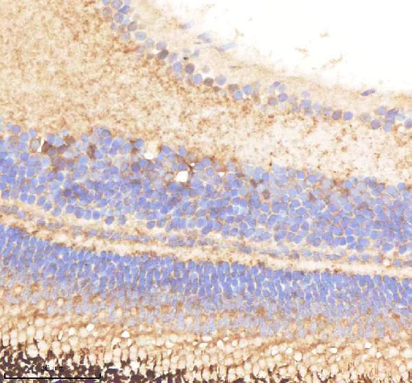
IHC analysis of PABPC4 using anti-PABPC4 antibody (AZA0A8M9PE33).
PABPC4 was detected in a paraffin-embedded section of zebrafish eye tissue. Heat mediated antigen retrieval was performed in EDTA buffer (pH 8.0, epitope retrieval solution). The tissue section was blocked with 10% goat serum. The tissue section was then incubated with 2 μg/ml rabbit anti-PABPC4 Antibody (AZA0A8M9PE33) overnight at 4°C. Peroxidase Conjugated Goat Anti-rabbit IgG was used as secondary antibody and incubated for 30 minutes at 37°C. The tissue section was developed using HRP Conjugated Rabbit IgG Super Vision Assay Kit (Catalog # SV0002) with DAB as the chromogen.

Click image to see more details
IHC analysis of PABPC4 using anti-PABPC4 antibody (AZA0A8M9PE33).
PABPC4 was detected in a paraffin-embedded section of zebrafish brain tissue. Heat mediated antigen retrieval was performed in EDTA buffer (pH 8.0, epitope retrieval solution). The tissue section was blocked with 10% goat serum. The tissue section was then incubated with 2 μg/ml rabbit anti-PABPC4 Antibody (AZA0A8M9PE33) overnight at 4°C. Peroxidase Conjugated Goat Anti-rabbit IgG was used as secondary antibody and incubated for 30 minutes at 37°C. The tissue section was developed using HRP Conjugated Rabbit IgG Super Vision Assay Kit (Catalog # SV0002) with DAB as the chromogen.

Click image to see more details
IHC analysis of PABPC4 using anti-PABPC4 antibody (AZA0A8M9PE33).
PABPC4 was detected in a paraffin-embedded section of zebrafish muscle tissue. Heat mediated antigen retrieval was performed in EDTA buffer (pH 8.0, epitope retrieval solution). The tissue section was blocked with 10% goat serum. The tissue section was then incubated with 2 μg/ml rabbit anti-PABPC4 Antibody (AZA0A8M9PE33) overnight at 4°C. Peroxidase Conjugated Goat Anti-rabbit IgG was used as secondary antibody and incubated for 30 minutes at 37°C. The tissue section was developed using HRP Conjugated Rabbit IgG Super Vision Assay Kit (Catalog # SV0002) with DAB as the chromogen.
Specific Publications For Anti-Zebrafish PABPC4 Antibody (AZA0A8M9PE33)
Loading publications
Recommended Resources
Here are featured tools and databases that you might find useful.
- Boster's Pathways Library
- Protein Databases
- Bioscience Research Protocol Resources
- Data Processing & Analysis Software
- Photo Editing Software
- Scientific Literature Resources
- Research Paper Management Tools
- Molecular Biology Software
- Primer Design Tools
- Bioinformatics Tools
- Phylogenetic Tree Analysis
Customer Reviews
Have you used Anti-Zebrafish PABPC4 Antibody?
Share your experimental results or join a short interview to earn up to $1,000 in product credits or other rewards.
0 Reviews For Anti-Zebrafish PABPC4 Antibody
Customer Q&As
Have a question?
Find answers in Q&As, reviews.
Can't find your answer?
Submit your question
