This website uses cookies to ensure you get the best experience on our website.
- Table of Contents
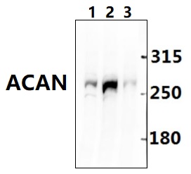
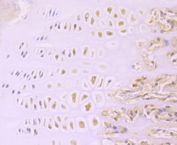
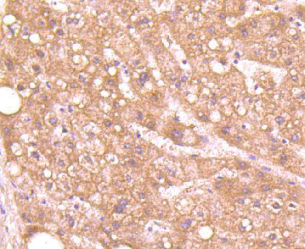
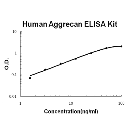
Facts about Aggrecan core protein.
It binds avidly to hyaluronic acid via an N-terminal globular region. .
Human | |
---|---|
Gene Name: | ACAN |
Uniprot: | P16112 |
Entrez: | 176 |
Belongs to: |
---|
aggrecan/versican proteoglycan family |
AGC1SEDK; aggrecan core protein; aggrecan; Cartilage-specific proteoglycan core protein; Chondroitin sulfate proteoglycan 1; Chondroitin sulfate proteoglycan core protein 1; CSPCP; CSPG1aggrecan 1; CSPGCP; large aggregating proteoglycan; MSK16AGCAN
Mass (kDA):
261.329 kDA
Human | |
---|---|
Location: | 15q26.1 |
Sequence: | 15; NC_000015.10 (88803436..88875354) |
Restricted to cartilages.
Secreted, extracellular space, extracellular matrix.
The ACAN Marker is an innovative way to assess the cellular phenotype of any sample. This highly specific marker allows scientists to measure the levels of protein in biological samples that are complex. It is highly useful for any scientist worldwide and is especially beneficial when working on samples of specific species. If the results are used in research, scientists are able to submit their findings for credit for their work. The ACAN Marker is highly effective in many applications such as the identification of tissues, cells and organs.
One of the benefits of the ACAN Marker is its ability to read text aloud. Simply plug in a pair headphones and you will be able to hear the text read aloud. You can also choose the language of your choice. ScanMarker is an excellent tool for readers and students. It can save you time and effort by scanning printed text and displaying it in any program on your computer. The buffer allows you to scan a portion of a page simultaneously, so you can do more in one step.
The pen scans an entire line of text within one second. It converts the text into a RAW image using the aid of Text Recognition Technology. After you've scanned documents, you can upload it to many platforms. This allows you to read the text, create summary documents, and then share them with others. The Scanmarker can also translate text between 40 languages, and read notes using Text to Speak technology.
Multiplex IHC is a revolutionary IHC technique allows multiple markers to be simultaneously labeled within one cell compartment. Multiplex IHC is especially beneficial in immuno-oncology as it permits researchers to measure the positivity of every single cell, and also reports staining intensities metrics across multiple markers. This technique gives a complete picture of cell structure and spatial arrangement, which can increase the precision of your findings.
It is necessary to collect tissue samples from the organ you want to study and fix them before staining. After fixing, you can conduct IHC on the fixed tissue or fix them in an ice bath. Optimizing your antibody binding or detection methods is essential for reproducible IHC workflows. To ensure the highest degree of reproducibility, you should always check the mIF results on tissues, cell lines, and TMAs, in accordance with the manufacturer's recommendations. You should also confirm the staining results using Western Blot analysis.
IHC can be used to determine the presence of organisms within cytological preparations, fluids, or sputum. This is accomplished by staining them with monoclonal antibody. This method is particularly effective to identify pneumocystis within patients with immunodeficiency as it could prompt immediate treatment. IHC also can help determine the role of genes using a monoclonal antibody. It can be used to detect apoptotic pathway or pro-apoptotic pathways.
To avoid this issue, IHC should be used with a charged slide. Use slides coated with APES. Because the ACAN marker is a sensitive marker, it's vital that tissue is prepared for IHC. Incorrectly prepared specimens may result in inconsistent staining results. You'll need to attach the adhesive for the protein-based section to the slide. The slide will not adhere to the surface if it isn't.
Another advantage of multiplex IHC is the higher reproducibility. These methods have lower coefficients of variation (CV) and are more reliable than other IHC-based tests. Therefore, they are suitable for translational applications in future. This technique is compatible with conventional darkfield and brightfield microscopes. It can be difficult to see colocalized biomarkers in cancerous cells, however.
IHC has a lot of applications, from developing drugs to diagnosing cancer. IHC can be used to determine specific tumor markers. It also assists in detecting up-regulation and down-regulation in disease-specific markers. In this procedure, the patient's tumor cells are stained with a particular protein. This allows for a precise diagnosis and classification. With a few simple steps, IHC can be applied for Continuing Education Credits.
Flow cytometry is an effective tool that has applications across the sciences. It determines the concentrations of particles and cells in the samples. Boster Bio provides monoclonal as in addition to polyclonal antibodies that target the target molecule. The high-affinity antibodies of Boster Bio have been the subject of numerous citations and continue to be highly referenced in the present. Continue reading to discover more about the numerous benefits of Boster's primary antibodies that are high affinity.
Single B cell screening can make high-affinity primary antibodies within only a few days. The process begins with the isolation of viable B cells from humans in convalescence and animals. High-throughput single-cell sorting determines the candidate antibodies. The next step is gene expression and evaluation of the potential antibodies are conducted. The aim is to determine the most effective antibodies against the antigen targeted. The aim of this process is to identify antigen-specific antibodies quickly and efficiently method.
There are a variety of steps involved in the production of high-affinity antibody using animal spleens. The first step is to immunize animals. conducted with purified hPCSK9. If the mice's antibodies are at one million/lbthey are killed. The spleen cells could then be used to create hybridoma. The hybridoma clone containing m5E12 was isolated by protein A affinity chromatography and further characterized using SDS-PAGE as well as Shodex PROTEIN KW-802.5.
This strain produces antibodies that are very specific against SARS-CoV-2. They neutralize and recognize SARS-CoV-2 spike proteins. The bivalent antibodies were evaluated using flow cytometry against biotinylated SARS-CoV-2 RBD immobilized on magnetic beads. The results of three independent experiments are reported. This analysis provides information on the specificity and specificity of these antibodies.
The discovery of a murine anti-microbial antibody (mAb) that had low immunogenicity resulted in the creation of the high-affinity, high-affinity h5E12scFv. The h5E12scFv gene was back-mutated with alanine scanning mutational analysis. Then, the optimized scFvs were transformed to form full-length IgG and fusionbed with human IgG1 constant region.
Further tests were conducted using differential scanning fluorimetry in order to determine the stability of the secondary antibodies. The h2SCF values for three antibodies against which we tested were determined and the secondary antibody's molarity determined. These values were adapted to the Fc concentration of each antibody to ensure that the molarity of the primary and secondary antibodies was within acceptable limits. Additionally, all of the h2ScFv antibodies are cross-affinity to the specific antigens targeted by the antibody.
The antibody-antigen interaction will be dominated high-affinity LCDR3 and HCDR3. One-step PCR technology was utilized to make the high-affinity LCDR3 and HCDR3 antibodies. During the process the antibody and the antigen of the target were immobilized onto a three-dimensional array of streptavidin Dynabeads. The beads were then washed twice using PBSB and blocked with 10% milk.
PMID: 1985970 by Doege K.J., et al. Complete coding sequence and deduced primary structure of the human cartilage large aggregating proteoglycan, aggrecan. Human-specific repeats, and additional alternatively spliced forms.
PMID: 7574678 by Ilic M.Z., et al. Catabolism of aggrecan by explant cultures of human articular cartilage in the presence of retinoic acid.