This website uses cookies to ensure you get the best experience on our website.
- Table of Contents
2 Citations 4 Q&As
Facts about Anterior gradient protein 2 homolog.

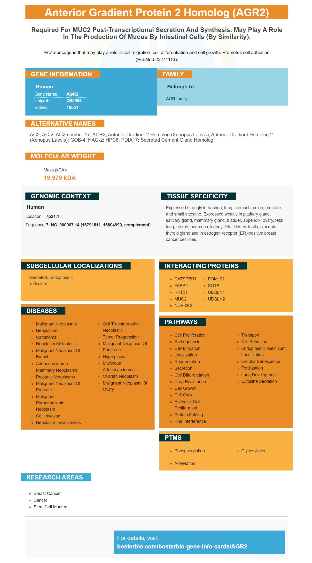
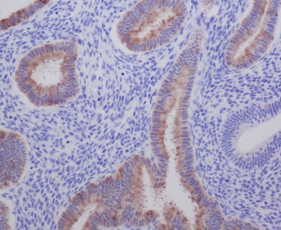
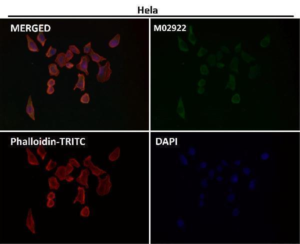
Proto-oncogene that may play a role in cell migration, cell differentiation and cell growth. Promotes cell adhesion (PubMed:23274113).
| Human | |
|---|---|
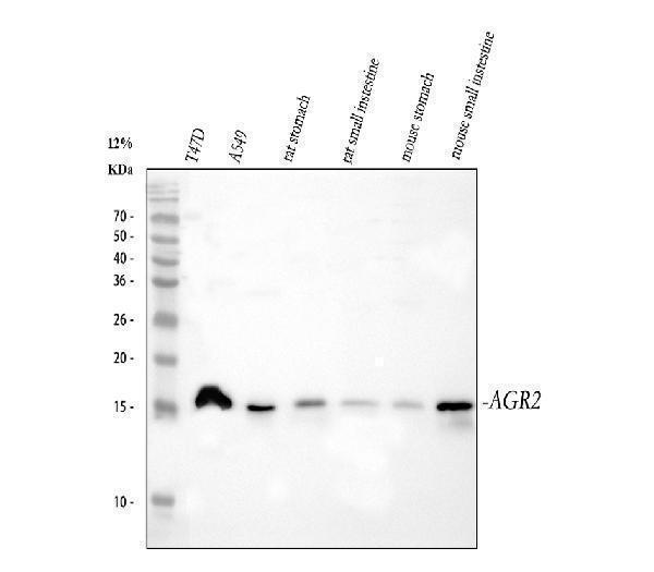
| Gene Name: | AGR2 |
| Uniprot: | O95994 |
| Entrez: | 10551 |

| Belongs to: |
|---|
| AGR family |

AG2; AG-2; AG2member 17; AGR2; anterior gradient 2 homolog (Xenopus laevis); anterior gradient homolog 2 (Xenopus laevis); GOB-4; hAG-2; HPC8; PDIA17; secreted cement gland homolog
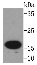
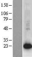
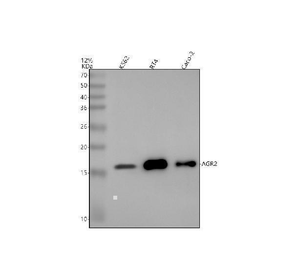
Mass (kDA):
19.979 kDA

| Human | |
|---|---|
| Location: | 7p21.1 |
| Sequence: | 7; NC_000007.14 (16791811..16804999, complement) |
Expressed strongly in trachea, lung, stomach, colon, prostate and small intestine. Expressed weakly in pituitary gland, salivary gland, mammary gland, bladder, appendix, ovary, fetal lung, uterus, pancreas, kidney, fetal kidney, testis, placenta, thyroid gland and in estrogen receptor (ER)-positive breast cancer cell lines.
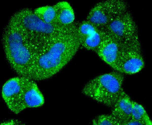
Secreted. Endoplasmic reticulum.


PMID: 9790916 by Thompson D.A., et al. hAG-2, the human homologue of the Xenopus laevis cement gland gene XAG-2, is coexpressed with estrogen receptor in breast cancer cell lines.
PMID: 12592373 by Fletcher G.C., et al. hAG-2 and hAG-3, human homologues of genes involved in differentiation, are associated with oestrogen receptor-positive breast tumours and interact with metastasis gene C4.4a and dystroglycan.