This website uses cookies to ensure you get the best experience on our website.
- Table of Contents
1 Citations 5 Q&As
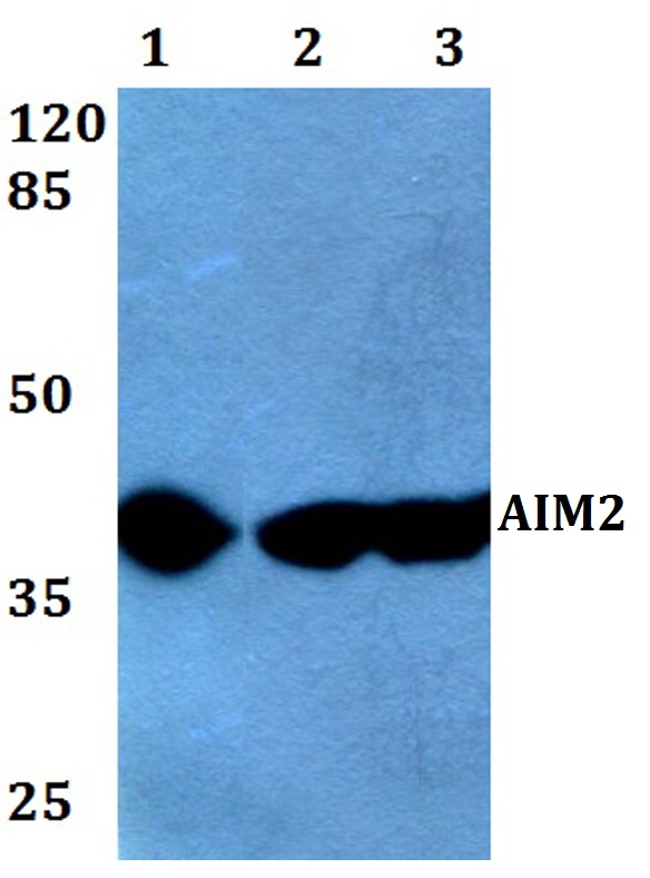
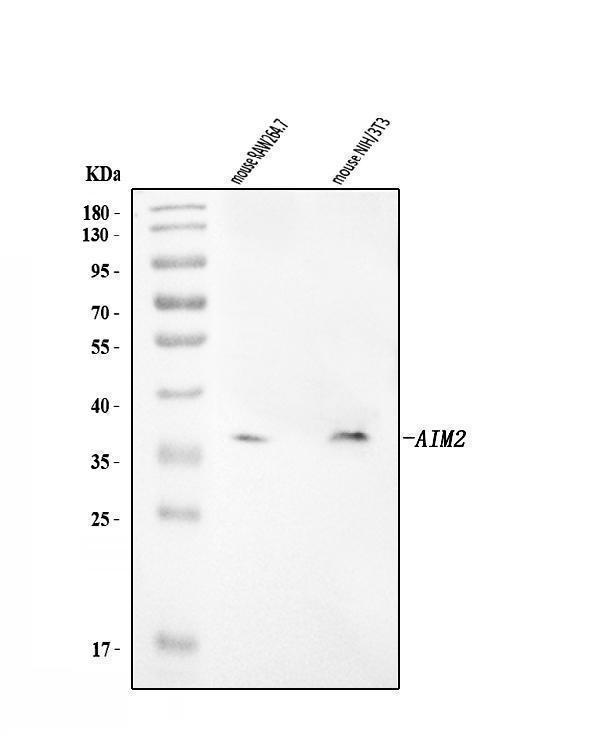
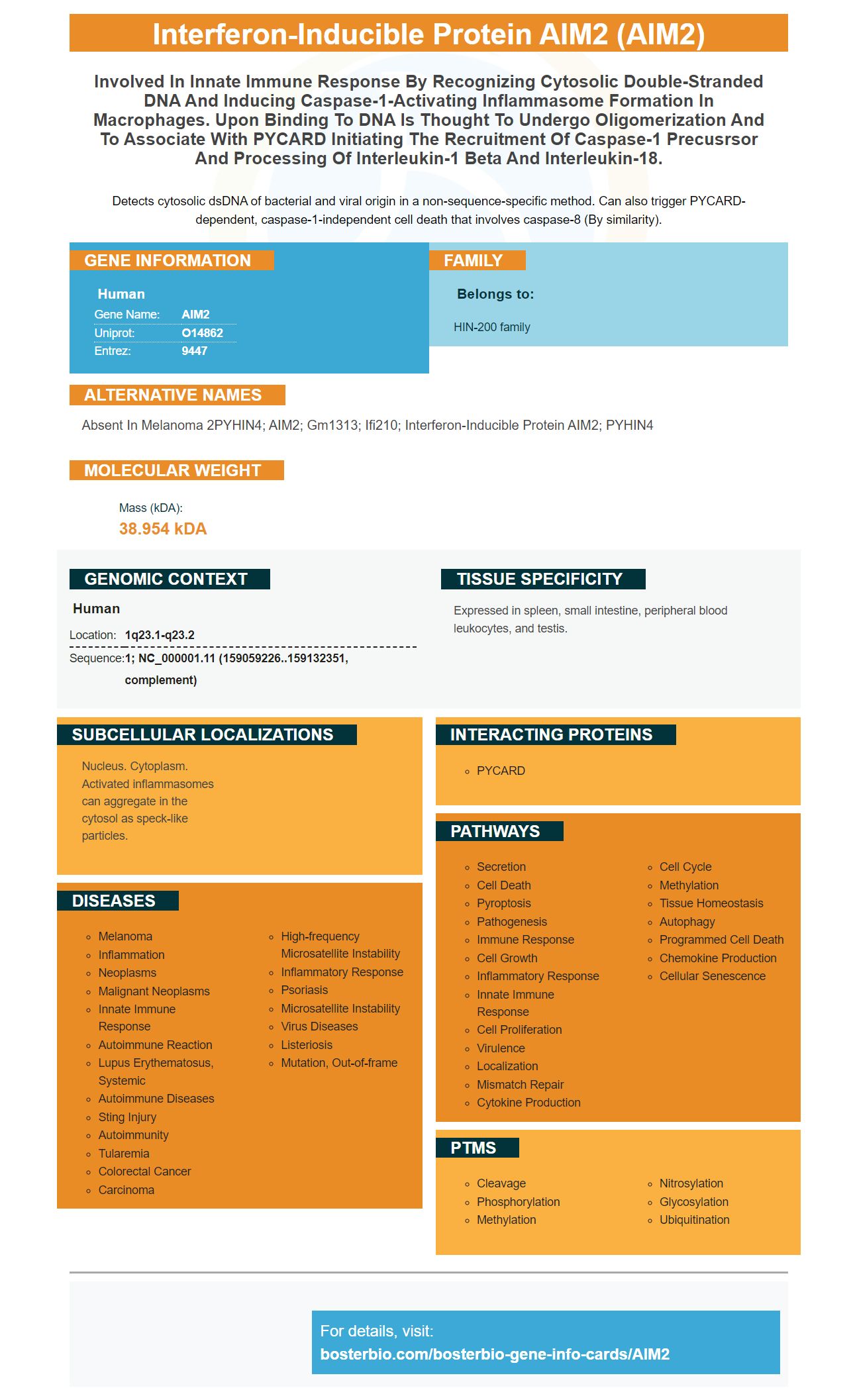
Facts about Interferon-inducible protein AIM2.
Detects cytosolic dsDNA of bacterial and viral origin in a non-sequence-specific method. Can also trigger PYCARD-dependent, caspase-1-independent cell death that involves caspase-8 (By similarity).
Human | |
---|---|
Gene Name: | AIM2 |
Uniprot: | O14862 |
Entrez: | 9447 |
Belongs to: |
---|
HIN-200 family |
absent in melanoma 2PYHIN4; AIM2; Gm1313; Ifi210; interferon-inducible protein AIM2; PYHIN4
Mass (kDA):
38.954 kDA
Human | |
---|---|
Location: | 1q23.1-q23.2 |
Sequence: | 1; NC_000001.11 (159059226..159132351, complement) |
Expressed in spleen, small intestine, peripheral blood leukocytes, and testis.
Nucleus. Cytoplasm. Activated inflammasomes can aggregate in the cytosol as speck-like particles.
Are you a researcher looking for high-affinity primary antibodies? If so, you've come to the right place. This article will go over Boster Bio's high-affinity antibodies and their application in ELISA and immunohistochemistry. Read on to learn more! Listed below are some of the best uses of the AIM2 marker antibodies. Listed below are just some of the many applications for Boster Bio's high-affinity primary antibodies.
Boster Bio manufactures quality primary and secondary antibodies for ELISA and WB. These products are validated and tested for ELISA and WB applications. Boster Bio offers rabbit polyclonal antibodies and free secondary antibodies with every purchase of a primary antibody. Boster Bio's range also includes antibodies for mouse and human samples, as well as controls and lysates.
Boster Bio's high-affinity antibodies are made with the AIM2 marker, a unique marker which distinguishes high-affinity primary antibodies from low-affinity ones. The AIM2 marker is the most widely used marker for human monoclonal antibodies. It allows for the precise measurement of specific analytes in crude preparations. High-affinity primary antibodies can be used for ELISA analysis.
It is imperative to properly document and describe antibodies in research. Antibodies are precious resources in the scientific community and if their controls are not properly documented, mistakes will continue to occur. Boster Bio's high-affinity primary antibodies use the AIM2 marker to minimize these problems. This marker is also important for the quality of the research performed using the antibodies. The quality of Boster Bio's antibodies is assured by its high quality and competitive prices.
Immunocytochemistry relies on primary antibodies that recognize a specific antigen with high affinity. The antigen-antigen molecule must not be too small to induce a robust immune response. The haptens and antigen-antibody chemistry become important as antigens are reduced in size. Consequently, the affinity of primary antibodies increases with the increasing concentration of the antibodies.
Primary antibodies are often labeled to distinguish them from secondary antibodies. The latter can cause cross-reactivity with the primary antibody. When secondary antibodies are used with the primary antibody, a nonspecific signal can result. Dual labeling of primary antibodies with primary antibodies also increases sensitivity. In addition, dual-antibody detection is highly sensitive, but requires more work and optimization.
AIM2 is a marker found on the surface of certain cells. It is used to distinguish a cell surface from a non-cellular environment. Antibodies are made from the immune system of animals. Their building blocks are proteins with complementary-determining regions. The definition of complementarity-determining regions is complex, but you can read more about them on Wikipedia. In the process of producing polyclonal primary antibodies, scientists first immunize host animals with antigens and then extract their sera and eggs.
Antibodies are selected on the basis of their specificity to epitopes on a larger antigen panel. The antibody specificity depends on the relative affinity of the antibody for related epitopes and cross-reactivity can result from polyspecificity or conserved interactions with the antigens presented in the application. An interesting and challenging application of immunohistochemistry is the use of microarrays of fixed tissues for global protein expression mapping.
As with any other laboratory procedure, the results of immunohistochemistry are dependent on the correct procedures and interpretation. The results of immunohistochemistry must be interpreted by a qualified pathologist and should not be based solely on the staining. The nature of the tissue under study and any interference between false positive and negative results are essential considerations. To improve the quality of immunohistochemistry, research must determine the true positive and negative reaction sites in a tissue sample.
Molecular analysis of endogenous Ngfr expression in the mouse has been achieved by high throughput immunohistochemistry using tissue microarrays. Example data from immunohistochemistry of human lymphoid tissue demonstrate the staining of cell surface receptor CD5, in the salivary gland, and in developing nerve tracts in an E14.5 embryo. Examples of nuclear staining of transcription factor ELF1 in the liver and uterus are also included.
Monoclonal anti-peptide antibodies have been raised against several synthetic peptides for the purpose of immunohistochemical study of neuronal histamine. These antibodies are suitable for use in immunohistochemistry as they react with epitopes that resist formalin fixation and wax embedding. The antibodies have excellent specificity and can be used in a variety of diagnostic techniques. And they are highly effective in identifying neuronal markers.
CA 125 is associated with epithelial gynecological tumors. Although there are few publications describing the use of CA 125 antibodies in immunohistochemistry, the antibody has shown excellent performance in formalin-fixed paraffin-embedded tissues. In addition to cancer cells, lung, breast, conjunctiva, and prostate glandular epithelium are all positive for this immunoreactive protein. Other areas that CA 125 has been associated with include the skin, esophagus, and fetal serosal linings of body cavities.
The latest WHO classification of lymphoma includes 33 subtypes, which vary in treatment and prognosis. However, diagnosis is difficult, resulting in a complicated immunohistochemical scheme for lymphoma. The CD20 and CD79a antibodies must be positive in both cases to determine B-cell lymphoma. CD3 expression and ALK translocations are also required for B-cell lymphoma diagnosis.
ELISA is an enzyme-linked immunosorbent assay (ELISA) that detects antibodies present in a sample. It can detect various types of antibodies, including antigens and hormones from pregnancy. ELISA can also be used to detect particular antibodies and determine their concentration. It is an important tool for detecting drugs and determining positive or negative status of diseases. Listed below are several applications of ELISA.
ELISA is used for detection of illicit and pharmaceutical drugs in the urine and serum. It also detects human chorionic gonadotrophin, or hCG, in urine. Pregnant women produce more hCG in urine, which can be detected using ELISA. ELISA can also be used to detect platelet antibodies in serum. This test is ideal for home use because it can be conducted without complex laboratory facilities.
ELISA uses an enzyme to convert the substance of interest into a coloured product. Spectrophotometry allows measurement of the colour. For example, an ELISA test for pregnancy uses purified HCG linked to an enzyme. The enzyme binds to the solid surface, and the more of the substance of interest, the more it reacts. Moreover, the less enzyme is linked to the substrate, the less the reaction occurs. Thus, ELISA is an accurate diagnostic tool.
Another application of ELISA is in the diagnosis of NVD. The disease is spread among birds and can infect humans. Among the different strains, velogenic is the most deadly. The other strains are mesogenic and lentogenic. ELISA is used to monitor the presence of NDV in a population, coordinating vaccination programs, and identifying infected flocks. These applications have numerous uses and are discussed in Practical Skills in Biomolecular Sciences.
The use of Mabs has extended the field of ELISA. For instance, one can use monoclonal antibodies to detect rock shrimp in meat samples. Under optimal conditions, detection of rock shrimp proteins is possible even at very low levels. Monoclonal antibodies can be used for adulteration detection in a variety of other applications, including identifying fish species or detecting tainted seafood products. For instance, in the case of adulteration, it is difficult to identify a piece of fish by physical appearance.
FPIA on MTPs is a less sensitive method of assaying asaP1. However, the detection limit is higher, and the lower limit is 14 times lower than the upper limit of ELISA. ELISA takes 20 hours to complete, while FPIA only takes ten minutes to complete. These two methods are often used for the same purpose. There are some similarities between the two methods, however, and they have several advantages.
ELISA is a less expensive method of platelet antibody detection compared to other commonly used methods. Its low-cost nature makes it particularly popular in the food industry. In fact, ELISA can detect low-level allergen contaminants that other methods cannot detect. It can also detect oils and other substances in the food supply. Consequently, it has numerous applications. This is an indispensable tool for the food industry. But if you're not sure whether ELISA is right for your needs, consider doing a simple test for free-from contaminants.
PMID: 9242382 by DeYoung K.L., et al. Cloning a novel member of the human interferon-inducible gene family associated with control of tumorigenicity in a model of human melanoma.
PMID: 15582594 by Cresswell K.S., et al. Biochemical and growth regulatory activities of the HIN-200 family member and putative tumor suppressor protein, AIM2.
*More publications can be found for each product on its corresponding product page