ALB / Albumin · IHC design guide

Design Immunohistochemistry for ALB

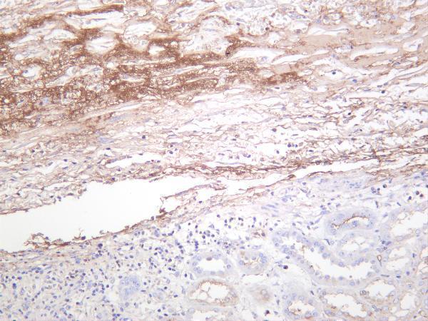
Plan chromogenic IHC-P for ALB using the catalog antibody's formaldehyde-fixed, paraffin-section validation (datasheet A01245). Interpret plasma and extracellular matrix staining with the tissue profile, bearing in mind that secreted ALB protein and RNA locations can differ (HPA tissue IHC).

Evidence assembled Oct 2026 · For research use; verify linked source records and product datasheet before use
Immunohistochemistry protocol sheet for ALB (IHC for ALB): expected localisation Plasma and extracellular matrix (HPA tissue IHC), antibody A01245, validated IHC image, and IHC protocol steps
Printable ALB IHC protocol sheet — expected localisation Plasma and extracellular matrix (HPA tissue IHC), antibody A01245, controls and protocol steps. Open the full ALB IHC guide →

ALB Immunohistochemistry Experimental Design Guide

Expected localisation, validated protocols, controls and antibodies — the at-a-glance facts below, then the full design guide.

Must know before staining
Expected localisation Plasma and extracellular matrix (HPA tissue IHC)
Staining pattern Plasma/ECM positivity; Bowman's capsule medium (HPA tissue IHC)
Antigen retrieval Citrate pH 6 HIER, heat-mediated (datasheet A01245)
Positive control ⓘ Kidney
Negative control ⓘ Adipose tissue+4 more · see all
Important caveats
Reasons your staining may differ from the expected pattern.
Fixation Formaldehyde-fixed paraffin sections (selected-SKU IHC image A01245); keep fixation consistent (standard IHC practice; not target-specific)
Caveat Secreted ALB may stain away from its RNA source (HPA tissue IHC)
Regulation Liver-enriched RNA expression (HPA RNA)
Isoform / epitope 3 isoforms; aa 1–24 removed during processing; verify epitope (UniProt)
Section 1

Recommended ALB IHC & IF Protocols

Compare the catalog antibody’s citrate pH 6 IHC-P protocol (datasheet A01245) with published ALB IHC methods for Japanese quail UVJ mucosa and liver sections (PMC7063080; PMC13149365).

Recommended immunohistochemistry (IHC-P) protocol parameters
SampleFormaldehyde-fixed, paraffin-embedded human liver tissue (datasheet A01245)
FixationImage formalin-fixed; duration unreported (datasheet A01245); verify before use.
Sectioning4–5 µm sections on charged slides (standard)
DeparaffinisationXylene, graded ethanol series to water (standard)
Antigen retrievalHeat retrieval: Citrate pH 6 (datasheet A01245); 20 min, 95–100 °C (standard)
Peroxidase block3% H2O2, 10 min, room temperature (standard)
Blocking10% serum (datasheet A01245)
Primary antibodyChicken anti-ALB, 10-20 μg/mL (datasheet A01245)
Primary incubationOvernight at 4 °C (standard)
DetectionHRP-polymer secondary, DAB chromogen 5–10 min (standard)
CounterstainHematoxylin, blue, dehydrate and mount (standard)
Expected resultALB-positive staining in bowman's capsule of kidney (HPA tissue IHC: Medium). HPA tissue profile: Positivity in plasma and extracellular matrix. No signal in the no-primary control.
💡Decision noteTry heat-mediated citrate pH 6 retrieval first (datasheet A01245); neither published ALB IHC excerpt specifies a retrieval method (PMC7063080; PMC13149365).
Section 2

What Is the Expected ALB Staining Pattern?

ALB is secreted and has no transmembrane segment (UniProt P02768: subcellular location and topology). In tissue IHC, expect signal in plasma and extracellular matrix, with medium staining reported at the kidney’s Bowman’s capsule (HPA: tissue IHC). HPA does not establish a generally positive cell type; its Enhanced reliability note cautions that secreted protein and tissue RNA can occur in different locations (HPA: reliability description).

What am I looking at on my slide?
Chromogenic signal outlines extracellular material or appears in plasma, with medium signal at Bowman’s capsule.This fits the reported tissue pattern (HPA: positivity in plasma and extracellular matrix; kidney Bowman’s capsule, Medium). Score the compartment and structure rather than treating every nearby cell as ALB positive (HPA: tissue IHC).
Strong, sharply nuclear or membrane-restricted staining dominates the section.Treat this as a questionable ALB pattern: ALB is secreted and has no transmembrane segment (UniProt P02768: location and topology). Review morphology and controls before assigning a cellular location; the supplied tissue IHC profile reports extracellular positivity (HPA: tissue IHC).
Adipocytes or other cells listed as negative show a distinct positive deposit.Check for cross-reactivity or endogenous detection activity before calling these cells positive (general IHC practice). HPA reports adipocytes in adipose tissue and breast, among other listed cell populations, as Not detected (HPA: tissue IHC).
A weak, diffuse deposit covers positive and negative regions alike.A uniform haze cannot distinguish the reported extracellular ALB pattern from background (HPA: tissue IHC; general IHC practice). Compare a no-primary control and inspect blocking, washing and detection conditions (general IHC practice).
No signal appears at Bowman’s capsule or in expected extracellular material.A blank result needs an assay check before biological interpretation: HPA reports medium staining at kidney Bowman’s capsule and positivity in plasma and extracellular matrix (HPA: tissue IHC). Confirm that the control contains the relevant structure, then review the antibody and detection workflow (general IHC practice).
💡Expected ALB appearanceCall a result consistent with ALB when chromogenic signal follows plasma or extracellular matrix and reaches medium intensity at kidney Bowman’s capsule; isolated strong nuclear signal or staining of HPA-listed negative cells warrants investigation (HPA: tissue IHC; UniProt P02768: secreted location).
How each factor affects the staining
Secretion and tissue RNAALB is secreted (UniProt P02768: subcellular location). HPA reports liver-enriched RNA but warns that the tissue location of RNA and secreted protein may differ; RNA enrichment alone does not establish a liver-cell IHC pattern (HPA: RNA specificity and reliability description).
Processing and epitope coverageThe precursor has a signal peptide at residues 1–18, a propeptide at 19–24, and a mature albumin chain at 25–609 (UniProt P02768: processing). Antibody recognition of a particular segment cannot be inferred without an epitope specification (general IHC interpretation).
Isoforms and antibody validationUniProt lists 3 ALB isoforms (UniProt P02768: isoforms). HPA rates HPA031024 and HPA031025 Enhanced for IHC, and CAB006262 Supported; these ratings do not specify which isoforms each antibody recognizes (HPA: antibody validation; UniProt P02768: isoforms).
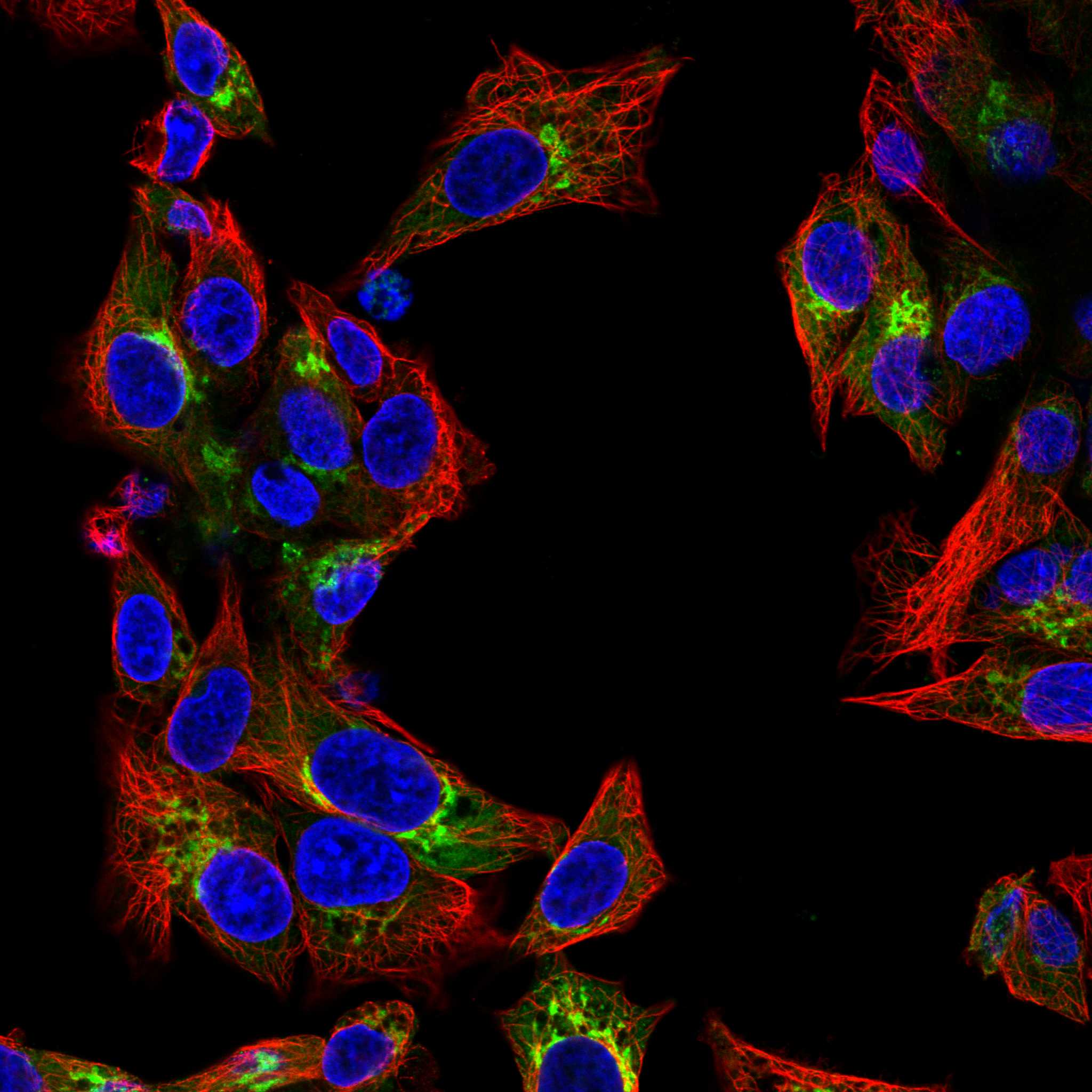
IF/ICC Q: What if intracellular ALB is visible?A: HPA’s ICC-IF summary places ALB in the endoplasmic reticulum and Golgi apparatus, with images from Hep-G2 and U2OS (HPA: subcellular ICC-IF). Interpret that observation in the IF/ICC assay context; tissue IHC reports plasma and extracellular matrix positivity (HPA: tissue IHC).
Antigen retrievalRetrieval conditions are an assay variable in paraffin-section IHC (general IHC practice). The supplied HPA and UniProt records give no ALB-specific retrieval or fixation-sensitivity result, so a weak stain cannot be attributed to those effects from this evidence alone (HPA: tissue IHC; UniProt P02768).
Why is my staining missing, weak or wrong?
SituationLikely causeNext action
Bowman’s capsule is blank in an intended positive section.The control may lack the relevant structure, or the assay may have failed; HPA reports Medium staining specifically at Bowman’s capsule (HPA: kidney tissue IHC).Verify the structure on the counterstained section, then check antibody use, retrieval and detection against the assay’s validated instructions (general IHC practice).
All compartments show similar pale chromogen.Nonspecific detection or inadequate washing may obscure the extracellular pattern (general IHC practice; HPA: tissue IHC profile).Compare a no-primary control, inspect wash and blocking steps, and adjust detection conditions under the assay’s controls (general IHC practice).
Signal appears in an HPA-listed negative cell population.Cross-reactivity or endogenous detection activity is possible; the listed population is Not detected in HPA tissue IHC (HPA: tissue IHC; general IHC practice).Check a no-primary control; if using enzyme detection, check the matching endogenous-enzyme control, then reassess the antibody result (general IHC practice).
Staining is predominantly nuclear or follows cell membranes.That distribution conflicts with ALB’s secreted, non-transmembrane annotation and the reported extracellular tissue profile (UniProt P02768: location and topology; HPA: tissue IHC).Recheck compartment assignment against morphology and controls before recording ALB-positive cells (general IHC practice).
A liver section is assumed to be an IHC positive control solely because ALB RNA is enriched there.Liver-enriched RNA does not specify a liver-cell protein staining pattern for this secreted protein (HPA: RNA specificity and reliability description).Use the documented Bowman’s capsule signal and extracellular pattern as interpretation anchors, while verifying that the chosen control contains the relevant structures (HPA: tissue IHC; general IHC practice).
IHC and ICC-IF appear to place ALB in different compartments.HPA reports extracellular tissue IHC positivity and endoplasmic reticulum/Golgi localization in ICC-IF (HPA: tissue IHC; subcellular ICC-IF).Record each result with its assay and compartment; evaluate tissue IHC against the extracellular profile and ICC-IF against the reported intracellular locations (HPA: tissue IHC; subcellular ICC-IF).

Sample controls for ALB IHC & IF

🧪Run kidney first and look for staining in Bowman's capsule cells (HPA: Kidney, Bowman's capsule, Medium). Use adipose tissue adipocytes as the negative tissue (HPA: Adipose tissue, adipocytes, Not detected); on the kidney slide, internal negative cells should show no specific signal above the no-primary background, but HPA does not identify a particular negative kidney cell type.
Positive control tissue: Kidney (Bowman's capsule, HPA Medium)
Negative control tissue: Adipose tissue (HPA Not detected)
ICC-IF cell lines (HPA subcellular resource): HPA ICC-IF images show ALB in Hep-G2, U2OS, with annotated localisation: Endoplasmic reticulum (approved), Golgi apparatus (approved) (HPA subcellular).
Technical controls: Include a no-primary (secondary-only) control, a host- and immunoglobulin-class-matched isotype control or matched nonimmune IgG for a polyclonal primary, and an ALB knockout sample or immunizing-peptide competition if available (standard IHC practice). Quench endogenous peroxidase and compare staining with the no-primary slide when using HRP detection (selected A01245 tissue-IHC caption: HRP secondary; standard IHC practice).
⚠️Feasibility: The selected A01245 paraffin-section caption reports formaldehyde fixation and heat-mediated citrate retrieval at pH 6, but gives no target-specific fixation window or evidence that retrieval is required (selected A01245 tissue-IHC caption). Paraffin IHC is directly illustrated; HPA also reports ICC-IF images in Hep-G2 and U2OS, but these sources do not establish that frozen sections or IF are easier (selected A01245 tissue-IHC caption; HPA: subcellular). Because ALB is secreted and present in plasma, extracellular or vascular signal can complicate assignment of kidney staining to cells (UniProt P02768: Secreted, tissue specificity Plasma).

HPA tissue IHC evidence for ALB

Comprehensive Human Protein Atlas IHC scoring per tissue (reliability: Enhanced — At least one protein variant secreted, tissue location of RNA and protein might differ and correlation is complex.). Rows are taken directly from the HPA tissue chart — click any row's HPA link to view the source.

Positive expression · recommended positive controls

TissueCell typeLevelEvidenceSource
Kidney Bowman's capsule Medium Protein (IHC) HPA →

Undetected expression · recommended negative controls

TissueCell typeLevelEvidenceSource
Adipose tissue Adipocytes Not detected Protein (IHC) HPA →
Adrenal gland Glandular cells Not detected Protein (IHC) HPA →
Appendix Glandular cells Not detected Protein (IHC) HPA →
Bone marrow Hematopoietic cells Not detected Protein (IHC) HPA →
Breast Adipocytes Not detected Protein (IHC) HPA →
Section 3

Advanced ALB IHC Tips

Troubleshoot ALB staining in paraffin-section chromogenic IHC by checking retrieval, compartment, extracellular signal, and controls (datasheet A01245; UniProt P02768; HPA tissue IHC).

What retrieval should I use when ALB staining is weak in paraffin sections?
Start with heat-mediated citrate buffer at pH 6 for paraffin-section ALB IHC (datasheet A01245). The catalog image used this retrieval with 2 µg/ml primary antibody, so repeat those conditions before changing several variables together (datasheet A01245; standard IHC practice). If staining remains weak, compare retrieval heating and cooling settings on adjacent sections while holding antibody concentration and detection constant (standard IHC practice). Check tissue morphology alongside signal, because excessive heating can damage sections and complicate compartment scoring (standard IHC practice). Include the same positive control in each run so a retrieval change can be distinguished from run variation (standard IHC practice).
How should I troubleshoot fixation-related variation in ALB IHC?
ALB-specific sensitivity to fixation duration or fixative composition is unknown from the supplied evidence; treat fixation effects as an experimental variable (supplied evidence). Record the fixative and processing history for each paraffin block, then compare sections with similar handling before attributing intensity differences to biology (standard IHC practice). Use the documented citrate retrieval at pH 6 and 2 µg/ml primary antibody as consistent starting conditions (datasheet A01245). Inspect morphology and section adhesion when staining varies, since processing damage can make chromogenic scoring unreliable (standard IHC practice). Report any fixation comparison as a local validation result, rather than an established ALB-specific effect (standard IHC practice).
Should ALB appear inside cells or in extracellular spaces?
Expect substantial extracellular or plasma-associated staining: ALB is secreted, and tissue IHC reports positivity in plasma and extracellular matrix (UniProt P02768; HPA tissue IHC). Intracellular signal may reflect secretory trafficking; IF localisation includes endoplasmic reticulum and Golgi apparatus (HPA subcellular). In paraffin sections, score intracellular and extracellular deposits separately, because a stained space does not identify its producer (standard IHC practice; UniProt P02768). Liver-enriched RNA supports liver as a useful expression context, while medium staining in kidney Bowman’s capsule shows why location alone needs careful interpretation (HPA tissue IHC). Compare stained structures with morphology and a negative reagent control before assigning cellular localisation (standard IHC practice).
Could processing or ALB isoforms explain different staining patterns?
ALB has 3 listed isoforms and a precursor whose signal peptide and propeptide precede the mature albumin chain (UniProt P02768). The supplied antibody evidence does not map its epitope, so isoform recognition and sensitivity to precursor processing remain unestablished (supplied evidence). Albumin also has annotated glycosylation sites and modified residues; their effect on this antibody’s tissue staining has not been shown (UniProt P02768; supplied evidence). Check the antibody’s immunogen and epitope documentation before interpreting a compartment difference as isoform-specific detection (standard IHC practice). Keep retrieval and antibody concentration fixed while comparing sections, so technical variation does not masquerade as epitope biology (standard IHC practice).
How can I examine ALB by IF alongside a cell-type marker?
For IF, multiplex ALB with a marker of the expected cell type and examine whether intracellular signal follows the secretory pathway (HPA subcellular; standard IF practice). HPA reports ALB in endoplasmic reticulum and Golgi apparatus, while tissue IHC also finds extracellular signal (HPA subcellular; HPA tissue IHC). Choose spectrally separated fluorophores and assess tissue autofluorescence in an unstained section, favouring a channel with lower background for the weaker target (standard IF practice). ALB has no transmembrane segment; permeabilise when testing intracellular epitopes, but assess extracellular staining without requiring membrane access (UniProt P02768; standard IF practice). Include single-stain controls to distinguish marker overlap from spectral bleed-through (standard IF practice).
What should I check when ALB IHC shows diffuse brown background?
First compare the stained section with a no-primary control to identify detection-system or endogenous-enzyme signal (standard IHC practice). Apply a peroxidase block for chromogenic HRP detection and optimise washes before raising the primary concentration (standard IHC practice). The catalog image used 10% serum blocking for 1 h, 2 µg/ml primary antibody, and an HRP secondary at 1:250 (datasheet A01245). Diffuse extracellular staining can still represent albumin, because ALB is secreted and tissue IHC reports plasma and extracellular-matrix positivity (UniProt P02768; HPA tissue IHC). Judge background against morphology, edge staining, and the no-primary control rather than colour intensity alone (standard IHC practice).
How should I quantify ALB when staining is both cellular and extracellular? ⚠ ANSWER MARKED FOR VERIFICATION
Define separate regions for cellular staining and extracellular or plasma-associated deposits before measuring chromogenic signal (HPA tissue IHC; standard IHC practice). For cells, report the percentage positive and an H-score of 0–300 when intensity grades are reproducible (standard IHC practice). For extracellular signal, report positive area or optical density per mm² of evaluable tissue rather than assigning deposits to nearby cells (standard IHC practice). Normalise cellular counts to the relevant cell population and extracellular measurements to analysed tissue area, excluding folds and necrosis consistently (standard IHC practice). Keep retrieval, imaging, threshold, and control sections consistent across samples before comparing groups (standard IHC practice).
How can I distinguish genuine ALB staining from artefact?
A plausible ALB pattern includes extracellular or plasma-associated signal, while intracellular signal should be evaluated against secretory-pathway localisation (UniProt P02768; HPA tissue IHC; HPA subcellular). Do not infer local synthesis from extracellular staining alone, especially where tissue and RNA locations can differ for a secreted protein (HPA tissue IHC). Compare suspected positive cells with morphology and an appropriate cell-type marker when identity matters (standard IHC practice). Discount signal confined to section edges, folds, or necrotic areas until it reproduces in intact tissue (standard IHC practice). A no-primary control and peroxidase block help identify endogenous-enzyme or detection artefacts in chromogenic IHC (standard IHC practice).
Boster reagents

Best ALB / Albumin IHC Antibodies

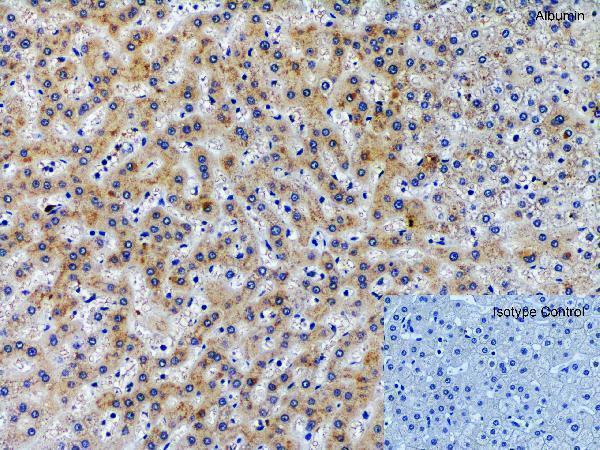
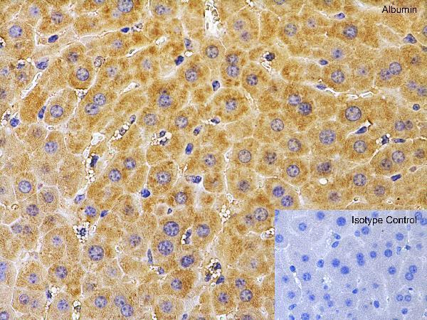
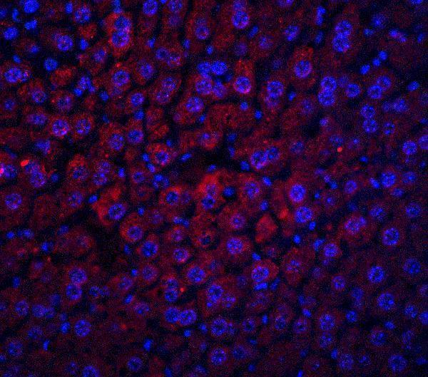
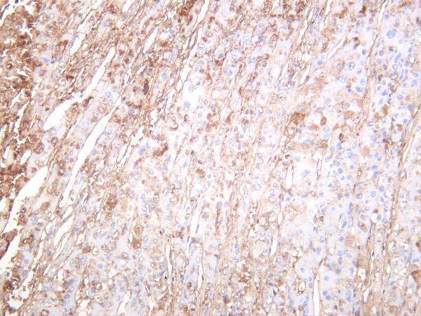
ALB IHC images cover human, mouse and rat liver, human hepatocellular carcinoma, and human and rat kidney (A01245 and M01245-4 IHC captions); mouse-liver IF is also shown (A01245 IF caption).

Real IHC data Immunohistochemistry Validation of Albumin in Human Liver Immunohistochemical analysis of paraffin-embedded human liver tissue using anti-albumin antibody (A01245) at 2 μg/ml. Tissue was fixed with formaldehyde and blocked with 10% serum for 1 h at RT; antigen retrieval was by heat mediation with a citrate buffer (pH6). Samples were incubated with primary antibody overnight at 4 ˚C. A goat anti- chicken IgY (HRP) at 1/250 was used as secondary. Counter stained with Hematoxylin.
Anti-Albumin Antibody
Cat # A01245
Real IHC data Human hepatocellular carcinoma was stained with anti-Albumin rabbit antibody
Anti-Albumin Rabbit Monoclonal Antibody
Cat # M01245-4
Real IHC data Immunohistochemical analysis of paraffin-embedded human liver, using Serum Albumin Antibody.
Anti-Albumin Rabbit Monoclonal Antibody
Cat # M01245

A01245 shows paraffin-section IHC in human, mouse and rat liver (A01245 IHC captions), while M01245 shows paraffin-section IHC in human liver (M01245 IHC caption). M01245-4 shows IHC in human hepatocellular carcinoma and human and rat kidney (M01245-4 IHC captions).

Which to pick: Choose A01245 for cross-species liver IHC: its chicken polyclonal antibody has human, mouse and rat paraffin-section images, whose captions report formaldehyde fixation and citrate pH 6 retrieval (A01245 catalog and IHC captions). For IF with an image, choose A01245 for mouse liver (A01245 IF caption); for ICC, consider A01245-4, which lists ICC but supplies no ICC image (A01245-4 catalog). For rabbit monoclonal tissue IHC, M01245 shows paraffin-section human liver and M01245-4 shows human hepatocellular carcinoma and human and rat kidney; fixation is unreported in their captions (M01245 and M01245-4 catalog and IHC captions).

Each figure is that product's own IHC / IF validation image from its datasheet.

References

  1. UniProt Consortium. UniProt entry P02768 (ALBU_HUMAN, Albumin).
  2. Human Protein Atlas. ALB tissue IHC expression (reliability: Enhanced).
  3. Human Protein Atlas. ALB subcellular location (ICC-IF): Localized to the Golgi apparatus and endoplasmic reticulum..
  4. Human Protein Atlas. ALB antibody validation summary (3 antibodies).
  5. Combined analysis of albumin in situ hybridisation and C reactive protein immunohistochemistry for the diagnosis of intrahepatic cholangiocarcinoma: towards a molecular classification paradigm. Journal of clinical pathology 2025 — PMC12015024.
  6. Expression of Transferrin and Albumin in the Sperm-Storage Tubules of Japanese Quail and their Possible Involvement in Long-Term Sperm Storage. The journal of poultry science 2020 — PMC7063080.
  7. Synergistic amelioration of cholestatic liver fibrosis by combined total astragalus saponins and AAV8.Numb-Exon3 through modulation of hepatic progenitor cell differentiation. Frontiers in pharmacology 2026 — PMC13149365.
  8. Nrf2 induces malignant transformation of hepatic progenitor cells by inducing β-catenin expression. Redox biology 2022 — PMC9618468.
  9. PubMed PMID:6171778 — UniProt-cited evidence.
  10. PubMed PMID:6275391 — UniProt-cited evidence.
  11. PubMed PMID:3009475 — UniProt-cited evidence.