This website uses cookies to ensure you get the best experience on our website.
- Table of Contents
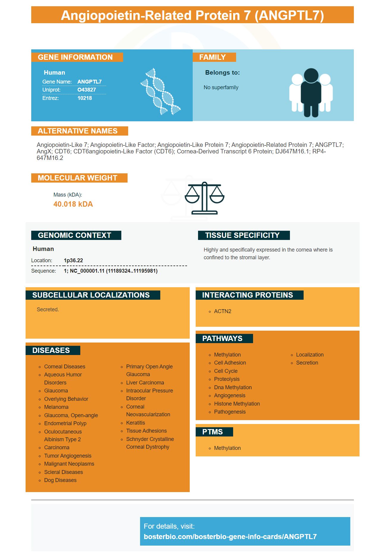
Facts about Angiopoietin-related protein 7.
Human | |
---|---|
Gene Name: | ANGPTL7 |
Uniprot: | O43827 |
Entrez: | 10218 |
Belongs to: |
---|
No superfamily |
angiopoietin-like 7; Angiopoietin-like factor; Angiopoietin-like Protein 7; angiopoietin-related protein 7; ANGPTL7; AngX; CDT6; CDT6angiopoietin-like factor (CDT6); Cornea-derived transcript 6 protein; dJ647M16.1; RP4-647M16.2
Mass (kDA):
40.018 kDA
Human | |
---|---|
Location: | 1p36.22 |
Sequence: | 1; NC_000001.11 (11189324..11195981) |
Highly and specifically expressed in the cornea where is confined to the stromal layer.
Secreted.
You've found the right place if you're searching for a protein that modulates ECM of the TM. This article will describe how Angptl7 works to regulate MMP mediated ECM turnover, which is vital for the regeneration and maintenance of tissue. Its uses are wide-ranging and can help your research in several fields.
Angptl7/CDT6 makes up an essential part of the TM’s extracellular matrix (ECM). It is thought to influence fibronectin matrix assembly, a key organizer and signaling molecule of the ECM. An immunohistochemistry test has shown that ANGPTL7/CDT6 are expressed in the TM at lower levels than in the cornea. However, this result may not be conclusive as the Angptl7/CDT6 siRNA-treated eye showed a higher MYOC staining intensity than the control.
Overexpression of ANGPTL7/CDT6 has no effect on tumor growth in immunocompetent mice, indicating that ANGPTL7/CDT6 may directly regulate the production and configuration of ECM proteins. However, in vivo studies are necessary to confirm these findings in other species. The tumor growth rate is not affected by Angptl7/CDT6 expression in human fibroblasts.
Six ECM proteins were less likely to be expressed in HTM69 cells when ANGPTL7/CDT6 was overexpressed. The expression of fibronectin was decreased 2.3-fold in mice with higher levels of Angptl7/CDT6. Versican was not affected by 786 -fold overexpression, while collagen type IA1 (and type IV) expression decreased by 1.2 fold.
Deduced angptl7 like genes exist in humans. However their corresponding proteins found in cephalochordates (lamprey) and invertebrates share highest sequence similarities with vertebrate ANGPTLs. Angptl7 has the most conserved FReD region. The unique N-glycosylation site in the deduced protein of cephalochordates or spotted gars is found in the deduced protein of cephalochordates.
ANGPTL7 is highly conserved in fish and humans, sharing 69 amino acid sequences. However, human ANGPTL7 is distinct from those in lizards, chicken, and other non-mammals. Non-mammals and fish lack Angptl8. This is because the human ANGPTL8 genetic code has been found in a wide range species.
ANGPTL family members play key roles in regulating skin regeneration. In fish, the expression of the human ANGPTL7 gene regulates skin regeneration. It is also associated with the initial stages of skin regeneration. Future research will shed light on the molecular foundation of skin regeneration. ANGPTL genes are also important for disease control in aquaculture.
The human TM consists primarily from beams of connective material that contain collagen and elastic fibers. These beams rest on a basement membrane. However, the outermost JCT is devoid of collagenous beams. It is characterized by cell layers that are immersed in an ECM fibril rich web. The adjacent SC acts as an endothelial conduit. Smooth muscle-specific, smooth proteins are expressed in TM cells.
TM cells are essential for maintaining ocular pressure homeostasis. The Rho/ROCK pathway activity causes increased permeability, and contraction of TM. Inhibition of the pathway increases the outflow facility of the TM and decreases the IOP. These results suggest Angptl7 modulates TM’s ECM for control of aqueous humour outflow.
Angptl7 a novel MMP inhibitor which modulates the activity MMPs. This inhibitor blocks the expression of MMP14 enzyme. MMP14 is an example of a membrane type (MT), MMP with intrinsic protease activity. It cleaves a variety of ECM components such as collagen IV, integrin Av subunit, transglutaminase, and others.
MMPs, zinc-dependent endopeptidases, can be secreted and diffused to their site of action. MMP-2, MMP-9 are the most well-studied MMPs. They degrade major ECM elements found in kidney tissue and other parts of the body. TACE inhibits MMP-2 activation, and deoxynucleotides block MMPs synthesis.
This inhibitor inhibits UB Branching Morphogenesis, a vital developmental process in a kidney. Furthermore, it suppresses the activity of MMP1-MMP, an inhibitor of MMP-mediated ECM turnover. ANGPTL7 has the potential for cancer patients and is a promising therapeutic candidate. However, more research is needed to confirm these findings.
MMPs are essential for normal kidney development. MMP1 plays a vital role in the formation of the kidney basement membrane. In addition, MMP2 regulates renal tubular cell migration and proliferation, and MT1 regulates both. The protein cleaves many ECM molecules, including collagen.
A high level of MMP expression can lead to poor prognosis and increased cancer aggressiveness. This enzyme plays a crucial role in basement membrane breakdown by invading cellular cells. Furthermore, it has been shown that increased MMP expression leads to aggressive stages of tumors. In the future, drug designers will likely use these enzyme inhibitors as therapeutic or anti-cancer agents. The inhibition of MMP-14 is the best treatment option for this enzyme.
Xu X is another inhibitor that inhibits MMP-2 Gelatinolysis by targeting exodomain/substrate interactions. Higashi S. designed it. J Biol Chem 288(13), 9066-9076. Hashimoto H. also studied its structure. He found it to be highly selective and powerful. Inhibition of MMP-2 expression in breast cancer cells is linked to inhibition of tumor development.
In addition, MMC induced expression of IL-8 and MCP-1 in corneal fibroblasts and induced protein secretion. MMC inhibits ECM destruction processes via mitogenactivated proteinkinases. These studies will be used to develop in vitro models of pathophysiology. In addition, MMC is a viable candidate for tissue engineering by self-assembly therapies.
Inflammatory arthritis in mice is caused by ANGPTL7 inhibition. In vitro studies in mice with ischemic arthritis have shown that this inhibitor promotes fibroblast survival. A mouse model for this inhibitor is in development. These findings are the first direct evidence that this inhibitor has therapeutic potential. In the meantime, ANGPTL7 is being explored as a therapeutic agent for cancer and other inflammatory diseases.
It is not clear what ANGPTL7 does in TM's ECM. However, it may be involved the regulation of lipids or glucose metabolism. These proteins are secreted in cells that express them. They can also be found in the circulatory system of living rats as well as humans. They have been implicated previously in the regulation of scar tissue formation and the development traumatic brain injuries.
The TM is a critical component of the human retina, regulating intraocular pressure, outflow facility, and ER structure. The cells in the TM phagocytose the debris through the outflow path, which helps maintain IOP. Although the true underlying pathogenesis of POAG remains unknown, several risk factors have been linked to the disease, including aging, family history, genetics, and hypertension. It has also been linked myopia. Furthermore, elevated IOP is a major risk factor for retinal ganglion cell death, which ultimately leads to progressive vision loss.
AngPTL7/CDT6 transcription has been shown reduce versican's expression in the TM. However it has been shown that its splicing wasoforms have been upregulated. Versican is a chondroitin Sulfate Glycan that binds with fibronectin and other ECM constituents. Versican has been implicated with signal transduction, cell attachment and contraction.
ANGPTL7/CDT6 silencing reduced collagen type IA1 in TM and fibronectin in TM. These decreases were statistically not significant. However, it is possible that these proteins are counterproductive to elevated IOP. Interestingly, MMP1 expression was upregulated in the fibronectin-reduced RNA samples. MMP1 is an interstitial collagenase which breaks down ECM collas. MMP1 was extensively studied in the trabecular network, but the gene also has increased outflow capabilities in organ culture.
AngPTL7 was previously identified as a chemokine receptor that regulates TM's ECM. This receptor is involved with signaling and localization TMSCs. In another study, TMSCs were treated with recombinant SDF1a or SDF1b. Neither antibody reduced SDF1 expression but increased TMSC attachments to TM-SDF1 treated TM cells.
In experiments to determine their roles in TMs, the experiments focused on the CDT6 protein and gene ANGPTL7. The levels of mRNA in mice expressing ANGPTL7/CDT6 were lower. The siRNA angPTL7/CDT6 also reduced mRNA levels by 30.3-fold in TM cells. Both CDT6 and ANGPTL7 siRNA were induced in HTM cells by DEX induction.
The iPSCTM-TM cell line has been successfully used to regenerate TM eyes cells. TM cellularity is declining with age. However this cell type can still be used to replace the degenerated TM during eye disease. The study involved the infusion into the eye of iPSCTM-TM cells. The transplanted cells maintained normal IOP levels, and promoted the proliferation endogenous TM cell.
AngPTL7/CDT6 has been inserted in the antisense orientation intron of MTOR gene. This makes it unlikely that MTOR gene transcription would affect ANGPTL7/CDT6. In contrast, the Tg-MYCY437H mice with mutations in myocilin were found to have increased endogenous TM cell proliferation after transplantation. These results support those of other studies.
PMID: 9727400 by Peek R., et al. Molecular cloning of a new angiopoietin-like factor from the human cornea.
PMID: 11426320 by Stover C., et al. The human gene for mannan-binding lectin-associated serine protease-2 (MASP-2), the effector component of the lectin route of complement activation, is part of a tightly linked gene cluster on chromosome 1p36.2-3.