This website uses cookies to ensure you get the best experience on our website.
- Table of Contents
3 Citations 6 Q&As


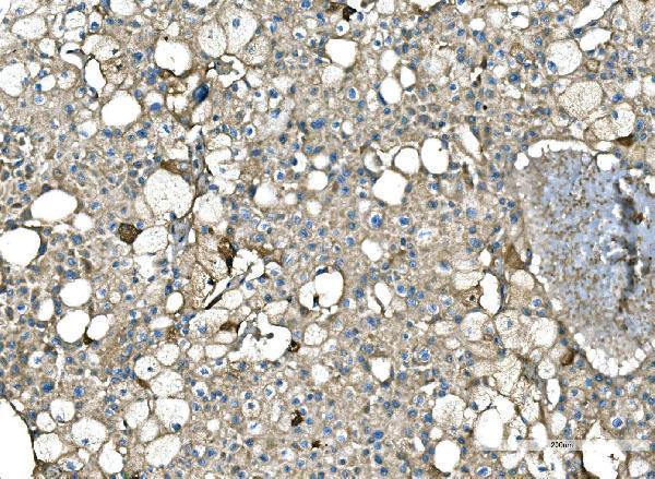
Facts about Serum amyloid P-component.

.
| Human | |
|---|---|
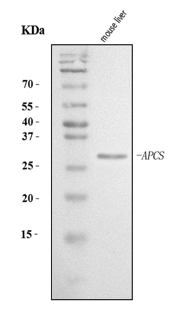
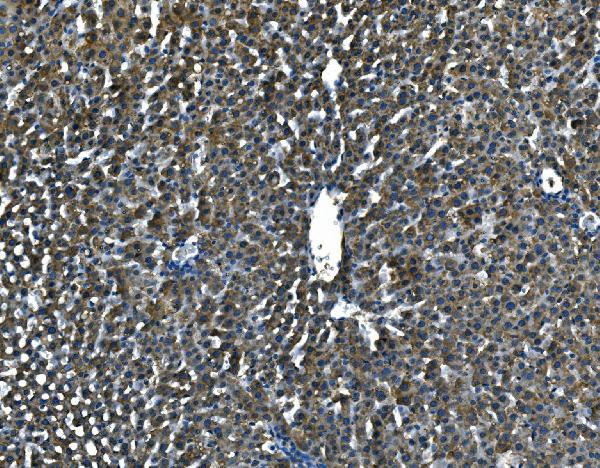
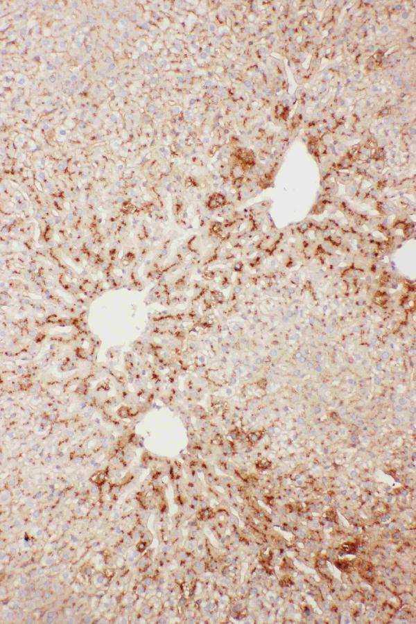
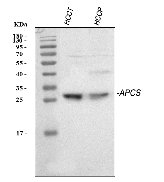
| Gene Name: | APCS |
| Uniprot: | P02743 |
| Entrez: | 325 |

| Belongs to: |
|---|
| pentraxin family |

9.5S alpha-1-glycoprotein; amyloid P component, serum; APCS; MGC88159; Pentraxin 2; PTX2; PTX2serum amyloid P-component; SAP; SAPpentaxin-related; Serum Amyloid P component
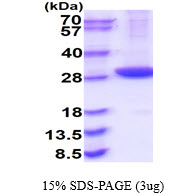
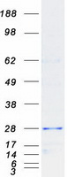
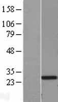
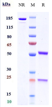
Mass (kDA):
25.387 kDA

| Human | |
|---|---|
| Location: | 1q23.2 |
| Sequence: | 1; NC_000001.11 (159587826..159588865) |
Found in serum and urine.
Secreted.


PMID: 2987268 by Mantzouranis E.C., et al. Human serum amyloid P component. cDNA isolation, complete sequence of pre-serum amyloid P component, and localization of the gene to chromosome 1.
PMID: 3029048 by Ohnishi S., et al. Isolation and characterization of the complete complementary and genomic DNA sequences of human serum amyloid P component.
*More publications can be found for each product on its corresponding product page