This website uses cookies to ensure you get the best experience on our website.
- Table of Contents
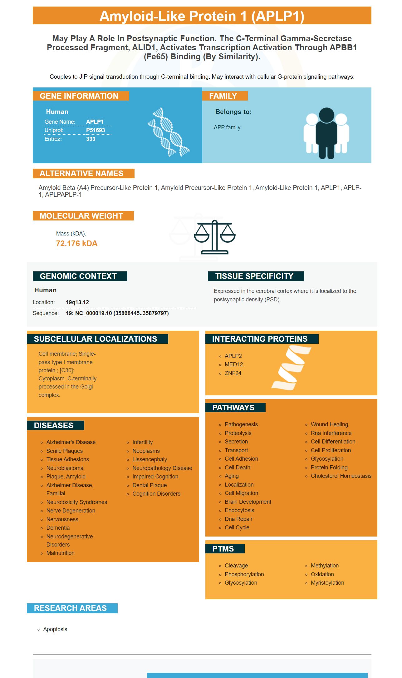
Facts about Amyloid-like protein 1.
Couples to JIP signal transduction through C-terminal binding. May interact with cellular G-protein signaling pathways.
Human | |
---|---|
Gene Name: | APLP1 |
Uniprot: | P51693 |
Entrez: | 333 |
Belongs to: |
---|
APP family |
amyloid beta (A4) precursor-like protein 1; amyloid precursor-like protein 1; amyloid-like protein 1; APLP1; APLP-1; APLPAPLP-1
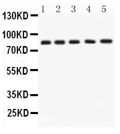
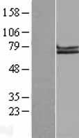
Mass (kDA):
72.176 kDA
Human | |
---|---|
Location: | 19q13.12 |
Sequence: | 19; NC_000019.10 (35868445..35879797) |
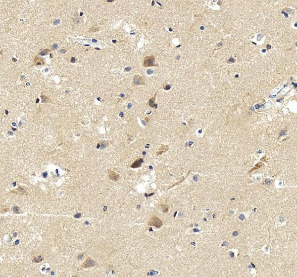
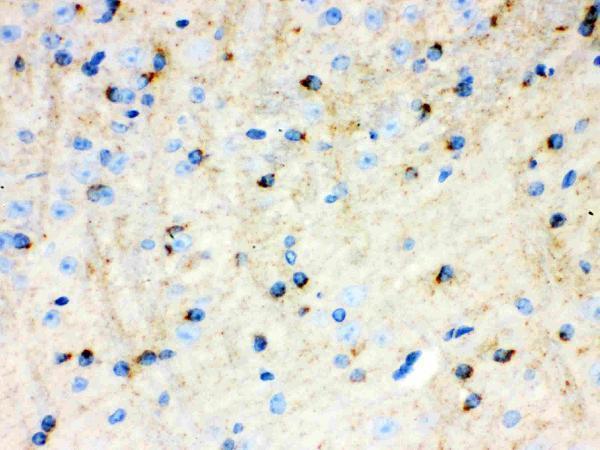
Expressed in the cerebral cortex where it is localized to the postsynaptic density (PSD).
Cell membrane; Single-pass type I membrane protein.; [C30]: Cytoplasm. C-terminally processed in the Golgi complex.
The APLP1 protein is a member of the family of proteins with conserved domains of homology. Despite its complexity, it is widely used in biomedical research, and its humanization and cloning possibilities are exciting and promising. But there are some problems that may arise from this protein. Fortunately, there are ways to prevent these problems and make your research more successful.
While APLP1 has no physiological role, its location on the chromosome is interesting given its relationship to Alzheimer's disease. The amyloid protein that is the hallmark of Alzheimer's disease is composed of the 39-43 amino acid bA4 peptide. APP is encoded by a gene on chromosome 21. Because the APLP1 marker is located on the same chromosome as APP, researchers can easily find out whether the two proteins are related.
A functional derivative of APLP1 is a modified or artificial protein with similar biological activities and immunological properties to APLP. It may not contain post-translational modifications necessary for its function. It may also include fragments, variants, analogues, or chemical derivatives. Its use is not limited to research purposes. Its application has been praised for its potential for gene therapy.
APLP1 is a membrane associated protein whose cDNA code encoding a family of proteins. The protein localizes in the golgi, where it is expressed. To find if the protein is expressed in a cell, it must be isolated from its host. The cDNA sequence of APLP1 can be obtained from bosterbio.
The APLP1 clone is located on human chromosome 19. The sequence is similar to that of the amyloid precursor protein, and thus can be used to identify APLP gene clones in research. It has also been shown to be compatible with several human clone sequences. It is important to note that there are a number of potential uses for APLP1 and other APLP markers in research.
APLP1 can be detected in adult human brains by hybridization with FB63. It can be used to detect APLP2 expression in fMRI scans. It is located on chromosome 19 and is suspected of being responsible for the late onset form of FAD. Further research is needed to determine whether APLP1 is a reliable marker. It is a versatile marker for a variety of biological studies, including tumorigenesis, gene therapy, and other diagnostic procedures.
APLP1 gene is a candidate for amplification by PCR. This marker contains an unfractionated set of oligonucleotide sequences that amplify a single oligonucleotide. The unfractionated set has the ability to clone genes that encode peptides. For these applications, APLP1 is an excellent choice.
APLP1 is a protein found on chromosomes of patients with cancer. In a study in which a patient's APLP expression is abnormal, an APLP antibody can be used to detect this protein. This antibody can be produced using a simple technique known to any ordinary laboratory. A fusion of two antibodies results in a fusion protein that is 24 kDa larger than b-galactosidase alone.
The antibodies of the present invention can be used in immunoassays for the detection of APLP in biological specimens. The APLP marker can be detected in a tissue section, serum, or other biological specimens by contacting the sample with an imaging-effective amount of the detecting label. The immunoassay can be sandwich or competitive and depends on the formation of an antibody-antigen immune complex.
Full-length wild-type APLP1 can be detected with a C-terminal short FLAG-tag antibody. Immunoprecipitation with a YFP-tagged APP enabled detection of derived sAPP*g, but it did not respond to g-secretase inhibitors. In contrast, a humanized antibody, 27576, failed to detect full-length sAPP*g.
The APLP1 TMS is a key factor in g-shedding and is decisive in membrane release. It converts the large type-I membrane protein APP into a direct g-secretase substrate. To understand the role of the APLP1 TMS in membrane release, we must understand how this protein is processed and humanized. Its role in the human APP-secretase pathway is unknown, but APLP1-derived p28 can act as a surrogate marker.
To isolate APLP1, GFP-tagged APLP1 was transfected into HEK293T cells. The cells were incubated with soluble matriptase and analyzed for APLP cleavage products. The mutant R124A was ineffective for matriptase cleavage, which indicates that Arg-124 is the primary cleavage site in APLP1. Matriptase cleaves in the E1 domain of APLP1.
APL1b peptides are non-amyloidogenic. They contain one peptide per kilobase, and are produced by a g-secretase enzyme. Despite their non-amyloidogenic properties, APLP1b peptides are not amyloidogenic and therefore not useful in predicting the progression of Alzheimer's disease.
The TMS region of APP and APLP1 contains four glycine residues. Glycine residues are associated with a higher degree of structural flexibility. In fact, APLP1 is capable of forming hinges and swivels. Therefore, it can be cloned using the Gly-cluster motif. It is important to remember that the TMS of APLP1 is a protein with a high glycine-to-hydrophobic ratio.
APLP1 coding sequences were subcloned in pCMV5 with a signal peptide and a mannose-6-phosphate receptor. Then, the resulting SP-GFP was produced by inserting a stop codon between GFP and APLP1. The cloned APLP1 cDNA was inserted into HEK293T cells to produce a protein that recognizes APLP1.
The APLP1 cDNA was subcloned into pcDNA3.1 Hygro(+). Using a QuikChange-II kit provided by Stratagene, we generated the FAD mutant PS1 gene by using wt PS1 cDNA as a template. HEK293 cells were cultured as described in Okochi et al., 2000b.
APLP1b28 has been detected by Simple WesternTM. This APLP1b labeled cell line has shown reactivity with human and mouse samples. AF3179 was used for reducing conditions. The antibody is highly specific for the APLP1b28 molecule, and has been validated as a candidate biomarker for Amyotrophic lateral sclerosis.
APLP1g is produced by a specific secretase. In this case, the cleavage of sAPLP1g takes place near or within the TMS. Moreover, a secretase inhibitor, ADAM10, blocks the cleavage of APLP1g. Therefore, it is important to develop a soluble inhibitor to inhibit this process.
APLP1 is an amyloid-like protein found in the central nervous system. This protein is highly susceptible to cleavage by secretases and generates multiple fragments in both extracellular and intracellular domains. It also contains regions for heparin and collagen binding. The human and mouse APLP1 proteins are 89% identical. This suggests that these proteins may be involved in neuronal development.
APLP1 is a membrane-associated glycoprotein with an alpha-adrenergic receptor binding. The gene is also homologous to the APP gene, which is implicated in the pathogenesis of Alzheimer's disease. The gene has 17 exons and has been mapped to chromosomal region 19q13.1. It is believed that the APLP1 gene is related to both Alzheimer's disease and brain compression.
PMID: 9428684 by Paliga K., et al. Human amyloid precursor-like protein 1 -- cDNA cloning, ectopic expression in COS-7 cells and identification of soluble forms in the cerebrospinal fluid.
PMID: 9521588 by Lenkkeri U., et al. Structure of the human amyloid-precursor-like protein gene APLP1 at 19q13.1.