Click image to see more details
-

-
-
-
-
+1
Product Info Summary
| SKU: | PB9476 |
|---|---|
| Size: | 100 μg/vial |
| Reactive Species: | Human, Mouse, Rat |
| Host: | Rabbit |
| Application: | Flow Cytometry, IHC, ICC, WB |
Customers Who Bought This Also Bought

Product info
Product Name
Anti-APLP1 Antibody Picoband®
SKU/Catalog Number
PB9476
PB0496 is an alternative SKU for this antibody, used in previous lots.
Size
100 μg/vial
Form
Lyophilized
Description
Boster Bio Anti-APLP1 Antibody Picoband® catalog # PB9476. Tested in Flow Cytometry, IHC, ICC, WB applications. This antibody reacts with Human, Mouse, Rat. The brand Picoband indicates this is a premium antibody that guarantees superior quality, high affinity, and strong signals with minimal background in Western blot applications. Only our best-performing antibodies are designated as Picoband, ensuring unmatched performance.
Storage & Handling
Store at -20˚C for one year from date of receipt. After reconstitution, at 4˚C for one month. It can also be aliquotted and stored frozen at -20˚C for six months. Avoid repeated freeze-thaw cycles.
Cite This Product
Anti-APLP1 Antibody Picoband® (Boster Biological Technology, Pleasanton CA, USA, Catalog # PB9476)
Host
Rabbit
Contents
Each vial contains antibody formulated with stabilizing components, 0.9 mg NaCl, 0.2 mg Na2HPO4, and 0.05 mg NaN3.
*This antibody is supplied in a stabilized formulation.
Compatibility with conjugation reactions depends on the chemistry of the conjugation method used.
For conjugation methods that are not compatible with the stabilizing components present in this formulation, a carrier-free antibody format is required.
Clonality
Polyclonal
Isotype
Rabbit IgG
Immunogen
A synthetic peptide corresponding to a sequence at the N-terminus of human APLP1, different from the related mouse sequence by three amino acids.
Cross-reactivity
No cross-reactivity with other proteins
Reactive Species
PB9476 is reactive to APLP1 in Human, Mouse, Rat
Observed Molecular Weight
85 kDa
Calculated molecular weight
72.2 kDa
Background of APLP1
Amyloid-precursor-like protein 1 (APLP1) is a membrane-associated glycoprotein, whose gene is homologous to the APP gene, which has been shown to be involved in the pathogenesis of Alzheimer's disease. APLP1 is predominantly expressed in brain, particularly in the cerebral cortex postsynaptic density. The human gene has been mapped to chromosomal region 19q13.1. The gene is 11.8 kb long and contains 17 exons. APLP1 has been considered a candidate gene for CNF. All exon regions of the gene were amplified by the polymerase chain reaction and sequenced from DNA of CNF patients. No differences were observed between CNF patients and controls, suggesting that mutations in APLP1 are not involved in the etiology of CNF.
Antibody Validation
Boster validates all antibodies on WB, IHC, ICC, Immunofluorescence, and ELISA with known positive control and negative samples to ensure specificity and high affinity, including thorough antibody incubations.
Application & Images
Applications
PB9476 is guaranteed for Flow Cytometry, IHC, ICC, WB Boster Guarantee
Recommend Dilution
| Application | Dilution | Species |
|---|---|---|
| Immunohistochemistry (Paraffin-embedded Section) | 0.5-1μg/ml | Mouse, Rat, Human |
| Western blot | 0.1-0.5μg/ml | Human, Rat |
| Immunohistochemistry (Frozen Section) | 0.5-1μg/ml | Human |
| Immunocytochemistry | 0.5-1μg/ml | Human |
| Flow Cytometry (Fixed) | 1-3μg/1x106 cells | Human |
Tested application
Suggested blocking solution with 5% non-fat milk or BSA; (*)Recommended protein loading: 20-40 µg per lane
Use TE buffer pH 9.0 for antigen retrieval; (*) citrate buffer pH 6.0 is an alternative.
Validation Images & Assay Conditions
Click image to see more details
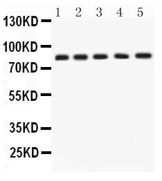
Western blot analysis of APLP1 using anti-APLP1 antibody (PB9476).
Electrophoresis was performed on a 5-20% SDS-PAGE gel at 70V (Stacking gel) / 90V (Resolving gel) for 2-3 hours. The sample well of each lane was loaded with 50ug of sample under reducing conditions.
Lane 1: Rat Brain Tissue Lysate,
Lane 2: Rat Testis Tissue Lysate,
Lane 3: SGC Whole Cell Lysate,
Lane 4: 22RV1 Whole Cell Lysate,
Lane 5: MCF-7 Whole Cell Lysate.
After Electrophoresis, proteins were transferred to a Nitrocellulose membrane at 150mA for 50-90 minutes. Blocked the membrane with 5% Non-fat Milk/ TBS for 1.5 hour at RT. The membrane was incubated with rabbit anti-APLP1 antigen affinity purified polyclonal antibody (Catalog # PB9476) at 0.5 μg/mL overnight at 4°C, then washed with TBS-0.1%Tween 3 times with 5 minutes each and probed with a goat anti-rabbit IgG-HRP secondary antibody at a dilution of 1:10000 for 1.5 hour at RT. The signal is developed using an Enhanced Chemiluminescent detection (ECL) kit (Catalog # EK1002) with Tanon 5200 system. A specific band was detected for APLP1 at approximately 85KD. The expected band size for APLP1 is at 72KD.

Click image to see more details
IHC analysis of APLP1 using anti-APLP1 antibody (PB9476).
APLP1 was detected in a paraffin-embedded section of human brain tissue. Heat mediated antigen retrieval was performed in EDTA buffer (pH 8.0, epitope retrieval solution). The tissue section was blocked with 10% goat serum. The tissue section was then incubated with 2 μg/ml rabbit anti-APLP1 Antibody (PB9476) overnight at 4°C. Peroxidase Conjugated Goat Anti-rabbit IgG was used as secondary antibody and incubated for 30 minutes at 37°C. The tissue section was developed using HRP Conjugated Rabbit IgG Super Vision Assay Kit (Catalog # SV0002) with DAB as the chromogen.

Click image to see more details
IHC analysis of APLP1 using anti-APLP1 antibody (PB9476).
APLP1 was detected in paraffin-embedded section of Mouse Brain Tissue. Heat mediated antigen retrieval was performed in citrate buffer (pH6, epitope retrieval solution) for 20 mins. The tissue section was blocked with 10% goat serum. The tissue section was then incubated with 1μg/ml rabbit anti-APLP1 Antibody (PB9476) overnight at 4°C. Biotinylated goat anti-rabbit IgG was used as secondary antibody and incubated for 30 minutes at 37°C. The tissue section was developed using Strepavidin-Biotin-Complex (SABC)(Catalog # SA1022) with DAB as the chromogen.

Click image to see more details
IHC analysis of APLP1 using anti-APLP1 antibody (PB9476).
APLP1 was detected in paraffin-embedded section of Rat Brain Tissue. Heat mediated antigen retrieval was performed in citrate buffer (pH6, epitope retrieval solution) for 20 mins. The tissue section was blocked with 10% goat serum. The tissue section was then incubated with 1μg/ml rabbit anti-APLP1 Antibody (PB9476) overnight at 4°C. Biotinylated goat anti-rabbit IgG was used as secondary antibody and incubated for 30 minutes at 37°C. The tissue section was developed using Strepavidin-Biotin-Complex (SABC)(Catalog # SA1022) with DAB as the chromogen.

Click image to see more details
Flow Cytometry analysis of SiHa cells using anti-APLP1 antibody (PB9476).
Overlay histogram showing SiHa cells stained with PB9476 (Blue line). To facilitate intracellular staining, cells were fixed with 4% paraformaldehyde and permeabilized with permeabilization buffer. The cells were blocked with 10% normal goat serum. And then incubated with rabbit anti-APLP1 Antibody (PB9476,1μg/1x106 cells) for 30 min at 20°C. DyLight®488 conjugated goat anti-rabbit IgG (BA1127, 5-10μg/1x106 cells) was used as secondary antibody for 30 minutes at 20°C. Isotype control antibody (Green line) was rabbit IgG (1μg/1x106) used under the same conditions. Unlabelled sample without incubation with primary antibody and secondary antibody (Red line) was used as a blank control.
Specific Publications For Anti-APLP1 Antibody Picoband® (PB9476)
Loading publications
Recommended Resources
Here are featured tools and databases that you might find useful.
- Boster's Pathways Library
- Protein Databases
- Bioscience Research Protocol Resources
- Data Processing & Analysis Software
- Photo Editing Software
- Scientific Literature Resources
- Research Paper Management Tools
- Molecular Biology Software
- Primer Design Tools
- Bioinformatics Tools
- Phylogenetic Tree Analysis
Customer Reviews
Have you used Anti-APLP1 Antibody Picoband®?
Share your experimental results or join a short interview to earn up to $1,000 in product credits or other rewards.
0 Reviews For Anti-APLP1 Antibody Picoband®
Customer Q&As
Have a question?
Find answers in Q&As, reviews.
Can't find your answer?
Submit your question
6 Customer Q&As for Anti-APLP1 Antibody Picoband®
Question
Is this PB9476 anti-APLP1 antibody reactive to the isotypes of APLP1?
Verified Customer
Verified customer
Asked: 2020-04-13
Answer
The immunogen of PB9476 anti-APLP1 antibody is A synthetic peptide corresponding to a sequence at the N-terminus of human APLP1 (82-112aa RRCLRDPQRVLEYCRQMYPELQIARVEQATQ), different from the related mouse sequence by three amino acids. Could you tell me which isotype you are interested in so I can help see if the immunogen is part of this isotype?
Boster Scientific Support
Answered: 2020-04-13
Question
We are currently using anti-APLP1 antibody PB9476 for mouse tissue, and we are satisfied with the Flow Cytometry results. The species of reactivity given in the datasheet says human, mouse, rat. Is it true that the antibody can work on feline tissues as well?
Verified Customer
Verified customer
Asked: 2020-03-04
Answer
The anti-APLP1 antibody (PB9476) has not been validated for cross reactivity specifically with feline tissues, though there is a good chance of cross reactivity. We have an innovator award program that if you test this antibody and show it works in feline you can get your next antibody for free. Please contact me if I can help you with anything.
Boster Scientific Support
Answered: 2020-03-04
Question
Is a blocking peptide available for product anti-APLP1 antibody (PB9476)?
Verified Customer
Verified customer
Asked: 2020-01-07
Answer
We do provide the blocking peptide for product anti-APLP1 antibody (PB9476). If you would like to place an order for it please contact support@bosterbio.com and make a special request.
Boster Scientific Support
Answered: 2020-01-07
Question
I see that the anti-APLP1 antibody PB9476 works with Flow Cytometry, what is the protocol used to produce the result images on the product page?
Verified Customer
Verified customer
Asked: 2019-05-03
Answer
You can find protocols for Flow Cytometry on the "support/technical resources" section of our navigation menu. If you have any further questions, please send an email to support@bosterbio.com
Boster Scientific Support
Answered: 2019-05-03
Question
Please see the WB image, lot number and protocol we used for cerebrospinal fluid using anti-APLP1 antibody PB9476. Please let me know if you require anything else.
Verified Customer
Verified customer
Asked: 2018-11-21
Answer
Thank you very much for the data. Our lab team are working to resolve this as quickly as possible, and we appreciate your patience and understanding! You have provided everything we needed. Please let me know if there is anything you need in the meantime.
Boster Scientific Support
Answered: 2018-11-21
Question
Will anti-APLP1 antibody PB9476 work on bovine Flow Cytometry with cerebrospinal fluid?
D. Jha
Verified customer
Asked: 2017-02-14
Answer
Our lab technicians have not tested anti-APLP1 antibody PB9476 on bovine. You can run a BLAST between bovine and the immunogen sequence of anti-APLP1 antibody PB9476 to see if they may cross-react. If the sequence homology is close, then you can perform a pilot test. Keep in mind that since we have not validated bovine samples, this use of the antibody is not covered by our guarantee. However we have an innovator award program that if you test this antibody and show it works in bovine cerebrospinal fluid in Flow Cytometry, you can get your next antibody for free.
Boster Scientific Support
Answered: 2017-02-14
