This website uses cookies to ensure you get the best experience on our website.


- Table of Contents
Plan chromogenic APOL1 IHC on paraffin sections with the IHC-validated antibody at 0.5–1 µg/mL (datasheet A01841-1). Assess plasma staining (HPA tissue IHC) and interpret cellular signal alongside APOL1 secretion (UniProt).

Expected localisation, validated protocols, controls and antibodies — the at-a-glance facts below, then the full design guide.
| Expected localisation | Plasma staining (HPA tissue IHC); secreted (UniProt) | |
| Staining pattern | Distinct positivity in plasma (HPA tissue IHC) | |
| Antigen retrieval | Citrate pH 6 HIER, heat-mediated (datasheet A01841-1) | |
| Positive control | Duodenum+1 more · see all | |
| Negative control | Adipose tissue+4 more · see all |
| Fixation | Keep fixation consistent across paraffin sections. (standard IHC practice; not target-specific) | |
| Caveat | Secreted APOL1 may contribute to plasma staining (UniProt) | |
| Regulation | Liver-enhanced RNA; staining may differ (HPA tissue RNA + IHC) | |
| Isoform / epitope | 3 isoforms; check epitopes against mature chain 28–398 (UniProt) |
The catalog antibody’s IHC-P protocol is followed by four published APOL1 chromogenic IHC protocols covering human choroid plexus, arterial sections and mouse tissues (datasheet A01841-1; PMC12236173; PMC11722280; PMC8651054; PMC9591058).
| Sample | Paraffin-embedded human liver cancer tissues; fixative not specified (datasheet A01841-1) |
| Fixation | Image fixative and duration unreported (datasheet A01841-1); verify before use. |
| Sectioning | 4–5 µm sections on charged slides (standard) |
| Deparaffinisation | Xylene, graded ethanol series to water (standard) |
| Antigen retrieval | Heat retrieval: Citrate pH 6, 20 min (datasheet A01841-1) |
| Peroxidase block | 3% H2O2, 10 min, room temperature (standard) |
| Blocking | 10% goat serum (datasheet A01841-1) |
| Primary antibody | Rabbit anti-APOL1, 0.5-1μg/ml (datasheet A01841-1) |
| Primary incubation | Overnight at 4 °C (datasheet A01841-1) |
| Detection | Streptavidin-biotin complex (SABC), DAB chromogen (datasheet A01841-1) |
| Counterstain | Hematoxylin, blue, dehydrate and mount (standard) |
| Expected result | APOL1-positive staining in enterocytes of duodenum (HPA tissue IHC: Medium). HPA tissue profile: Distinct positivity in plasma. No signal in the no-primary control. |
APOL1 is secreted and has no annotated transmembrane segment (UniProt O14791 topology). In tissue IHC, expect distinct plasma positivity and medium staining in duodenal enterocytes and testicular spermatogonia (HPA tissue IHC). HPA rates the tissue pattern Supported, while warning that secretion complicates agreement between tissue RNA and protein and that presumed off-target binding was disregarded (HPA tissue IHC).
| Distinct plasma staining, with medium staining in duodenal enterocytes or testicular spermatogonia (HPA tissue IHC). | This matches the reported IHC pattern (HPA tissue IHC). Because APOL1 is secreted and occurs in plasma HDL, extracellular signal does not by itself identify the cells that produced it (UniProt O14791 tissue specificity; HPA tissue IHC caveat). |
| Predominantly nuclear staining in an otherwise positive section. | A nuclear pattern does not fit the secreted protein annotation or HPA's membrane/secreted summary (UniProt O14791 subcellular location; HPA subcellular). Treat it as suspect and check a negative reagent control and an independently validated antibody before assigning APOL1 localisation (general IHC practice). |
| Strong cellular staining in adipocytes or bronchial respiratory epithelial cells. | Those cell types were reported as Not detected (HPA tissue IHC). Consider antibody cross-reactivity or endogenous chromogen-generating activity, especially if a negative reagent control also stains (HPA tissue IHC off-target caveat; general chromogenic IHC practice). |
| Broad, poorly defined colour across cells and surrounding tissue. | This is difficult to score against HPA's distinct plasma pattern and specified medium-positive cell populations (HPA tissue IHC). Compare with a negative reagent control, then review blocking, washing and antibody concentration as general IHC background controls (general IHC practice). |
| No staining in duodenal enterocytes, testicular spermatogonia or plasma. | The result conflicts with the reported medium cellular staining and distinct plasma positivity (HPA tissue IHC). First check whether the expected structures are present; then review reagent performance and the detection workflow before calling APOL1 absent (general IHC practice). |
| Secretion and tissue attribution (UniProt O14791; HPA tissue IHC). | APOL1 occurs in plasma on APOA-I-containing HDL, while HPA reports distinct plasma positivity (UniProt O14791 tissue specificity; HPA tissue IHC). A positive extracellular compartment cannot establish local synthesis; HPA explicitly cautions that tissue RNA and protein locations may differ (HPA tissue IHC). |
| Cell-specific reference pattern (HPA tissue IHC). | Enterocytes and spermatogonia are Medium; adipocytes and bronchial respiratory epithelial cells are Not detected (HPA tissue IHC). Use the named cell populations when judging a slide, rather than treating every cell in a named tissue as an expected positive (HPA tissue IHC). |
| Antibody validation and apparent off-target signal (HPA antibodies; HPA tissue IHC). | Two listed antibodies have Supported IHC status, and HPA says presumed off-target binding was observed and disregarded (HPA antibodies; HPA tissue IHC). Supported is a useful comparison point, but an unexpected compartment or cell population still needs a specificity check (general IHC practice). |
| Processing and sequence variation (UniProt O14791). | The annotated signal peptide spans residues 1–27, the mature chain spans 28–398, and three isoforms are listed (UniProt O14791 processing; isoforms). Epitope coverage cannot be inferred without the antibody's immunogen or epitope information (general antibody interpretation practice). |
| Limits of IF localisation evidence (HPA subcellular). | HPA summarizes APOL1 as Membrane, Secreted, but supplies no main location or ICC-IF image-bearing cell line (HPA subcellular). Treat a precise IF compartment assignment as provisional; UniProt also annotates APOL1 as secreted with no transmembrane segment (UniProt O14791 topology). |
| Situation | Likely cause | Next action |
|---|---|---|
| No signal in a section expected to contain positive cells. | The expected cells may be absent, or an IHC reagent or detection step may have failed (HPA tissue IHC; general IHC practice). | Confirm enterocytes or spermatogonia are present, then check an established positive section and each detection reagent (HPA tissue IHC; general IHC practice). |
| Plasma stains, but the intended cellular population does not. | Plasma APOL1 can be present independently of detectable local cellular staining (UniProt O14791 tissue specificity; HPA tissue IHC caveat). | Score plasma and cells separately; verify the sampled cells against HPA's Medium enterocyte or spermatogonia observations (HPA tissue IHC). |
| Strong signal appears in an HPA Not detected cell type. | Possible off-target binding or endogenous detection activity; HPA reports presumed off-target binding in its tissue assessment (HPA tissue IHC; general chromogenic IHC practice). | Run a negative reagent control and compare with an independently validated antibody; review endogenous activity blocking if the control stains (general IHC practice). |
| The whole section has diffuse chromogenic background. | Nonspecific reagent binding or incomplete washing can obscure the distinct pattern reported by HPA (general IHC practice; HPA tissue IHC). | Inspect the negative reagent control, then review blocking, washes and antibody concentration using the assay's validated instructions (general IHC practice). |
| Signal is mainly nuclear. | That compartment lacks support in the supplied APOL1 localisation records (UniProt O14791 subcellular location; HPA subcellular). | Check controls and compare another Supported IHC antibody before interpreting nuclear colour as APOL1 (HPA antibodies; general IHC practice). |
| What should an IF/ICC signal mean? | HPA lists Membrane, Secreted but gives no main subcellular location or ICC-IF image-bearing cell line (HPA subcellular). | Describe the observed IF compartment cautiously and confirm specificity with controls; do not treat membrane-only fluorescence as an established APOL1 reference pattern (HPA subcellular; general IF practice). |
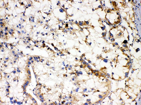
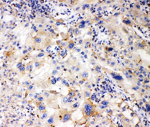
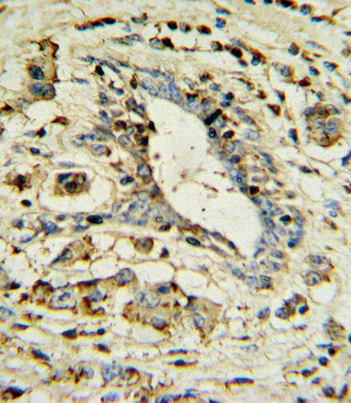


Comprehensive Human Protein Atlas IHC scoring per tissue (reliability: Supported — At least one protein variant secreted, tissue location of RNA and protein might differ and correlation is complex. Presumed off target binding observed and disregarded.). Rows are taken directly from the HPA tissue chart — click any row's HPA link to view the source.
| Tissue | Cell type | Level | Evidence | Source |
|---|---|---|---|---|
| Duodenum | Enterocytes | Medium | Protein (IHC) | HPA → |
| Testis | Spermatogonia cells | Medium | Protein (IHC) | HPA → |
| Tissue | Cell type | Level | Evidence | Source |
|---|---|---|---|---|
| Adipose tissue | Adipocytes | Not detected | Protein (IHC) | HPA → |
| Adrenal gland | Glandular cells | Not detected | Protein (IHC) | HPA → |
| Appendix | Endocrine cells | Not detected | Protein (IHC) | HPA → |
| Bone marrow | Hematopoietic cells | Not detected | Protein (IHC) | HPA → |
| Breast | Adipocytes | Not detected | Protein (IHC) | HPA → |
Troubleshoot APOL1 staining in paraffin sections by checking retrieval, detection, and compartment-specific controls before scoring chromogenic signal.
The catalog provides human APOL1 paraffin-section IHC images (catalog image captions) and a human-reactive ICC/IF antibody without an IF image (catalog applications; IF image alts).
A01841-1 has an IHC image from a paraffin-embedded human liver cancer section; its fixative is unreported (A01841-1 image caption). A01841-2 has an IHC image from formalin-fixed, paraffin-embedded human breast carcinoma (A01841-2 image caption).
Which to pick: For tissue IHC, choose A01841-1 for the documented paraffin-section preparation (A01841-1 image caption) or A01841-2 for the documented paraffin-section, paraffin-embedded preparation (A01841-2 image caption). For IF/ICC, M01841-1 is a human-reactive rabbit monoclonal, clone 19A39, with ICC/IF listed but no IF image supplied (M01841-1 catalog reactivity, host, clone, applications, IF image alts). None is documented for another species; all three list human reactivity only (catalog reactivity). The selected A01841-1 tissue-IHC caption documents paraffin sections, but does not specify the fixative (selected-SKU IHC image A01841-1).