This website uses cookies to ensure you get the best experience on our website.


- Table of Contents
This guide helps plan chromogenic ARPC5 IHC in paraffin sections using the reported cytoplasmic tissue pattern (HPA tissue IHC). Testis elongated or late spermatids provide a high-staining reference, but interpret antibody specificity cautiously because the HPA antibody may target more than one gene (HPA tissue IHC).

Expected localisation, validated protocols, controls and antibodies — the at-a-glance facts below, then the full design guide.
| Expected localisation | Cytoplasmic tissue staining (HPA tissue IHC); nuclear location annotated (UniProt) | |
| Staining pattern | Several tissue cell types show general cytoplasmic staining (HPA tissue IHC) | |
| Antigen retrieval | Tris-EDTA pH 9.0 HIER, 95–98 °C, 20 min (rule: nuclear antigen) | |
| Positive control | Testis+4 more · see all | |
| Negative control | Adipose tissue+4 more · see all |
| Fixation | Keep fixation consistent across paraffin sections. (standard IHC practice; not target-specific) | |
| Caveat | Staining may include a target from another gene (HPA tissue IHC) | |
| Regulation | No expression regulator annotated (UniProt) | |
| Isoform / epitope | 2 isoforms; epitope coverage is unspecified (UniProt) |
The catalog antibody’s IHC-P protocol is supplemented by two published ARPC5 protocols using paraffin sections (PMC9485570; PMC3584917).
| Sample | Paraffin-embedded human colon tissue; fixative not specified (datasheet M02096) |
| Fixation | Image fixative and duration unreported (datasheet M02096); verify before use. |
| Sectioning | 4–5 µm sections on charged slides (standard) |
| Deparaffinisation | Xylene, graded ethanol series to water (standard) |
| Antigen retrieval | Heat-induced epitope retrieval in Tris-EDTA buffer, pH 9.0, 20 min at 95–98 °C (standard rule: nuclear antigen) |
| Peroxidase block | 3% H2O2, 10 min, room temperature (standard) |
| Blocking | 10% normal serum of the secondary host, 30 min, room temperature (standard) |
| Primary antibody | Rabbit monoclonal (clone CCD-1) anti-ARPC5, 1:50 (datasheet M02096) |
| Primary incubation | Overnight at 4 °C (standard) |
| Detection | HRP-polymer secondary, DAB chromogen 5–10 min (standard) |
| Counterstain | Hematoxylin, blue, dehydrate and mount (standard) |
| Expected result | ARPC5-positive staining in elongated or late spermatids of testis (HPA tissue IHC: High). HPA tissue profile: General cytoplasmic expression in several different tissue types. No signal in the no-primary control. |
ARPC5 is associated with the cytoplasmic cytoskeleton, cell projections and nucleus, and has no transmembrane segment (UniProt O15511). In paraffin section IHC, expect predominantly cytoplasmic staining across several tissues, with high staining in testicular elongated or late spermatids and medium staining in selected glandular, respiratory epithelial and neuronal cells (HPA tissue IHC). Interpret specificity cautiously: HPA rates its tissue staining Approved but reports low agreement with RNA data and cautions that the antibody targets protein from more than one gene (HPA tissue IHC).
| Cytoplasmic chromogen in elongated or late spermatids; weaker staining in other documented positive cells. | This fits the strongest listed testis signal (High) and the broader cytoplasmic profile. Adrenal, appendix and breast glandular cells, bronchial respiratory epithelium, and listed neuronal cells are Medium comparators (HPA tissue IHC). |
| Predominantly crisp nuclear staining with little cytoplasmic signal, or a uniform outline of every cell. | Reassess specificity and detection: HPA describes general cytoplasmic tissue staining (HPA tissue IHC). UniProt also lists the nucleus, so nuclear signal alone cannot be dismissed as impossible; ARPC5 has no transmembrane segment, although ICC-IF reports membrane and junction staining (UniProt O15511; HPA ICC-IF). |
| Strong signal in a listed undetected cell type, such as adipocytes or lymph node germinal center cells. | That result conflicts with those cell-specific HPA observations (HPA tissue IHC). Consider antibody cross-reactivity or endogenous detection activity, then compare an omission control and tissue morphology (standard IHC practice). HPA's multi-gene targeting caution makes a positive stain insufficient for ARPC5-specific attribution (HPA tissue IHC). |
| Diffuse color across cells, stroma and empty areas, with poorly defined cell boundaries. | Treat this as background until controls support cellular staining; excess primary or detection reagent, incomplete blocking or washing, and chromogen deposits are general IHC possibilities (standard IHC practice). HPA's cell-specific intensity categories do not validate a diffuse haze (HPA tissue IHC). |
| No signal in elongated or late spermatids while the section and counterstain remain interpretable. | An absent signal in this High reference cell population warrants a run check before calling another tissue negative (HPA tissue IHC; standard IHC practice). Check the antibody's documented IHC-P conditions, detection reagents and control section; no ARPC5-specific retrieval or fixation effect is supplied (standard IHC practice). |
| Tissue and cell selection | Compare like cell populations: HPA records High elongated or late spermatids, Medium adrenal, appendix and breast glandular cells, and undetected adipocytes, among other entries. A negative call applies to the specified cell type, not every cell in its tissue (HPA tissue IHC). |
| IHC evidence and specificity | The tissue profile is Approved, while HPA reports low staining–RNA consistency and possible recognition of protein from more than one gene. HPA022013 is Approved for IHC; that status supports use of its observations but does not prove every stained structure contains ARPC5 (HPA tissue IHC; HPA antibodies). |
| Compartment and topology | UniProt lists cytoplasm, cytoskeleton, cell projection and nucleus, with no transmembrane segment (UniProt O15511). HPA's tissue IHC summary is generally cytoplasmic; use that section-level pattern as the primary visual comparator (HPA tissue IHC). |
| Isoforms and processing | UniProt lists 2 isoforms, no signal peptide or propeptide, and a chain beginning at residue 2 (UniProt O15511). Epitope coverage across isoforms is not supplied; do not infer an isoform-specific stain or a shed extracellular pool from this record (UniProt O15511). |
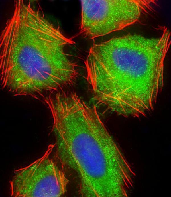
| IF/ICC: what pattern should I expect? | HPA ICC-IF reports vesicles, plasma membrane, cell junctions and cytosol, with images in A-431, U-251MG and U2OS cells (HPA ICC-IF). This is a separate imaging context: those locations do not establish an IHC-P membrane pattern, and HPA031972 has Uncertain ICC status (HPA antibodies). |
| Situation | Likely cause | Next action |
|---|---|---|
| All tissues, including testis, appear negative. | The positive reference may have failed, or the IHC detection run may have failed (HPA tissue IHC; standard IHC practice). | Inspect elongated or late spermatids on the control section, then check the documented IHC-P antibody conditions and detection reagents. Optimize retrieval against a positive control only as a general IHC step; ARPC5-specific retrieval sensitivity is unreported (HPA tissue IHC; standard IHC practice). |
| Weak signal in a tissue expected to be medium. | HPA's Medium designation is cell-specific, and weak contrast can also reflect a general staining-run problem (HPA tissue IHC; standard IHC practice). | Identify the relevant glandular, respiratory epithelial or neuronal population before scoring; compare a same-run testis control and review antibody concentration, detection and counterstain using documented IHC-P conditions (HPA tissue IHC; standard IHC practice). |
| Strong color appears in listed undetected cells. | Non-specific binding or endogenous detection activity may contribute; HPA also cautions about multi-gene targeting (standard IHC practice; HPA tissue IHC). | Verify the cell identity, review primary-omission and detection controls, and apply appropriate endogenous activity blocking for the detection chemistry. Do not assign the stain to ARPC5 solely from color intensity (standard IHC practice; HPA tissue IHC). |
| Brown haze or deposits obscure cellular boundaries. | Background binding, inadequate washing or chromogen precipitation are general IHC causes (standard IHC practice). | Compare the primary-omission control; review blocking, wash steps, reagent concentration and chromogen development. Score only localized cellular signal after the background is resolved (standard IHC practice). |
| Signal is mainly nuclear or outlines cell borders. | A mismatch with HPA's general cytoplasmic IHC profile needs review; UniProt permits nuclear localization, and HPA ICC-IF reports membrane and junction locations in cells (HPA tissue IHC; UniProt O15511; HPA ICC-IF). | Confirm morphology and compartment against controls and the cytoplasmic positive reference. Treat the ICC-IF pattern as context, not proof of the same paraffin-section distribution (standard IHC practice; HPA tissue IHC; HPA ICC-IF). |
| A weak or absent result is being called an ARPC5-negative tissue. | HPA reports low tissue RNA specificity and lists staining by specific cell population; its IHC reliability note also limits certainty (HPA tissue IHC). | Record the tissue, cell type, compartment and intensity, then compare a same-run positive reference. Report an undetected stain as an assay observation rather than proof that the tissue lacks ARPC5 (HPA tissue IHC; standard IHC practice). |



Comprehensive Human Protein Atlas IHC scoring per tissue (reliability: Approved — Low consistency between antibody staining and RNA expression data. Caution, targets protein from more than one gene.). Rows are taken directly from the HPA tissue chart — click any row's HPA link to view the source.
| Tissue | Cell type | Level | Evidence | Source |
|---|---|---|---|---|
| Testis | Elongated or late spermatids | High | Protein (IHC) | HPA → |
| Adrenal gland | Glandular cells | Medium | Protein (IHC) | HPA → |
| Appendix | Glandular cells | Medium | Protein (IHC) | HPA → |
| Breast | Glandular cells | Medium | Protein (IHC) | HPA → |
| Bronchus | Respiratory epithelial cells | Medium | Protein (IHC) | HPA → |
| Tissue | Cell type | Level | Evidence | Source |
|---|---|---|---|---|
| Adipose tissue | Adipocytes | Not detected | Protein (IHC) | HPA → |
| Cervix | Glandular cells | Not detected | Protein (IHC) | HPA → |
| Esophagus | Squamous epithelial cells | Not detected | Protein (IHC) | HPA → |
| Lymph node | Germinal center cells | Not detected | Protein (IHC) | HPA → |
| Oral mucosa | Squamous epithelial cells | Not detected | Protein (IHC) | HPA → |
Troubleshoot ARPC5 staining in paraffin section chromogenic IHC by checking retrieval, compartment, specificity and cell level scoring before interpreting intensity.
The catalog includes ARPC5 antibodies for paraffin-section IHC and cellular IF, with images from human colon and U-2 OS cells, respectively (M02096 IHC caption; M02096-1 IF caption).
M02096 has an IHC image from paraffin-embedded human colon; its listed reactivity is human, mouse and rat (M02096 IHC caption; catalog applications/reactivity). M02096-1 has an IF image from paraformaldehyde-fixed, permeabilized human U-2 OS cells; its listed reactivity is human, mouse and rat (M02096-1 IF caption; catalog applications/reactivity).
Which to pick: Choose monoclonal M02096 for tissue IHC: its image documents human paraffin sections, but does not report the fixative (M02096 IHC caption; catalog clone CCD-1). Choose M02096-1 for cellular IF because its image documents that application; its catalog lists IF, but not ICC (M02096-1 IF caption; catalog applications). For work across human, mouse and rat, M02096 lists IHC and IF and all three species, although its IHC image documents human tissue only (M02096 catalog applications/reactivity; M02096 IHC caption).