This website uses cookies to ensure you get the best experience on our website.


- Table of Contents
Plan BCL10 chromogenic IHC around the cytoplasmic staining seen in most cell types (HPA tissue IHC). This guide covers fixation, retrieval, detection and interpretation, including the possibility of nuclear staining in some tumor cells (UniProt).

Expected localisation, validated protocols, controls and antibodies — the at-a-glance facts below, then the full design guide.
| Expected localisation | Cytoplasmic staining in most cell types (HPA tissue IHC) | |
| Staining pattern | Cytoplasmic signal in most cell types (HPA tissue IHC) | |
| Antigen retrieval | EDTA pH 8.0 HIER, heat-mediated (datasheet PB9525) | |
| Positive control | Adrenal gland+4 more · see all | |
| Negative control | Adipose tissue+4 more · see all |
| Fixation | Keep fixation consistent across sections. (standard IHC practice; not target-specific) | |
| Caveat | Some tumor cells may show nuclear BCL10 (UniProt) | |
| Regulation | Ubiquitous tissue expression (UniProt) | |
| Isoform / epitope | No annotated isoforms; one 1–233 chain (UniProt) |
Start with the catalog antibody’s EDTA pH 8.0 protocol (datasheet: PB9525); the four published IHC protocols below provide tissue specific comparisons (PMC5662601; PMC8375138; PMC12421401; PMC9312497).
| Sample | Paraffin-embedded human lymph node carcinoma tissue; fixative not specified (datasheet PB9525) |
| Fixation | Image fixative and duration unreported (datasheet PB9525); verify before use. |
| Sectioning | 4–5 µm sections on charged slides (standard) |
| Deparaffinisation | Xylene, graded ethanol series to water (standard) |
| Antigen retrieval | Heat retrieval: EDTA pH 8.0 (datasheet PB9525); 20 min, 95–100 °C (standard) |
| Peroxidase block | 3% H2O2, 10 min, room temperature (standard) |
| Blocking | 10% goat serum (datasheet PB9525) |
| Primary antibody | Rabbit anti-BCL10, 2-5 μg/ml (datasheet PB9525) |
| Primary incubation | Overnight at 4 °C (datasheet PB9525) |
| Detection | HRP-conjugated secondary, DAB chromogen (datasheet PB9525) |
| Counterstain | Hematoxylin, blue, dehydrate and mount (standard) |
| Expected result | BCL10-positive staining in glandular cells of adrenal gland (HPA tissue IHC: Medium). HPA tissue profile: Cytoplasmic expression in most cell types. No signal in the no-primary control. |
BCL10 should stain mainly in the cytoplasm, sometimes with a compact perinuclear pattern; it has no transmembrane segment (UniProt O95999, subcellular location and topology). HPA reports cytoplasmic expression in most cell types and medium staining in selected glandular and epithelial cells (HPA tissue IHC). Its tissue IHC reliability is Approved, with medium consistency between staining and RNA expression (HPA tissue IHC).
| Cytoplasmic staining in appendix glandular cells or bronchial respiratory epithelial cells, with identifiable cell borders. | This matches the reported medium staining in those cells (HPA tissue IHC) and the principal cytoplasmic location (UniProt O95999). Score staining in the named cells; surrounding unstained cells do not invalidate a positive result (general IHC practice). |
| Predominantly nuclear staining in ordinary tissue, with little or no cytoplasmic staining. | Review as a possible localisation artefact because the expected tissue pattern is cytoplasmic (HPA tissue IHC). Nuclear signal alone is not conclusive: UniProt reports BCL10 in nuclei of several tumor cell types, and HPA reports an additional nucleoplasmic ICC-IF location (UniProt O95999; HPA subcellular). |
| Strong staining in adipocytes or cardiomyocytes while the expected positive cells are weak. | HPA lists adipocytes in adipose tissue and cardiomyocytes in heart muscle as not detected (HPA tissue IHC). Treat discordant staining as possible cross-reactivity or endogenous detection activity (general IHC practice); first check whether the signal persists when primary antibody is omitted. |
| Uniform chromogen haze across cells, extracellular areas and the slide background. | This distribution does not resemble the reported cell-associated cytoplasmic pattern (HPA tissue IHC). Consider nonspecific reagent binding, inadequate washing or endogenous detection activity (general IHC practice); a stained region alone does not establish BCL10 localisation. |
| No staining in appendix glandular cells on a run intended to include a positive control. | Those cells have reported medium staining (HPA tissue IHC). An absent control signal makes a negative study section difficult to interpret (general IHC practice). Check section integrity and the assay steps before calling the study specimen BCL10-negative. |
| Cell and compartment choice | Use a named HPA-positive cell population when judging localisation: appendix glandular cells and bronchial respiratory epithelial cells are medium, whereas adipocytes in adipose tissue are not detected (HPA tissue IHC). The tissue label alone is less precise than the cell label. |
| Perinuclear pattern and assembly | A compact or filamentous perinuclear appearance is described for BCL10 (UniProt O95999, subcellular location). UniProt also describes CARD-dependent BCL10 oligomerization (UniProt O95999, subunit); morphology alone cannot establish pathway activation (general IHC interpretation). |
| Antibody evidence | HPA lists IHC as Approved for HPA017925 and CAB001944; it lists ICC as Supported for HPA018953 (HPA antibodies). These are application-specific statuses, and the supplied record does not establish that an ICC-tested antibody performs in paraffin IHC. |
| Strength of tissue evidence | HPA describes the tissue IHC profile as cytoplasmic in most cell types and rates its agreement with RNA data as medium (HPA tissue IHC). Use the reported levels as comparison points, not absolute thresholds for every specimen (general IHC interpretation). |
| Detection system | Chromogenic signal can arise from endogenous tissue activity or nonspecific reagent binding (general IHC practice). Primary-omission and detection-only controls help separate those signals from antibody-dependent staining; neither control establishes molecular specificity by itself (general IHC practice). |
| Situation | Likely cause | Next action |
|---|---|---|
| Positive-control tissue is blank. | A missed assay step, weak detection or unsuitable control section can produce no signal (general IHC practice). | Repeat with a documented positive cell population, such as appendix glandular cells with reported medium staining (HPA tissue IHC). Verify retrieval and detection steps against the IHC-validated antibody's instructions (general IHC practice). |
| Positive cells show diffuse nuclear signal without the expected cytoplasm. | The distribution differs from the principal tissue pattern (HPA tissue IHC), though nuclear localisation has been reported in some tumor cells (UniProt O95999). | Compare cytoplasmic and nuclear signal in a known-positive control, inspect morphology and run a primary-omission control (general IHC practice). Record a genuine nuclear component separately instead of forcing a cytoplasmic score. |
| Adipocytes stain strongly. | Adipocytes in adipose tissue are listed as not detected (HPA tissue IHC); cross-reactivity or endogenous detection activity is possible (general IHC practice). | Check a primary-omission control and the detection system, then compare staining in a named HPA-positive population (general IHC practice; HPA tissue IHC). Do not use adipocyte colour alone as evidence of BCL10 expression. |
| Background obscures cell boundaries. | Nonspecific binding, residual detection reagents or inadequate washing can obscure a cell-associated pattern (general IHC practice). | Inspect an unstained section and a primary-omission control; review blocking, washing and chromogen development for the chosen detection system (general IHC practice). Score only where cell boundaries and cytoplasmic signal remain interpretable. |
| A low-staining cell population appears negative. | HPA reports low staining in cerebral cortex neuronal cells and pancreatic exocrine glandular cells (HPA tissue IHC). Weak signal may be difficult to distinguish from assay background (general IHC practice). | Interpret alongside a reported medium positive cell population and a negative control (HPA tissue IHC; general IHC practice). Avoid declaring assay failure from a low-staining population alone. |
| IF/ICC Q: Should nucleoplasmic fluorescence replace the IHC expectation? | HPA reports cytosol as the main ICC-IF location and nucleoplasm as an additional location (HPA subcellular); its tissue IHC profile is cytoplasmic (HPA tissue IHC). | A: Interpret IF/ICC on its own guide page and record both compartments when visible (HPA subcellular). For paraffin IHC, judge the tissue pattern against the cytoplasmic IHC evidence (HPA tissue IHC). |

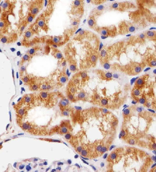
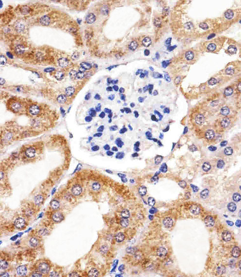

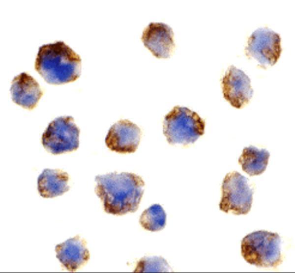
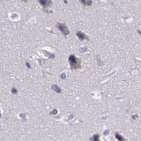

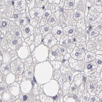


Comprehensive Human Protein Atlas IHC scoring per tissue (reliability: Approved — Medium consistency between antibody staining and RNA expression data.). Rows are taken directly from the HPA tissue chart — click any row's HPA link to view the source.
| Tissue | Cell type | Level | Evidence | Source |
|---|---|---|---|---|
| Adrenal gland | Glandular cells | Medium | Protein (IHC) | HPA → |
| Appendix | Glandular cells | Medium | Protein (IHC) | HPA → |
| Bronchus | Respiratory epithelial cells | Medium | Protein (IHC) | HPA → |
| Cervix | Glandular cells | Medium | Protein (IHC) | HPA → |
| Colon | Endothelial cells | Medium | Protein (IHC) | HPA → |
| Tissue | Cell type | Level | Evidence | Source |
|---|---|---|---|---|
| Adipose tissue | Adipocytes | Not detected | Protein (IHC) | HPA → |
| Bone marrow | Hematopoietic cells | Not detected | Protein (IHC) | HPA → |
| Breast | Adipocytes | Not detected | Protein (IHC) | HPA → |
| Caudate | Glial cells | Not detected | Protein (IHC) | HPA → |
| Cerebellum | Cells in granular layer | Not detected | Protein (IHC) | HPA → |
Troubleshoot BCL10 staining in paraffin sections by checking retrieval, compartment, controls and cell-specific scoring before interpreting chromogenic signal.
Anti-BCL10 antibodies have IHC images from human paraffin tissue sections (PB9525, M01616, M01616-1: image captions) and IF/ICC images from Raji cells (A01616: image captions).
PB9525 shows human lymph node carcinoma IHC, while M01616 shows human kidney IHC (PB9525, M01616: image captions). M01616-1 shows human breast cancer IHC, and A01616 shows Raji cell ICC and IF (M01616-1, A01616: image captions).
Which to pick: For paraffin section IHC, choose PB9525 for its documented EDTA pH 8.0 retrieval and 2 μg/ml primary incubation (PB9525: image caption), or M01616 for its 1:25 human kidney example (M01616: image caption); M01616-1 also has a paraffin section image from human breast cancer (M01616-1: image caption). For IF/ICC, choose A01616 because it has Raji cell ICC and IF images; it also has the broadest listed species reactivity—human, mouse and rat (A01616: image captions and catalog applications/reactivity). The paraffin captions do not report a fixative (PB9525, M01616, M01616-1: image captions).