CASP1 / Caspase-1 · IHC design guide

Design Immunohistochemistry for CASP1

Plan chromogenic CASP1 IHC on paraffin sections using lung macrophages or small-intestinal glandular cells as positive references (HPA tissue IHC). Interpret predominantly cytoplasmic staining (HPA tissue IHC) with attention to CASP1 cleavage and the antibody’s epitope region (UniProt).

Evidence assembled Oct 2026 · For research use; verify linked source records and product datasheet before use
Immunohistochemistry protocol sheet for CASP1 (IHC for CASP1): expected localisation Predominantly cytoplasmic; occasional nuclear staining (HPA tissue IHC), antibody M00048-3, validated IHC image, and IHC protocol steps
Printable CASP1 IHC protocol sheet — expected localisation Predominantly cytoplasmic; occasional nuclear staining (HPA tissue IHC), antibody M00048-3, controls and protocol steps. Open the full CASP1 IHC guide →

CASP1 Immunohistochemistry Experimental Design Guide

Expected localisation, validated protocols, controls and antibodies — the at-a-glance facts below, then the full design guide.

Must know before staining
Expected localisation Predominantly cytoplasmic; occasional nuclear staining (HPA tissue IHC)
Staining pattern Lung macrophages and intestinal glands: cytoplasmic, sometimes nuclear (HPA tissue IHC)
Antigen retrieval Citrate pH 6.0 HIER, 95–98 °C, 20 min (rule: cytoplasmic / membrane antigen)
Positive control ⓘ Duodenum+4 more · see all
Negative control ⓘ Adipose tissue+4 more · see all
Important caveats
Reasons your staining may differ from the expected pattern.
Fixation Keep formalin fixation consistent across sections (standard IHC practice; not target-specific); Matched tissue-IHC evidence does not establish the fixation claim. Validate the specimen-specific method before use. (selected-SKU IHC image M00048-3)
Caveat Activation cleaves CASP1, so epitope position may affect staining (UniProt)
Regulation Inflammasomes activate CASP1 (UniProt)
Isoform / epitope 5 isoforms; cleavage yields p20 and p10, so epitope region matters (UniProt)
Section 1

Recommended CASP1 IHC & IF Protocols

Compare the catalog antibody’s IHC-P protocol with four published CASP1 IHC methods (PMC9174061; PMC5503597; PMC7202523; PMC9663582).

Recommended immunohistochemistry (IHC-P) protocol parameters
SampleTissue sections; selected-image fixative not specified (standard IHC workflow)
FixationImage fixative and duration unreported (datasheet M00048-3); verify before use.
Sectioning4–5 µm sections on charged slides (standard)
DeparaffinisationXylene, graded ethanol series to water (standard)
Antigen retrievalHeat-induced epitope retrieval in citrate buffer, pH 6.0, 20 min at 95–98 °C (standard rule: cytoplasmic / membrane antigen)
Peroxidase block3% H2O2, 10 min, room temperature (standard)
Blocking10% normal serum of the secondary host, 30 min, room temperature (standard)
Primary antibodyRabbit anti-CASP1, 1:200-1:1000 (datasheet M00048-3)
Primary incubationOvernight at 4 °C (standard)
DetectionHRP-polymer secondary, DAB chromogen 5–10 min (standard)
CounterstainHematoxylin, blue, dehydrate and mount (standard)
Expected resultCASP1-positive staining in glandular cells of duodenum (HPA tissue IHC: High). HPA tissue profile: Ubiquitous cytoplasmic expression with varying intensity that in cases was combined with nuclear expression. No signal in the no-primary control.
💡Decision noteStart with citrate pH 6.0 retrieval at 95–98 °C for 20 min (page retrieval rule); compare the published steamer method if needed (PMC7202523).
Section 2

What Is the Expected CASP1 Staining Pattern?

CASP1 stains cytoplasm in lung macrophages and gut glands (HPA: Approved, medium consistency; UniProt: no TM).

What am I looking at on my slide?
Cytoplasm stains in lung macrophages (HPA: High).Matches the observed tissue pattern (HPA: High in lung macrophages).
Nuclei stain without cytoplasm (HPA: cytoplasm predominates).Atypical; assess for artifact (HPA tissue IHC; standard IHC practice).
Adipocytes stain strongly (HPA: Not detected).Consider cross-reactivity or endogenous enzyme signal (HPA tissue IHC; standard IHC practice).
Uniform brown haze obscures cells (standard IHC practice).Likely background rather than cell-linked staining (HPA tissue IHC; standard IHC practice).
Lung macrophages show no signal (HPA: High).Unexpected in a positive tissue; check assay performance (HPA tissue IHC; standard IHC practice).
💡Expected CASP1 appearanceStrong macrophage cytoplasm is positive (HPA: lung High); uniform haze suggests background (standard IHC practice).
How each factor affects the staining
Tissue choice (HPA tissue IHC)Lung macrophages and gut glands stain High; adipocytes are not detected (HPA tissue IHC).
Processing (UniProt P29466)CASP1 yields p20/p10; staining alone cannot identify the detected form without epitope data (UniProt P29466).
Isoforms (UniProt P29466)Five isoforms are listed; antibody coverage requires epitope data (UniProt P29466).
Why is my staining missing, weak or wrong?
SituationLikely causeNext action
No stain in lung macrophages (HPA: High).Assay sensitivity may be low (standard IHC practice).Check positive control, retrieval and antibody dilution (standard IHC practice).
Diffuse brown haze (standard IHC practice).Nonspecific binding or endogenous enzyme activity (standard IHC practice).Review blocking, washes and detection controls (standard IHC practice).
Nuclear-only signal (HPA: cytoplasm predominates).Possible artifact; some nuclear staining occurs (HPA tissue IHC).Compare a cytoplasmic positive control and no-primary control (standard IHC practice).
Adipocytes stain (HPA: Not detected).Possible cross-reactivity or endogenous activity (HPA tissue IHC; standard IHC practice).Check matched negative tissue and no-primary control (standard IHC practice).
Patchy stain across a section (standard IHC practice).Uneven retrieval or reagent coverage (standard IHC practice).Standardize retrieval and reagent coverage (standard IHC practice).
IF/ICC shows nuclear signal (HPA ICC-IF).Nucleoplasm is uncertain; cytosol is supported (HPA ICC-IF).Use cytosol as the localization reference in the separate IF/ICC guide (HPA ICC-IF).

Sample controls for CASP1 IHC & IF

🧪Run lung first and score macrophages for CASP1 staining (HPA: High in lung macrophages). Use caudate glial cells as a negative tissue control (HPA: Not detected in caudate glial cells); on the lung slide, compare macrophages with nearby non-macrophage cells for background staining rather than assuming every other cell is CASP1-negative (HPA: High in lung macrophages).
Positive control tissue: Duodenum (Glandular cells, HPA High)
Negative control tissue: Adipose tissue (HPA Not detected)
ICC-IF cell lines (HPA subcellular resource): HPA ICC-IF images show CASP1 in A-431, U-251MG, U2OS, with annotated localisation: Cytosol (supported) (HPA subcellular).
Technical controls: Include a no-primary, secondary-only control and a concentration-matched isotype control matched to the primary antibody’s host and clonality; the captioned antibody is rabbit-derived (caption: rabbit antibody). Process CASP1-knockout material alongside the sections as a biological negative, and check lung macrophages for endogenous peroxidase activity or pigment that could mimic chromogenic signal (HPA: High in lung macrophages; standard IHC practice).
⚠️Feasibility: A target-specific fixation window or fixation effect is unreported, and the selected M00048-3 tissue-IHC caption does not state its fixative (caption: fixative unreported). Retrieval dependency is unreported; optimize antigen retrieval empirically for the IHC-validated antibody (standard IHC practice). The supplied evidence does not establish that frozen sections or IF are easier than paraffin IHC; in lung, macrophage pigment can complicate chromogenic scoring (HPA: High in lung macrophages; standard IHC practice).

HPA tissue IHC evidence for CASP1

Comprehensive Human Protein Atlas IHC scoring per tissue (reliability: Approved — Medium consistency between antibody staining and RNA expression data.). Rows are taken directly from the HPA tissue chart — click any row's HPA link to view the source.

Positive expression · recommended positive controls

TissueCell typeLevelEvidenceSource
Duodenum Glandular cells High Protein (IHC) HPA →
Gallbladder Glandular cells High Protein (IHC) HPA →
Lung Macrophages High Protein (IHC) HPA →
Rectum Glandular cells High Protein (IHC) HPA →
Small intestine Glandular cells High Protein (IHC) HPA →

Undetected expression · recommended negative controls

TissueCell typeLevelEvidenceSource
Adipose tissue Adipocytes Not detected Protein (IHC) HPA →
Adrenal gland Glandular cells Not detected Protein (IHC) HPA →
Breast Adipocytes Not detected Protein (IHC) HPA →
Caudate Glial cells Not detected Protein (IHC) HPA →
Hippocampus Glial cells Not detected Protein (IHC) HPA →
Section 3

Advanced CASP1 IHC Tips

Troubleshoot CASP1 staining in paraffin sections by checking retrieval, compartment, cell type, controls, and whether the signal reflects protein abundance or activation.

What should I change when CASP1 staining is weak after antigen retrieval?
Use citrate pH 6.0 HIER at 95–98 °C for 20 min for paraffin-section CASP1 IHC (page retrieval rule: cytoplasmic / membrane antigen). Compare sections processed with the same cooling, washing, antibody incubation, and chromogen development before changing retrieval, because those steps also affect signal (standard IHC practice). Check a section containing an expected positive cell population, such as lung macrophages, alongside the weak specimen (HPA: High in lung macrophages). If staining remains weak, vary retrieval time on matched sections while keeping the citrate buffer and pH 6.0 fixed, and watch for tissue damage or rising background (standard IHC practice).
Could fixation explain weak or uneven CASP1 staining?
CASP1-specific sensitivity to fixation is unknown from the supplied evidence, so do not assign weak staining to a particular fixative or fixation duration (supplied CASP1 evidence: no target-specific fixation comparison). The selected product image shows stained human colon carcinoma but does not state its fixative (caption: M00048-3). Record each specimen’s fixative and processing history, then compare sections with matched retrieval, antibody incubation, and detection conditions (standard IHC practice). Include an expected positive cell population and a no-primary control in the same run; persistent differences between processing batches can guide a controlled fixation study without proving a CASP1-specific mechanism (standard IHC practice; HPA: High in lung macrophages).
Where should convincing CASP1 staining appear in tissue sections?
Assess cytoplasmic staining first, since CASP1 is annotated in the cytoplasm and at the cell membrane, while HPA reports mainly cytosolic localisation (UniProt P29466: subcellular location; HPA: cytosol supported). HPA tissue IHC describes cytoplasmic expression that sometimes accompanies nuclear staining, so isolated nuclear signal warrants closer scrutiny (HPA: tissue IHC profile). Compare the pattern with morphology and cell identity: lung macrophages and intestinal glandular cells are documented high-staining populations (HPA: High in lung macrophages, duodenal and small-intestinal glandular cells). Use a no-primary section and inspect section edges before accepting diffuse staining across compartments as specific (standard IHC practice).
Can this stain distinguish pro-CASP1, processed CASP1, or splice isoforms?
Do not infer the detected molecular form from chromogenic staining alone; the supplied evidence does not map the catalog antibody’s epitope (supplied antibody evidence: epitope unspecified). CASP1 has 5 listed isoforms, and its precursor is processed into p20 and p10 chains, so epitope location matters when interpreting what the antibody could detect (UniProt P29466: isoforms and processing). Request epitope or immunogen information before claiming recognition of the propeptide, a processed chain, or a particular isoform (standard antibody validation practice; UniProt P29466: processing). Interpret positive IHC as CASP1 immunoreactivity unless form-specific validation is available, and keep that distinction explicit in figure labels (standard IHC interpretation practice).
How can I cross-check CASP1 localisation by multiplex IF?
On the separate IF/ICC guide, pair CASP1 with a validated macrophage marker when examining lung macrophages, an expected high-staining population (HPA: High in lung macrophages; standard IF practice). Choose fluorophores after examining unstained tissue for autofluorescence, and place the weaker signal in a cleaner detection channel (standard IF practice). CASP1 is mainly cytosolic, so permeabilisation must allow antibody access to intracellular epitopes; optimise its strength against morphology and the marker signal (HPA: cytosol supported; standard IF practice). Include single-stain and no-primary controls to check spectral spillover and background, and assess cell-level overlap rather than assuming adjacent signals arise in the same cell (standard IF practice).
How do I reduce diffuse or misleading CASP1 chromogenic background?
Run a no-primary control and inspect pigment, folds, section edges, and necrotic regions before changing the CASP1 antibody conditions (standard IHC practice). For peroxidase-based chromogenic detection, block endogenous peroxidase and compare background after the same DAB development time across sections (standard IHC practice). Titrate primary antibody and detection reagent on matched sections, using consistent washes and a tissue containing an expected positive population to preserve interpretable signal (standard IHC practice; HPA: High in lung macrophages). Because HPA describes widespread cytoplasmic staining with variable intensity, require a reproducible cell-associated pattern above control background rather than treating every brown area as positive (HPA: tissue IHC profile; standard IHC practice).
What should I score when comparing CASP1 across specimens? ⚠ ANSWER MARKED FOR VERIFICATION
Define the cell population and compartment before scoring, because CASP1 staining varies by tissue and is predominantly cytoplasmic (HPA: tissue IHC profile). For a defined population, report the percentage of positive cells or an H-score using preset intensity categories; for discrete positive cells, report density per mm² of evaluable tissue (standard IHC quantification practice). Normalise counts to the number of eligible cells or evaluable area, and exclude folds, necrosis, and empty spaces consistently (standard IHC quantification practice). Apply identical thresholds and chromogen development conditions across specimens, and report macrophages separately from glandular cells when both are present (standard IHC practice; HPA: High in lung macrophages and intestinal glandular cells).
How can I tell true CASP1 signal from artefact or activation?
Favor reproducible, cell-associated cytoplasmic staining in an expected population over isolated signal at section edges or in necrotic material (HPA: tissue IHC profile and High in lung macrophages; standard IHC practice). Investigate purely nuclear staining because HPA reports nucleoplasmic localisation as uncertain, even though some tissue samples show combined nuclear and cytoplasmic staining (HPA: subcellular summary and tissue IHC profile). Compare suspicious brown deposits with a no-primary control and the endogenous peroxidase block to identify detection-related artefact (standard IHC practice). CASP1 immunoreactivity alone does not establish inflammasome activation or cleavage of IL1B, IL18, or GSDMD; those claims require separate functional evidence (UniProt P29466: function and processing).
Boster reagents

Best CASP1 / Caspase-1 IHC Antibodies

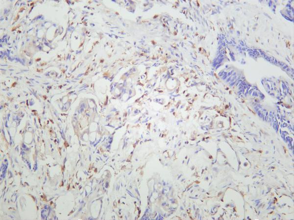
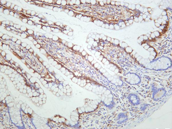
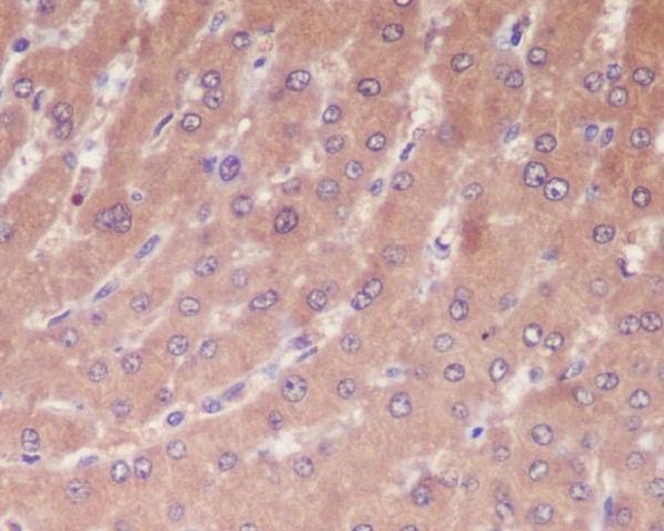
The catalog shows real IHC images for human colon carcinoma, liver, and mammary cancer tissue; one antibody also lists IF and human, mouse, and rat reactivity (catalog image captions; applications/reactivity).

Real IHC data Human colon carcinoma was stained with anti-Caspase-1 rabbit antibody
Anti-Caspase-1 Rabbit Monoclonal Antibody
Cat # M00048-3
Real IHC data Immunohistochemical analysis of paraffin-embedded human liver, using Caspase-1 Antibody.
Anti-Caspase-1 CASP1 Rabbit Monoclonal Antibody
Cat # M00048
Real IHC data Anti-Caspase-1(P20) antibody, PA1440, IHC(P) IHC(P): Human Mammary Cancer Tissue
Anti-Caspase-1(P20)/CASP1 Antibody ®
Cat # PA1440
Real IHC data Anti-Caspase-1(P10) antibody, PA1522, IHC(P) IHC(P): Human Mammary Cancer Tissue
Anti-Caspase-1(P10)/CASP1 Antibody ®
Cat # PA1522

M00048-3 has a human colon carcinoma IHC image and lists IF plus human, mouse, and rat reactivity; M00048 has a paraffin-embedded human liver IHC image and lists human and rat reactivity (catalog image captions; applications/reactivity). PA1440 and PA1522 each have a human mammary cancer IHC-P image and list human reactivity (catalog image captions; reactivity).

Which to pick: For tissue IHC, choose M00048 for the pictured paraffin-embedded human liver sample; its fixative is unreported (M00048 image caption). For IF/ICC planning, choose M00048-3 because IF is listed, although no IF image or ICC validation is supplied (M00048-3 applications; image captions). M00048-3 is also the cross-species choice because it is a rabbit monoclonal listing human, mouse, and rat reactivity; its IHC image shows human colon carcinoma without reporting section processing or fixative (M00048-3 catalog entry; image caption).

Each figure is that product's own IHC / IF validation image from its datasheet.

References

  1. UniProt Consortium. UniProt entry P29466 (CASP1_HUMAN, Caspase-1).
  2. Human Protein Atlas. CASP1 tissue IHC expression (reliability: Approved).
  3. Human Protein Atlas. CASP1 subcellular location (ICC-IF): Mainly localized to the cytosol. In addition localized to the nucleoplasm..
  4. Human Protein Atlas. CASP1 antibody validation summary (2 antibodies).
  5. Comprehensive Analysis of Prognostic Value and Immune Infiltration of NLRC4 and CASP1 in Colorectal Cancer. International journal of general medicine 2022 — PMC9174061.
  6. Cytochrome P450 1B1 inhibition suppresses tumorigenicity of prostate cancer via caspase-1 activation. Oncotarget 2017 — PMC5503597.
  7. Protective effects of autophagy and NFE2L2 on reactive oxygen species-induced pyroptosis of human nucleus pulposus cells. Aging 2020 — PMC7202523.
  8. Significance of elevated serum and hepatic NOD-like receptor pyrin domain containing 3 (NLRP3) in hepatitis C virus-related liver disease. Scientific reports 2022 — PMC9663582.
  9. PubMed PMID:1574116 — UniProt-cited evidence.
  10. PubMed PMID:1373520 — UniProt-cited evidence.
  11. PubMed PMID:7876192 — UniProt-cited evidence.